Burn Injuries

Published on 25/03/2015 by admin

Filed under Pediatrics

Last modified 22/04/2025

Print this page

rate 1 star rate 2 star rate 3 star rate 4 star rate 5 star
Your rating: none, Average: 0 (0 votes)

This article have been viewed 4135 times

Chapter 68 Burn Injuries

Burns are a leading cause of unintentional death in children, second only to motor vehicle crashes. There has been a decline in the incidence of burn injury requiring medical care over the last decade. This decline has coincided with a stronger focus on burn treatment and prevention, increased fire and burn prevention education, greater availability of regional treatment centers, widespread use of smoke detectors, greater regulation of consumer products and occupational safety, and societal changes such as reductions in smoking and alcohol abuse.

Epidemiology

Approximately 1.2 million people in the USA require medical care for burn injuries each year, with 51,000 requiring hospitalization. Approximately 30-40% of these patients are younger than 15 yr, with an average age of 32 mo. Fires are a major cause of mortality in children, accounting for up to 34% of fatal injuries in those younger than 16 yr. Scald burns account for 85% of total injuries and are most prevalent in children younger than 4 yr. Although the incidence of hot water scalding has been reduced by legislation requiring new water heaters to be preset at 120°F, scald injury remains the leading cause of hospitalization for burns. Steam inhalation used as a home remedy to treat respiratory infections is another potential cause of burns. Flame burns account for 13%; the remaining are electrical and chemical burns. Clothing ignition events have declined since passage of the Federal Flammable Fabric Act requiring sleepwear to be flame-retardant; however, the U.S. Consumer Product Safety Commission has voted to relax the existing children’s sleepwear flammability standard. Approximately 18% of burns are the result of child abuse (usually scalds), making it important to assess the pattern and site of injury and their consistency with the patient history (Chapter 37). Friction burns from treadmills are also a problem. Hands are the most commonly injured sites, with deep 2nd-degree friction injury sometimes associated with fractures of the fingers. Anoxia, not the actual burn, is a major cause of morbidity and mortality in house fires.

Review of the history usually shows a common pattern: scald burns to the side of the face, neck, and arm if liquid is pulled from a table or stove; burns in the pant leg area if clothing ignites; burns in a splash pattern from cooking; and burns on the palm of the hand from contact with a hot stove. However, “glove or stocking” burns of the hands and feet, single-area deep burns on the trunk, buttocks, or back, and small, full-thickness burns (cigarette burns) in young children should raise the suspicion of child abuse (Chapter 37).

Burn care involves a range of activities: prevention, acute care and resuscitation, wound management, pain relief, reconstruction, rehabilitation, and psychosocial adjustment. Children with massive burns require early and appropriate psychologic and social support as well as resuscitation. Surgical debridement, wound closure, and rehabilitative efforts should be instituted concurrently to promote optimal rehabilitation. Aggressive surgical removal of devitalized tissue, infection control, and judicious use of antibiotics, as well as early nutrition and cautious use of intubation and mechanical ventilation, are necessary to maximize survival. Children who have sustained burn injuries differ in appearance from their peers, necessitating supportive efforts for reentry to school and social and sporting activities.

Prevention

The aim of burn prevention is a continuing reduction in the number of serious burn injuries (Table 68-1). Effective first aid and triage can decrease both the extent (area) and the severity (depth) of injuries. The use of flame-retardant clothing and smoke detectors, control of hot water temperature (thermostat settings) within buildings, and prohibition of cigarette smoking have been partially successful in reducing the incidence of burn injuries. Treatment of children with significant burn injuries in dedicated burn centers facilitates medically effective care, improves survival, and leads to greater cost efficiency. Survival of at least 80% of patients with burns of 90% of the body surface area (BSA) is possible; the overall survival rate of children with burns of all sizes is 99%. Death is more likely in children with irreversible anoxic brain injury sustained at the time of the burn.

Acute Care, Resuscitation, and Assessment

Emergency Care

Life support measures are as follows (Table 68-3):

2 Ensure and maintain an adequate airway and provide humidified oxygen by mask or endotracheal intubation (Fig. 68-1). The latter may be needed in children who have facial burns or a burn sustained in an enclosed space, before facial or laryngeal edema becomes evident. If hypoxia or carbon monoxide poisoning is suspected, 100% oxygen should be used (Chapters 62 and 65).
4 Evaluate the child for associated injuries, which are common in patients with a history of high-tension electrical burn, especially if there has also been a fall from a height. Injuries to the spine, bones, and thoracic or intra-abdominal organs may occur (Chapter 66). Cervical spine precautions should be observed until this injury is ruled out. There is a very high risk of cardiac abnormalities, including ventricular tachycardia and ventricular fibrillation, resulting from conductivity of the high electric voltage. Cardiopulmonary resuscitation should be instituted promptly at the scene, and cardiac monitoring should be started upon the patient’s arrival at the emergency department (ED) (Chapter 62).
image

Figure 68-1 Algorithm for the primary survey of a major burn injury. O2, oxygen.

(From Hettiaratchy S, Papini R: Initial management of a major burn I: overview. BMJ 328:1555–1557, 2004.)

Classification of Burns

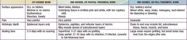
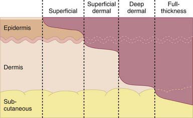
Proper triage and treatment of burn injury require assessment of the extent and depth of the injury (Table 68-4 and Fig. 68-2). 1st-degree burns involve only the epidermis and are characterized by swelling, erythema, and pain (similar to mild sunburn). Tissue damage is usually minimal, and there is no blistering. Pain resolves in 48-72 hr; in a small percentage of patients, the damaged epithelium peels off, leaving no residual scars.
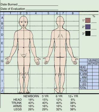
image

Figure 68-2 Diagram of the different burn depths.

(From Hettiaratchy S, Papini R: Initial management of a major burn II: assessment and resuscitation, BMJ 329:101–103, 2004.)

A 2nd-degree burn involves injury to the entire epidermis and a variable portion of the dermal layer (vesicle and blister formation are characteristic). A superficial 2nd-degree burn is extremely painful because a large number of remaining viable nerve endings is exposed. Superficial 2nd-degree burns heal in 7-14 days as the epithelium regenerates in the absence of infection. Midlevel to deep 2nd-degree burns also heal spontaneously if wounds are kept clean and infection-free. Pain is less than in more superficial burns because fewer nerve endings remain viable. Fluid losses and metabolic effects of deep dermal (2nd-degree) burns are essentially the same as those of 3rd-degree burns.

Full-thickness, or 3rd-degree, burns involve destruction of the entire epidermis and dermis, leaving no residual epidermal cells to repopulate the damaged area. The wound cannot epithelialize and can heal only by wound contraction or skin grafting. The absence of painful sensation and capillary filling demonstrates the loss of nerve and capillary elements.

Treatment

Acticoat Nonadherent dressing that delivers silver AQUACEL-Ag Absorptive hydro-fiber that delivers silver Various semipermeable membranes Provide vapor and bacterial barrier Various hydrocolloid dressings Various impregnated gauzes Provide barrier while allowing drainage

Burns to the palm with large blisters usually heal beneath the blisters; they should receive close follow-up on an outpatient basis. The great majority of superficial burns heal in 10-20 days. Deep 2nd-degree burns take longer to heal and may benefit from enzymatic debridement ointment application (collagenase ointment) applied daily on the wound, which aids in the removal of the dead tissue. These ointments should not be applied to the face to avoid the risk of getting them into the eyes.

The depth of scald injuries is difficult to assess early; conservative treatment is appropriate initially, with the depth of the area involved determined before grafting is attempted (Fig. 68-4). This approach obviates the risk of anesthesia and unnecessary grafting.

image

Figure 68-4 Tea scald over the chest and shoulder of a child showing heterogeneity of burn depth. D, deep; I, intermediate; S, superficial.

(From Enoch S, Roshan A, Shah M: Emergency and early management of burns and scalds, BMJ 338:937–941, 2009.)

Fluid Resuscitation

For most children, the Parkland formula is an appropriate starting guideline for fluid resuscitation (4 mL lactated Ringer solution /kg/% BSA burned). Half of the fluid is given over the 1st 8 hr, calculated from the time of onset of injury; the remaining fluid is given at an even rate over the next 16 hr. The rate of infusion is adjusted according to the patient’s response to therapy. Pulse and blood pressure should return to normal, and an adequate urine output (>1 mL/kg/hr in children; 0.5-10 mL/kg/hr in adolescents) should be accomplished by varying the IV infusion rate. Vital signs, acid-base balance, and mental status reflect the adequacy of resuscitation. Because of interstitial edema and sequestration of fluid in muscle cells, patients may gain up to 20% over baseline (pre-burn) body weight. Patients with burns of 30% of BSA require a large venous access (central venous line) to deliver the fluid required over the critical 1st 24 hr. Patients with burns of >60% of BSA may require a multilumen central venous catheter; these patients are best cared for in a specialized burn unit. In addition to fluid resuscitation, children should receive standard maintenance fluids (Chapter 53).

During the 2nd 24 hr after the burn, patients begin to reabsorb edema fluid and to experience diuresis. Half of the 1st day’s fluid requirement is infused as lactated Ringer solution in 5% dextrose. Children younger than 5 yr may require the addition of 5% dextrose in the 1st 24 hr of resuscitation. Controversy exists as to whether colloid should be provided in the early period of burn resuscitation. One preference is to use colloid replacement concurrently if the burn is >85% of total BSA. Colloid is usually instituted 8-24 hr after the burn injury. In children younger than 12 mo, sodium tolerance is limited; the volume and sodium concentration of the resuscitation solution should be decreased if the urinary sodium level is rising. The adequacy of resuscitation should be constantly assessed by means of vital signs as well as urine output, blood gas, hematocrit, and serum protein measurements. Some patients require arterial and central venous lines, particularly those undergoing multiple excision and grafting procedures, as needed, for monitoring and replacement purposes. Central venous pressure monitoring may be indicated to assess circulation in patients with hemodynamic or cardiopulmonary instability. Femoral vein cannulation is a safe access for fluid resuscitation, especially in infants and children. Burn patients who require frequent blood gas monitoring benefit from radial or femoral arterial catheterization.

Oral supplementation may start as early as 48 hr after burn. Milk formula, artificial feedings, homogenized milk, or soy-based products can be given by bolus or constant infusion through a nasogastric or small bowel feeding tube. As oral fluids are tolerated, IV fluids are decreased proportionately in an effort to keep the total fluid intake constant, particularly if pulmonary dysfunction is present.

A 5% albumin infusion may be used to maintain the serum albumin levels at a desired 2 g/dL. The following rates are effective: For burns of 30-50% of total BSA, 0.3 mL of 5% albumin/kg/% BSA burn is infused over 24 hr; for burns of 50-70% of total BSA, 0.4 mL/kg/% BSA burn is infused over 24 hr; and for burns of 70-100% of total BSA, 0.5 mL/kg/% BSA burn is infused over 24 hr. Infusion of packed red blood cells is recommended if the hematocrit falls to <24% (hemoglobin = 8 g/dL). Some authorities recommend treatment for hematocrit <30% or hemoglobin <10 g/dL in patients with systemic infection, hemoglobinopathy, cardiopulmonary disease, or anticipated (or ongoing) blood loss, if repeated excision and grafting of full-thickness burns is needed. Fresh frozen plasma is indicated if clinical and laboratory assessment shows a deficiency of clotting factors, a prothrombin level >1.5 times control, or a partial thromboplastin time >1.2 times control in children who are bleeding or are scheduled for an invasive procedure or a grafting procedure that could result in an estimated blood loss of more than half of blood volume. Fresh frozen plasma may be used for volume resuscitation within 72 hr of injury in patients younger than 2 yr with burns over 20% of BSA and associated inhalation injury.

Sodium supplementation may be required for children with burns of >20% of BSA if 0.5% silver nitrate solution is used as the topical antibacterial burn dressing. Sodium losses with silver nitrate therapy are regularly as high as 350 mmol/m2 burn surface area. Oral sodium chloride supplement of 4 g/m2 burn area/24 hr is usually well tolerated, divided into 4-6 equal doses to avoid osmotic diarrhea. The aim is to maintain serum sodium levels >130 mEq/L and urinary sodium concentration >30 mEq/L. IV potassium supplementation is supplied to maintain a serum potassium level >3 mEq/dL. Potassium losses may be significantly increased when 0.5% silver nitrate solution is used as the topical antibacterial agent or when aminoglycoside, diuretic, or amphotericin therapy is required.

Prevention of Infection and Surgical Management of the Burn Wound

Controversy exists over the prophylactic use of penicillin for all patients hospitalized with acute burn injury and the periodic replacement of central venous catheters to prevent infection. In some units, a 5-day course of penicillin therapy is used for all patients with acute burns; standard-dose crystalline penicillin is given orally or intravenously in 4 divided doses. Erythromycin may be used as an alternative in penicillin-allergic children. Other units have discontinued prophylactic use of penicillin therapy without an increase in the infection rate. Similarly, there is conflicting evidence as to whether relocation of the IV catheter every 48-72 hr decreases or increases the incidence of catheter-related sepsis. Some recommend that the central venous catheter be replaced and relocated every 5-7 days, even if the site is not inflamed and there is no suspicion of catheter-related sepsis.

Mortality related to burn injury is associated not with the toxic effect of thermally injured skin, but with the metabolic and bacterial consequences of a large open wound, reduction of the patient’s host resistance, and malnutrition. These abnormalities set the stage for life-threatening bacterial infection originating from the burn wound. Wound treatment and prevention of wound infection also promote early healing and improve aesthetic and functional outcomes. Topical treatment of the burn wound with 0.5% silver nitrate solution, silver sulfadiazine cream, or mafenide acetate (Sulfamylon) cream or topical solution aims at prevention of infection (Table 68-6). These 3 agents have tissue-penetrating capacity. Regardless of the choice of topical antimicrobial agent, it is essential that all 3rd-degree burn tissue be fully excised before bacterial colonization occurs and that the area be grafted as early as possible to prevent deep wound sepsis. Children with a burn of >30% of BSA should be housed in a bacteria-controlled nursing unit to prevent cross-contamination and to provide a temperature- and humidity-controlled environment to minimize hypermetabolism.

Table 68-6 TOPICAL AGENTS USED FOR BURNS

AGENT EFFECTIVENESS EASE OF USE
Silver sulfadiazine Broad spectrum Closed dressings
Silvadene cream Good penetration Changed twice daily
Residue must be washed off with each dressing change
Mafenide acetate Broad spectrum, including Pseudomonas

Rapid and deep wound penetration Residue must be washed off with each dressing changed 0.5% silver nitrate solution Bacteriostatic Closed bulky dressing soaked every 2 hr and changed once daily Broad spectrum, including some fungi Superficial penetration AQUACEL Ag Dressing impregnated with silver Applied directly to 2nd-degree burn; occlusive dressing kept for 10 days Accuzyme ointment Topical enzymatic debridement Applied daily

Deep 2nd-degree burns of >10% of BSA benefit from early excision and grafting. To improve outcome, sequential excision and grafting of 3rd-degree and deep 2nd-degree burns is required in children with large burns. Prompt excision with immediate wound closure is achieved with autografts, which are often meshed to increase the efficiency of coverings. Alternatives for wound closure, such as allografts, xenografts, and Integra (Integra LifeSciences) and other synthetic skin coverings (bilaminate membrane composed of a porous lattice of cross-linked chondroitin-6-sulfate engineered to induce neovascularization as it is biodegraded), may be important for wound coverage in patients with extensive injury to limit fluid, electrolyte, and protein losses and to reduce pain and minimize temperature loss. Epidermal cultured cells (autologous keratinocytes) are a costly alternative and are not always successful. An experienced burn team can safely carry out early-stage or total excision while burn fluid resuscitation continues. Important keys to success are: (1) accurate preoperative and intraoperative determination of burn depth, (2) the choice of excision area and appropriate timing, (3) control of intraoperative blood loss, (4) specific instrumentation, (5) the choice and use of perioperative antibiotics, and (6) the type of wound coverage chosen. This process can accomplish early coverage without the use of recombinant human growth hormone.

Nutritional Support

Supporting the increased energy requirements of a patient with a burn is a high priority. The burn injury produces a hypermetabolic response characterized by both protein and fat catabolism. Depending on the time since the burn, children with a burn of 40% of total BSA require basal energy expenditure (oxygen consumption) approximately 50-100% higher than predicted for their age. Early excision and grafting can decrease the energy requirement. Pain, anxiety, and immobilization increase the physiologic demands. Additional energy expenditure is caused by cold stress if environmental humidity and temperature are not controlled; this is especially true in young infants, in whom the large surface area–mass ratio allows proportionately greater heat loss than in adolescents and adults. Providing environmental temperatures of 28-33°C, adequate covering during transport and liberal use of analgesics and anxiolytics can decrease calorie demands. Special units to control ambient temperature and humidity may be necessary for children with large surface area burns. Appropriate sleep intervals are necessary and should be part of the regimen. Sepsis increases metabolic rates, and early enteral nutrition, initially with high-carbohydrate, high-protein caloric support (1,800 cal/m2/24 hr maintenance plus 2,200 cal/m2 of burn/24 hr) reduces metabolic stress.

The objective of caloric supplementation programs is to maintain body weight and minimize weight loss by meeting metabolic demands. This reduces the loss of lean body mass. Calories are provided at approximately 1.5 times the basal metabolic rate, with 3-4 g/kg of protein/day. The focus of nutritional therapy is to support and compensate for the metabolic needs. Multivitamins, particularly the B vitamin group, vitamin C, vitamin A, and zinc, are also necessary.

Alimentation should be started as soon as is practical, both enterally and parenterally, to meet all of the caloric needs and keep the gastrointestinal tract active and intact after the resuscitative phase. Patients with burns of >40% of total BSA need a flexible nasogastric or small bowel feeding tube to facilitate continuous delivery of calories without the risk of aspiration. To decrease the risk of infectious complications, parenteral nutrition is discontinued as soon as is practical, after delivery of sufficient enteral calories is established. Continuous gastrointestinal feeding is essential, even if feeding is interrupted, causing frequent visits to the operating room, until full grafting takes place. The use of anabolic agents (growth hormone, oxandrolone, low-dose insulin) or anticatabolic agents (propranolol) remains controversial, although β-blocking agents may reduce metabolic stress. Burn centers caring for large burns (>50% BSA, 3rd-degree) in patients who might be malnourished have used the anabolic steroid oxandrolone, at a dose of 0.1 to 0.2 mg/kg/day given orally, to promote better protein synthesis while the nutritional support by nasogastric feeding and IV hyperalimentation continues.

Topical Therapy

Topical therapy is widely used and is effective against most burn pathogens (see Table 68-6). A number of topical agents are used: 0.5% silver nitrate solution, sulfacetamide acetate cream or solution, silver sulfadiazine cream, and Accuzyme ointment or AQUACEL Ag+. Accuzyme is an enzymatic debridement agent and may cause a stinging feeling for 15 min after application. Preferences vary among burn units. Each topical agent has advantages and disadvantages in application, comfort, and bacteriostatic spectrum. Sulfacetamide acetate is a very effective broad-spectrum agent with the ability to diffuse through the burn eschar; it is the treatment of choice for injury to cartilaginous surfaces, such as the ears. The carbonic anhydrase inhibition activity of sulfacetamide may cause acid-base imbalance if large surface areas are treated, and adverse reactions to the sulfur-containing agents may produce transient leukopenia. This latter reaction is mostly noted with the use of silver sulfadiazine cream when applied over large surface areas in children younger than 5 yr. This phenomenon is transient, self-limiting, and reversible. No sulfa-containing agent should be used if the child has a history of sulfa allergies.

Inhalational Injury

Inhalational injury is serious in the infant and child, particularly if pre-existing pulmonary conditions are present (Chapter 65). Mortality estimates vary, depending on the criteria for diagnosis, but are 45-60% in adults; exact figures are not available in children. Evaluation aims at early identification of inhalation airway injuries. These may occur from (1) direct heat (greater problems with steam burns), (2) acute asphyxia, (3) carbon monoxide poisoning, and (4) toxic fumes, including cyanides from combustible plastics. Sulfur and nitrogen oxides and alkalis formed during the combustion of synthetic fabrics produce corrosive chemicals that may erode mucosa and cause significant tissue sloughing. Exposure to smoke may cause degradation of surfactant and decrease its production, resulting in atelectasis. Inhalation injury and burn injury are synergistic, and the combined effect can increase morbidity and mortality.

The pulmonary complications of burns and inhalation can be divided into 3 syndromes that have distinct clinical manifestations and temporal patterns:

Inhalation injury should be assessed from the evidence of obvious injury (swelling or carbonaceous material in the nasal passages), wheezing, crackles or poor air entry, and laboratory determinations of (HbCO) and arterial blood gases.

Treatment is initially focused on establishing and maintaining a patent airway through prompt and early nasotracheal or orotracheal intubation and adequate ventilation and oxygenation. Wheezing is common, and β-agonist aerosols or inhaled corticosteroids are useful. Aggressive pulmonary toilet and chest physiotherapy are necessary in patients with prolonged nasotracheal intubation or in the rare patient with a tracheotomy. An endotracheal tube can be maintained for months without the need for tracheostomy. If tracheotomy must be performed, it should be delayed until burns at and near the site have healed, and then it should be performed electively, with the child under anesthesia and the use of optimal tracheal positioning and hemostasis. In children with inhalation injury or burns of the face and neck, upper airway obstruction can develop rapidly; endotracheal intubation becomes a lifesaving intervention. Extubation should be delayed until the patient meets the accepted criteria for maintaining the airway.

Signs of CNS injury from hypoxemia due to asphyxia or carbon monoxide poisoning vary from irritability to depression. Carbon monoxide poisoning may be mild (<20% HbCO), with slight dyspnea, headache, nausea, and decreased visual acuity and higher cerebral functions; moderate (20-40% HbCO), with irritability, agitation, nausea, dimness of vision, impaired judgment, and rapid fatigue; or severe (40-60% HbCO), producing confusion, hallucination, ataxia, collapse, acidosis, and coma. Measurement of HbCO is important for diagnosis and treatment. The PaO2 value may be normal and the HbCO saturation values misleading because HbCO is not detected by the usual tests of oxygen saturation. Carbon monoxide poisoning is assumed until the tests are performed, and it is treated with 100% oxygen. Significant carbon monoxide poisoning requires hyperbaric oxygen therapy (Chapter 58).

Patients with severe inhalation injury or with other causes of respiratory deterioration that lead to acute respiratory distress syndrome who do not improve with conventional pressure-controlled ventilation (progressive oxygenation failure, as manifested by oxygen saturation <90% while receiving FIO2 of 0.9-1.0 and positive end-expiratory pressure of at least 12.5 cm H2O) may benefit from high-frequency ventilation or nitric oxide inhalation treatment. Nitric oxide usually is administered through the ventilator at 5 parts per million (ppm) and increased to 30 ppm. This method of therapy reduces the need for extracorporeal membrane oxygenation.

Pain Relief and Psychologic Adjustment

(See Chapter 71.)

It is important to provide adequate analgesia, anxiolytics, and psychologic support to reduce early metabolic stress, decrease the potential for post-traumatic stress syndrome, and allow future stabilization as well as physical and psychologic rehabilitation. Patients and family members require team support to work through the grieving process and accept long-term changes in appearance.

Children with burn injury show frequent and wide fluctuations in pain intensity. Appreciation of pain depends on the depth of the burn; the stage of healing; the patient’s age and stage of emotional development and cognition; the experience and efficiency of the treating team; the use of analgesics and other drugs; the patient’s pain threshold; and interpersonal and cultural factors. From the onset of treatment, preemptive pain control during dressing changes is of paramount importance. The use of a variety of nonpharmacologic interventions as well as pharmacologic agents must be reviewed throughout the treatment period. Opiate analgesia, prescribed in an adequate dose and timed to cover dressing changes, is essential to comfort management. A supportive person who is consistently present and “knows” the patient profile can integrate and encourage patient participation in burn care. The problem of undermedication is most prevalent in adolescents, in whom fear of drug dependence may inappropriately influence treatment. A related problem is that the child’s specific pain experience may be misinterpreted; for anxious patients, those who are confused and alone, or those with pre-existing emotional disorders, even small wounds may illicit intense pain. Anxiolytic medication added to the analgesic is usually helpful and has more than a synergistic effect. Equal attention is necessary to decrease stress in the intubated patient. Other modalities of pain and anxiety relief (relaxation techniques) can decrease the physiologic stress response. Oral morphine sulfate (immediate release) is recommended at a consistent schedule at a dose of 0.3-0.6 mg/kg every 4-6 hr initially and until wound cover is accomplished. Morphine sulfate IV bolus at a dose of 0.05-0.1 mg/kg maximum of 2-5 mg every 2 hr is administered. Morphine sulfate rectal suppositories may be useful at a dose of 0.3-0.6 mg/kg every 4 hr when oral administration is not possible. For anxiety, lorazepam is given on a consistent schedule, 0.05-0.1 mg/kg/dose every 6-8 hr. To control pain during a procedure (dressing change or debridement), oral morphine at a dose of 0.3-0.6 mg/kg is given 1-2 hr before the procedure and this is supplemented by a morphine IV bolus at a dose of 0.05-0.1 mg/kg given immediately before the procedure. Lorazepam at a dose of 0.04 mg/kg is given orally or intravenously, if necessary, for anxiety before the procedure. Midazolam (Versed) is also very useful for conscious sedation given at a dose of 0.01-0.02 mg/kg for nonintubated patients and 0.05-0.1 mg/kg for intubated patients, as an intravenous infusion or bolus, and may be repeated in 10 min. During the process of weaning from analgesics, the dose of oral opiates is reduced by 25% over 1-3 days, sometimes with the addition of acetaminophen as opiates are tapered. Antianxiety medications are tapered by reducing the dose of benzodiazepines at 25-50%/dose daily over 1-3 days.

For ventilated patients, pain control is accomplished by using morphine sulfate intermittently as an IV bolus at a dose of 0.05-0.1 mg/kg every 2 hr. Doses may need to be increased gradually, and some children may need continuous infusion; a starting dose of 0.05 mg/kg/hr given as an infusion is increased gradually as the need of the child changes. Naloxone is rarely needed but should be immediately available to reverse the effect of morphine, if necessary; if needed for an airway crisis, it should be given in a dose of 0.1 mg/kg up to a total of 2 mg, either intramuscularly or intravenously. For patients undergoing assisted respiration who require treatment of anxiety, midazolam is used as an intermittent IV bolus (0.04 mg/kg given by slow push every 4-6 hr) or as a continuous infusion. For intubated patients, opiates do not need to be discontinued during the process of weaning from the ventilator. Benzodiazepine should be reduced to approximately half the dose over 24-72 hr before extubation; too-rapid weaning from a benzodiazepine can lead to seizures.

There has been an expansion of the use of psychotropic medication in the care of children with burns, including prescription of selective serotonin reuptake inhibitors (SSRIs) as antidepressants, the use of haloperidol as a neuroleptic in the critical care setting and the treatment of post-traumatic stress disorder (PTSD) with benzodiazepines. Another area of expansion is the use of conscious sedation utilizing ketamine or propofol for major dressing changes.

School Reentry and Long-Term Outcome

It is best for the child to return to school immediately after discharge. Occasionally, a child may need to attend a few half-days (because of rehabilitation needs). It is important for the child to return to his or her normal routine of attending school and being with peers. Planning for a return to home and school often requires a school reentry program that is individualized to each child’s needs. For a school-aged child, planning for the return to school occurs simultaneously with planning for discharge. The hospital schoolteacher contacts the local school and plans the program with the school faculty, nurses, social workers, recreational/child life therapists, and rehabilitation therapists. This team should work with students and staff to ease anxiety, answer questions, and provide information. Burns and scars evoke fears in those who are not familiar with this type of injury and can result in a tendency to withdraw from or reject the burned child. A school reentry program should be appropriate to a child’s development and changing educational needs.

Major advances have made it possible to save the lives of children with massive burns; whereas some children have had lingering physical difficulties, most have a satisfactory quality of life. The comprehensive burn care that includes experienced multidisciplinary aftercare plays an important role in recovery. Long-term complications of burns are listed in Table 68-7.

Special Situations

Electrical Burns

There are 3 types of electrical burns. Minor electrical burns usually occur as a result of biting on an extension cord. These injuries produce localized burns to the mouth, which usually involve the portions of the upper and lower lips that come in contact with the extension cord. The injury may involve or spare the corners of the mouth. Because these are nonconductive injuries (do not extend beyond the site of injury), hospital admission is not necessary and care is focused on the area of the injury visible in the mouth. Treatment with topical antibiotic creams is sufficient until the patient is seen in a burn unit outpatient department or by a plastic surgeon.

A more serious category of electrical burn is the high-tension electrical wire burn, for which children must be admitted for observation, regardless of the extent of the surface area burn. Deep muscle injury is typical and cannot be readily assessed initially. These injuries result from high voltage (>1,000 V) and occur particularly at high-voltage installations, such as electric power stations or railroads; children climb an electric pole and touch an electric box out of curiosity or accidentally touch a high-tension electric wire. Such injuries have a mortality rate of 3-15% for children who arrive at the hospital for treatment. Survivors have a high rate of morbidity, including major limb amputations. Points of entry of current through the skin and the exit site show characteristic features consistent with current density and heat. The majority of entrance wounds involve the upper extremity, with small exit wounds in the lower extremity. The electrical path, from entrance to exit, takes the shortest distance between the 2 points and may produce injury in any organ or tissue in the path of the current. Multiple exit wounds in some patients attest to the possibility of several electrical pathways in the body, placing virtually any structure in the body at risk (Table 68-8). Damage to the abdominal viscera, thoracic structures, and the nervous system in areas remote from obvious extremity injury occurs and must be sought, particularly in injuries with multiple current pathways or those in which the victim falls from a high pole. Sometimes arcing occurs and results in concurrent flame burn and clothing fire. Cardiac abnormalities, manifested as ventricular fibrillation or cardiac arrest, are common; patients with high-tension electrical injury need cardiac monitoring until they are stable and have been fully assessed. Higher-risk patients have abnormal electrocardiographic findings and a history of loss of consciousness. Renal damage from deep muscle necrosis and subsequent myoglobinuria is another complication; such patients need forced alkaline diuresis to minimize renal damage. Aggressive removal of all dead and devitalized tissue, even with the risk of functional loss, remains the key to effective management of the electrically damaged extremity. Early debridement facilitates early closure of the wound. Damaged major vessels must be isolated and buried in a viable muscle to prevent exposure. Survival depends on immediate intensive care, whereas a functional result depends on long-term care and delayed reconstructive surgery.

Table 68-8 ELECTRICAL INJURY: CLINICAL CONSIDERATIONS

  CLINICAL MANIFESTATIONS MANAGEMENT
General Extricate the patient; perform ABCs of resuscitation; immobilize the spine
History: voltage, type of current
Complete blood count with platelets, electrolytes, blood urea nitrogen (BUN), creatinine, glucose
Cardiac Dysrhythmias: asystole, ventricular fibrillation, sinus tachycardia, sinus bradycardia, premature atrial contractions (PACs), premature ventricular contractions (PVCs), conduction defects, atrial fibrillation, ST-T wave changes

Creatinine phosphokinase with isoenzyme measurements if indicated Pulmonary Respiratory arrest, acute respiratory distress, aspiration syndrome Protect and maintain the airway Mechanical ventilation if indicated, chest radiograph, arterial blood gas levels Renal Acute renal failure, myoglobinuria Provide aggressive fluid management unless a central nervous system injury is present Maintain adequate urine output, >1 mL/kg/hr Consider central venous or pulmonary artery pressure monitoring Measure urine myoglobin; perform urinalysis; measure BUN, creatinine Neurologic Immediate: loss of consciousness, motor paralysis, visual disturbances, amnesia, agitation; intracranial hematoma

Secondary: pain, paraplegia, brachial plexus injury, syndrome of inappropriate antidiuretic hormone secretion (SIADH), autonomic disturbances, cerebral edema Consider spine radiographs, especially cervical Delayed: paralysis, seizures, headache, peripheral neuropathy CT scan of the brain if indicated Cutaneous/oral Oral commissure burns, tongue and dental injuries; skin burns resulting from ignition of clothes, entrance and exit burns, and arc burns Obtain a plastic surgery of ear, nose, and throat consultation if needed Abdominal Viscus perforation and solid organ damage; ileus; abdominal injury rare without visible abdominal burns Place a nasogastric tube if the patient has airway compromise or ileus Obtain SGOT (serum glutamate oxaloacetate transaminase or aspartate aminotransferase), SGPT (serum glutamate pyruvate transaminase, alanine aminotransferase), amylase, BUN, and creatinine measurements and, CT scans as indicated Musculoskeletal Compartment syndrome from subcutaneous necrosis limb edema and deep burns Monitor the patient for possible compartment syndrome Long bone fractures, spine injuries Obtain radiographs and orthopedic/general surgery consultations as indicated Ocular Visual changes, optic neuritis, cataracts, extraocular muscle paresis Obtain an ophthalmology consultation as indicated

Modified from Hall ML, Sills RM: Electrical and lightning injuries. In Barkin RM, editor: Pediatric emergency medicine, St Louis, 1997, Mosby, p 484.

Lightning burns occur when a high-voltage current directly strikes a person (most dangerous) or when the current strikes the ground or an adjacent (in-contact) object. A step voltage burn is observed when lightning strikes the ground and travels up one leg and down the other (the path of least resistance). Lightning burns depend on the current path, the type of clothing worn, the presence of metal, and cutaneous moisture. Entry, exit, and path lesions are possible; the prognosis is poorest for lesions of the head or legs. Internal organ injury along the path is common and does not relate to the severity of the cutaneous burn. Linear burns, usually 1st- or 2nd-degree, are in the locations where sweat is present. Feathering or an arborescent pattern is characteristic of lightning injury. Lightning may ignite clothing or produce serious cutaneous burns from heated metal in the clothing. Internal complications of lightning burns include cardiac arrest caused by asystole, transient hypertension, premature ventricular contractions, ventricular fibrillation, and myocardial ischemia. Most severe cardiac complications resolve if the patient is supported with cardiopulmonary resuscitation (Chapter 62). CNS complications include cerebral edema, hemorrhage, seizures, mood changes, depression, and paralysis of the lower extremities. Rhabdomyolysis and myoglobinuria (with possible renal failure) also occur.

Bibliography

Ali SN, O’Toole G, Tyler M. Milk bottle burns. J Burn Care Rehabil. 2004;25:461-462.

Arnoldo B, Purdue GF, Kowalske K, et al. Electrical injuries: a 20-year review. J Burn Care Rehabil. 2004;25:479-484.

Bracco D, Eggimann P. Prophylaxis with systemic antibiotics in patients with severe burns. BMJ. 2010;340:487-488.

Chen EH, Sareen A. Do children require ECG evaluation and inpatient telemetry after household electrical exposures? Ann Emerg Med. 2007;49:64-67.

Collier ML, Ward RS, Saffle JR, et al. Home treadmill friction injuries: a five-year review. J Burn Care Rehabil. 2004;25:441-444.

Enoch S, Roshan A, Shah M. Emergency and early management of burns and scalds. BMJ. 2009;338:937-941.

Garrel D. Burn scars: a new cause of vitamin D deficiency. Lancet. 2004;363:259-260.

Hart DW, Wolf SE, Ramzy PI, et al. Anabolic effects of oxandrolone after severe burn. Ann Surg. 2001;233:556-564.

Herndon DN, Hart DW, Wolf SE, et al. Reversal of catabolism by beta-blockade after severe burns. N Engl J Med. 2001;345:1223-1229.

Herndon DN, Tompkins RG. Support of the metabolic response to burn injury. Lancet. 2004;363:1895-1902.

Hohlfeld J, de Buys Roessingh A, Hiti-Burri N, et al. Tissue engineered fetal skin constructs for paediatric burns. Lancet. 2006;366:840-842.

Juurlink DN, Buckley NA, Stanbrook MB, et al: Hyperbaric oxygen for carbon monoxide poisoning, Cochrane Database Syst Rev (1):CD002041, 2005.

Kai-Yang L, Zhao-Fan X, Luo-Man Z, et al. Epidemiology of pediatric burns requiring hospitalization in China: a literature review of retrospective studies. Pediatrics. 2008;122:132-142.

Lowell G, Quinlan K, Gottleib LJ. Preventing unintentional scald burns: moving beyond tap water. Pediatrics. 2008;122:799-804.

Musgrave MA, Fingland R, Gomez M, et al. The use of inhaled nitric oxide as adjuvant therapy in patients with burn injuries and respiratory failure. J Burn Care Rehabil. 2000;21:551-557.

Orgill DP. Excision and skin grading of thermal burns. N Engl J Med. 2009;360:893-900.

Sanghavi P, Bhalla K, Das V. Fire-related deaths in India in 2001: a retrospective analysis of data. Lancet. 2009;373:1282-1288.

Sheridan RL. Thermal injuries. In: McInerny TK, Adam HM, Campbell DE, editors. American Academy of Pediatrics pediatric textbook of pediatric care. Elk Grove Village, IL: American Academy of Pediatrics, 2008.

Sheridan RL, Hinson M, Liang MH, et al. Long-term outcomes of children surviving massive burns. JAMA. 2000;283:69-73.

Singer AJ, Dagum AB. Current management of acute cutaneous wounds. N Engl J Med. 2008;359:1037-1046.

Stoddard FJ, Usher CT, Abrams AJ. Psychopharmacology in pediatric critical care. Child Adolesc Psychiatr Clin North Am. 2006;15:611-655.

Wiechman SA, Patterson DR. Psychosocial aspects of burn injuries. BMJ. 2004;329:391-393.

Volinsky J, Hanson J, Lustig J. Lightning burns. Arch Pediatr Adolesc Med. 1994;148:529-530.

Walker A. Emergency department management of house fire burns and carbon monoxide poisoning in children. Curr Opin Pediatr. 1996;8:239-242.

Wolf SE, Thomas SJ, Dasu MR, et al. Improved net protein balance, lean mass, and gene expression changes with oxandrolone treatment in the severely burned. Ann Surg. 2003;237:801. 10; discussion 810–811

Young AE, Thornton KL. Toxic shock syndrome in burns: diagnosis and management. Arch Dis Child Edu Pract Ed. 2007;92:ep97-ep100.