Bronchoscopy

Published on 12/06/2015 by admin

Filed under Pulmolory and Respiratory

Last modified 12/06/2015

Print this page

rate 1 star rate 2 star rate 3 star rate 4 star rate 5 star
Your rating: none, Average: 3.3 (22 votes)

This article have been viewed 6257 times

Bronchoscopy

Zaza Cohen

The word endoscopy (from Greek words endo, meaning “inside,” and skopeo, “to examine”) describes examination of the interior of a canal or a hollow viscus by means of a special instrument, such as an endoscope. Endoscopy is a somewhat general term that includes procedures, such as laryngoscopy, examination of the larynx; bronchoscopy, examination of the bronchi; gastroscopy, examination of the stomach; and colonoscopy, examination of the colon, among many others. Gustav Killian, widely regarded as the “father” of bronchoscopy, was first to visualize the bronchial tree through a hollow tube (bronchoscope) inserted in the patient’s larynx in 1897. Two major advances of the 20th century in the field of bronchoscopy were the addition of the suction channel by Chevalier Jackson and the use of fiberoptics for the light source by Shigeto Ikeda.

The early versions of bronchoscopy used straight pieces of open pipe, beveled on the end to prevent tearing the trachea while being forced into the anesthetized patient with the head tipped back as far as possible. A simple light shining down the tube allowed limited viewing of the trachea and mainstem bronchi. Although these rigid devices provided a limited view of the larger airways, they allowed removal of large volumes of blood, mucus, or foreign objects and biopsy of tumors in the upper airway. At the same time, mechanical ventilation was provided to ensure better gas exchange during the procedure. Those early rigid bronchoscopes have been replaced by more sophisticated rigid instruments with lights at the tips and valves to allow general anesthesia, oxygenation, suctioning, and biopsy. The new rigid bronchoscopes remain useful for a few problems, such as removal of difficult foreign bodies and management of severe bleeding, but well more than 90% of bronchoscopy procedures now are done with flexible fiberoptic bronchoscopes. Given that the emphasis of this text is on patient assessment, and that rigid bronchoscopy is relatively infrequently performed, the focus of this chapter is on flexible fiberoptic bronchoscopy.

The first application of flexible fiberoptics to the field of endoscopy was in 1957 and was initially applied to gastroscopes. As experience grew and the techniques improved, it was applied to the other disciplines within the field of endoscopy. In the 1960s, Shigeto Ikeda of Japan designed the first fiberoptic bronchoscope, which incorporated fiberoptics into a flexible tube suitable in size and length to enter the trachea and visualize the lower airway. The first bronchoscopy in the United States using flexible fiberoptic technique was done in the Mayo Clinic in 1969. The flexible fiberoptic bronchoscope has greatly expanded the practice of pulmonary medicine by allowing inspection of the airways, removal of many different foreign bodies, and biopsy of airway and peripheral lung tissue. Most of these activities can be performed in the outpatient setting with moderate sedation.

In most institutions, flexible bronchoscopy is performed by a properly trained and licensed physician (usually a pulmonologist, but sometimes a surgeon or a critical care physician) and one or two assistants, usually a respiratory therapist (RT) or a registered nurse (RN). For the remainder of this chapter, we refer to the physician performing the procedure as “the bronchoscopist” and the RN or the RT assisting as “the assistant.” In many teaching institutions, students, medical residents, or other trainees may also be present and participate in various parts of the procedure under the supervision of a licensed health care provider. Sometimes, a radiology technician is available to perform fluoroscopy (see the discussion on fluoroscopy later in this chapter or in Chapter 10), as well as a histology technician to evaluate the quality of the specimen from biopsy or brushings. RTs are educated and trained about flexible fiberoptic bronchoscopes because they often maintain the equipment and the materials needed for the procedure. The RT has become indispensable in helping the physician perform the procedure both in hospitals and outpatient clinics.

Characteristics and Capabilities of the Bronchoscope

The bronchoscopes available today serve a wide range of applications, from small-diameter scopes used for viewing the airways of infants and children to larger-diameter scopes with suction channels used to remove thick secretions from patients on ventilators in the intensive care unit (ICU), to scopes that incorporate ultrasound equipment for better visualization of the structures. Many bronchoscopes are now digital. Light is produced in an external light source and carried into the airways through a fiberoptic bundle. A digital video camera on the end of the bronchoscope acquires the images of the lighted airways, and the real-time images are displayed on a large video monitor. This allows easier and more hygienic manipulation of the bronchoscope and allows both still and video photographs of the procedure for documenting and teaching purposes.

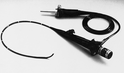
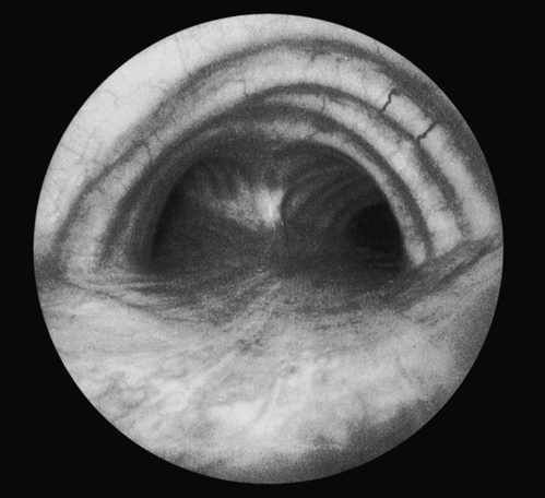
The standard flexible bronchoscope has an external diameter of 5.3 mm and a total length of 605 mm (Fig. 17-1). It can pass easily down the trachea (first-order bronchus) and into the right or left mainstem bronchus. From there, it can be advanced through most fourth-order bronchi and one third of all fifth-order bronchi (Fig. 17-2). Visualization of half of the lung’s sixth-order bronchi is possible. Ultrathin scopes with a 2.7-mm external diameter and 0.8-mm biopsy channels will pass down 3.5-mm or larger endotracheal tubes, making it easier to study and perform biopsies on infants and children who are intubated. One must keep in mind that even with such depth of visualization, bronchoscopy still allows examination of only a small percentage of all airways. For comparison, colonoscopy allows the entire length of the colon to be examined during the procedure.

The amount of tip angulation, which makes the scope directional, is crucial to the scope’s utility and ability to visualize hard-to-reach but important airways. Most bronchoscope tips can bend from the axial plane at least 130 degrees in one direction and 160 degrees in the other. Bronchoscopes flex less with a brush, forceps, or needle in place, and all bronchoscopes lose flexibility at the tip over time and with use.

A light source is the foundation for fiberoptic bronchoscopy. Light sources range in size from 2-inch battery cases that attach to the head of the bronchoscope to large units that deliver light at varied intensities, in different wavelengths, and in pulses for photography or other specialized jobs.

Biopsy devices can be passed down the scope and greatly extend the scope’s use. The devices include biting and grasping forceps, brushes (shielded and unshielded), sheathed needles, and sampling catheters. The biting forceps come in various types (biting cusps with smooth edges or serrated edges). Smooth-cusped flexible forceps can be used if the physician needs to obtain a piece of lung parenchyma during the procedure. If a large, dense (tough) lesion is seen in the larger airways, a serrated forceps capable of cutting tissue is preferred. Forceps are metallic, are easily visualized by radiographs, and can be directed to the desired area for biopsy of peripheral lung lesions using real-time radiography (fluoroscopy).

A hollow needle fixed to a long, small-lumen plastic tube can be passed down the scope to perform biopsy on lesions under the mucosa. Specimens usually contain a few cells, rather than a larger piece of actual tissue sample with preserved lung architecture that is obtained during forceps biopsy. Positive yields (obtaining diagnostic tissue samples) from submucosal lesions are less than those for large cancers growing in the larger airways.

Unshielded brushes are used to harvest cells from tumor masses, located both centrally and in the periphery. The double-shielded sterile brush is used to gather microbiologically important material to diagnose infections in the lung. Because the bronchoscope is passed into the lung by way of the nose or mouth, the entire scope immediately becomes contaminated with the patient’s upper-tract bacterial mix (normal flora). Thus, unshielded sampling may misrepresent the specific bacteria present in the lower lung and causing the lung infection. Sterile double-shielded brushes are passed through the entire length of the scope until the bronchoscopist sees that the tip is well in view into the airway of concern. The inner sheath is then advanced, followed by the wire brush that comes out of the sterile inner sheath. The sampling is done by gently brushing the airway. The wire is then drawn back into its sheath and pulled from the bronchoscope. The wire tip is cut off with sterile scissors, placed in a sterile container with sterile normal saline, and sent for microbiologic studies.

During the bronchoalveolar lavage (BAL), the bronchoscope is passed to the affected part of the lung, and the tip of the scope is positioned into a fourth-generation bronchus. A total of about 100 mL of normal saline is flushed through the scope’s suction channel in four or five increments of 20 to 30 mL to distend the distal bronchioles and fill the alveoli, thereby washing out samples of any microorganisms. A little more than half of the BAL fluid is usually suctioned back into a collection chamber, and the balance is absorbed by the lymphatic system. The lavage is typically done for diagnostic purposes: to look for infection (smears and cultures) or certain types of interstitial diseases (cell count and differential). In rare situations, such as patients with pulmonary alveolar proteinosis and highly selected patients with severe asthma, the lavage can be done for therapeutic purposes. The fluid is collected very carefully to avoid contamination from oral or upper airway secretions. In normal patients, the fluid is sterile and contains predominantly macrophages.

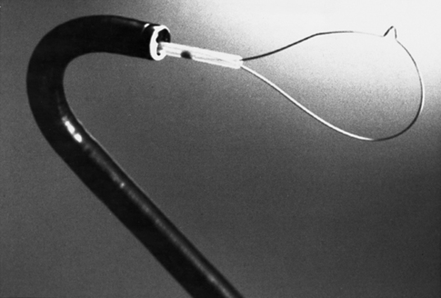
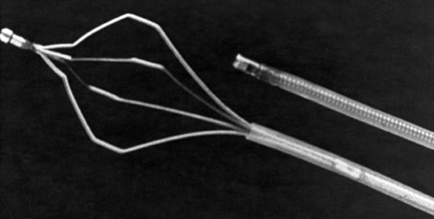
Patients accidentally inhale a remarkable variety of objects, including corn, peas, beans, peanuts, garlic cloves, teeth, dental crowns, dental fillings, dental drill bits, pills, coins and, beads. These objects, as well as mucous plugs or clots, can be retrieved with a bronchoscope. In some cases, these items or conditions do not show up on the chest radiograph, and their discovery by bronchoscopy is based on the patient’s history or is an unexpected discovery during an evaluation for chronic cough. Grasping forceps and snares can be used with the bronchoscope to retrieve foreign objects (Figs. 17-3 and 17-4). Grasping forceps are available with rubber tips for needle and nail removal; basket types for removal of marbles, seeds, and nuts; “pelican” types for removal of food particles; and W-shaped forceps for the removal of coins.

Lasers can be used to obliterate tumors obstructing large airways. Flexible quartz monofilaments pass through the bronchoscope and conduct a laser beam beyond the end of the scope. The bronchoscopist can direct the red “aiming dot” with precision, step on the foot pedal to activate the laser beam, and send a pulsed beam of tissue-vaporizing laser energy into the tumor. Safety is a major issue because lasers are more likely to ignite airway fires if the oxygen concentration is above 30%. Other specialty catheters available for use through a bronchoscope include suture cutters, magnetic extractors, and injector catheters to direct medications into an area of the lung. Coagulation electrodes and hot biopsy forceps are also available.

Interventional Bronchoscopy

The past two decades of the 20th century saw a great expansion in the types of secondary procedures performed during bronchoscopy, leading to the development of a new discipline interventional bronchoscopy, within the field of bronchoscopy. In a combined European Respiratory Society and American Thoracic Society (ERS/ATS) statement, interventional bronchoscopy was defined as “the art and science of medicine as related to the performance of diagnostic and invasive therapeutic procedures that require additional training and expertize beyond that required in a standard pulmonary medicine training programme.” The field of interventional bronchoscopy includes procedures such as autofluorescent bronchoscopy, endobronchial ultrasound (EBUS), laser bronchoscopy, endobronchial electrosurgery, electromagnetic navigation bronchoscopy (ENB), argon-plasma coagulation, bronchial thermoplasty, endobronchial cryotherapy, airway stent placement, endobronchial brachytherapy, photodynamic therapy, percutaneous tracheostomy placement, and endobronchial valve placement, and it is constantly expanding. A detailed description of these procedures is outside the scope of this chapter. A review of the aforementioned ERS/ATS statement is highly recommended for a better understanding of these procedures.

Indications for Bronchoscopy

There are two basic categories of indications for flexible fiberoptic bronchoscopy: diagnostic and therapeutic (Box 17-1). The flexible fiberoptic bronchoscope is used most often for diagnostic purposes, with therapeutic indications being less common. The most common indication for flexible bronchoscopy is to diagnose the cause of an abnormality seen on a chest radiograph. These abnormalities include infiltrates of any size or location, atelectasis, or mass lesions. These lesions are of particular concern if recently they have appeared on the chest radiograph, or if the patient has risk factors for malignancy, such as history of previous cancer diagnosis or history of smoking. Bronchoscopy may be indicated in the patient with a normal chest radiograph if the patient has symptoms of chronic cough, stridor, hoarseness, or history of choking.

Nodules and Masses

Radiologists and pulmonologists often distinguish between pulmonary nodules and masses. An abnormal solid structure in the pulmonary parenchyma that is smaller than 3 cm on a chest radiograph or computed tomography (CT) scan is typically called a nodule. Structures larger than 3 cm are referred to as masses. Rounded small lesions are often called coin lesions because their shadows resemble coins. The malignant potential of these lesions depends on their size, location, shape, rate of growth, and the patient’s history (smoking, weight loss, hemoptysis, history of cancer, or other risk factors for malignancy). Although most nodules and some masses turn out to be benign, a biopsy is often necessary for accurate diagnosis. The possibility of obtaining adequate tissue for diagnosis depends on the lesion size, its location (endobronchial or not, central vs. peripheral), and the expertise of the bronchoscopist.

Naturally, large lesions that are in the central airways are easy to find and excise for biopsy. However, coin lesions are often in the lung periphery and cannot be seen through a flexible bronchoscope. Fluoroscopy is used to aim the tip of the bronchoscope toward the lesion (see Chapter 10 on chest imaging for the utility and limitations of the fluoroscopy). Biopsy forceps can then be advanced into the appropriate subsegmental bronchus to reach the lesion and snip off pieces to be sent for analysis. New advanced bronchoscopy techniques such as ENB and EBUS increase the likelihood of obtaining adequate tissue sample. During ENB, the patient’s lungs and the lesion are mapped out using conventional CT scan, and then the software guides the bronchoscope toward the lesion even when the lesion cannot be directly visualized. During EBUS, the ultrasound probe that is mounted on a special bronchoscope guides the biopsy needle into the submucosal lymph nodes that cannot otherwise be seen.