[level-membership-for-opthalmology-category]14.1
Branch Retinal Vein Occlusion
Clinical Features:
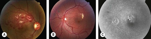
Patients may complain of visual blurring, distortion or metamorphopsia. On examination, intraretinal flame and blot shaped hemorrhages as seen in the territory of a dilated, tortuous retinal vein are present (Fig. 14.1.1). Given the distribution of retinal veins the accompanying pathology of BRVO almost never crosses the horizontal raphe. Cotton wool spots, retinal edema in the area drained by the occluded branch, collateral vessels and occasionally retinal neovascularization and vitreous hemorrhage may be seen. Vision loss is largely due to of retinal edema and may sometimes be secondary to retinal ischemia.
OCT Features:
OCT shows retinal thickening and edema limited to the retinal area drained by the obstructed vein (Figs 14.1.2 and 14.1.3). Macular thickness scans will show retinal thickening, usually confined to half the macula. Line scans through the macula show diffuse retinal edema and cystic (hyporeflective) spaces in the outer retina. Subretinal fluid may also be observed in severe cases. Hard exudates can be seen as small hyper-reflective intraretinal spots. Macular thickness scans are particularly valuable in monitoring edema over time and assessing the effects of treatment.
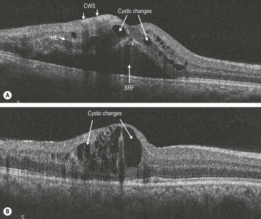
Figure 14.1.2 OCT scans through the macula of the patient (A) with diffuse retinal thickening and cystic changes (arrows). Note the cotton wool spots (CWS) (arrows) in the nerve fiber layer that cause shadowing of the layers beneath them. Some hard exudates (HE) are noted (arrow) as hyper-reflective clusters deeper within the retina and spanning several layers. SRF is also seen (arrow). (B) Note that only part of the retina is thickened unlike in CRVO.
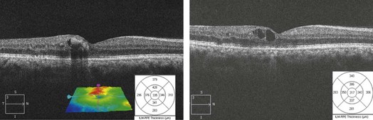
Figure 14.1.3 Successive OCT scans in the same patient after treatment with laser and anti-vascular endothelial growth factor therapy, with thinning of the retina seen on the line and macular thickness scans as well as a smaller geographic spread of the thickening seen on the retinal pigment membrane–internal limiting membrane overlay.
[/level-membership-for-opthalmology-category][not-level-membership-for-opthalmology-category]14.1
Branch Retinal Vein Occlusion
Clinical Features:
Patients may complain of visual blurring, distortion or metamorphopsia. On examination, intraretinal flame and blot shaped hemorrhages as seen in the territory of a dilated, tortuous retinal vein are present (Fig. 14.1.1
[/not-level-membership-for-opthalmology-category]
