[level-membership-for-opthalmology-category]15.1
Branch Retinal Artery Occlusion
Clinical Features:
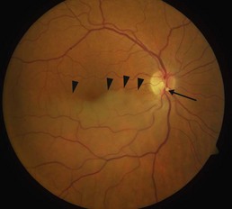
Retinal whitening is seen along the sector of retina that is supplied by the arterial branch from the central retinal artery (Fig. 15.1.1). The temporal hemisphere is most commonly affected. Emboli are often visualized at the site of blockage, which tends to be at a bifurcation point.
OCT Features:
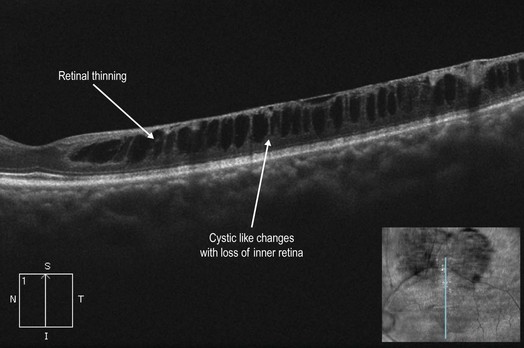
In the acute setting, there is intense hyper-reflectivity of the inner retinal layers, similar to that seen in CRAO (see Chapter 15.2), but limited to the sector of retina involved. Vertical, instead of horizontal, OCT cuts can help to make this distinction (Fig. 15.1.2). With time, the edema resolves leaving attenuation and atrophy of the inner retinal layers, which can appear as thinning or even schisis-like changes (Fig. 15.1.3).
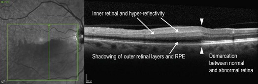
Figure 15.1.2 Acute branch retinal artery occlusion. OCT vertical cut shows inner retinal hyper-reflectivity and thickening only in the sectoral area of retina that is affected by the branch arterial occlusion (left of arrowheads). As with the case in central retinal artery occlusion, the inner hyper-reflectivity in the affected region causes shadowing of the outer layers, which attenuates the signal from the outer retina and retinal pigment epithelium (RPE).
Ancillary Testing:
Fluorescein angiography can help in securing the diagnosis by revealing a sectoral perfusion deficiency in the acute setting (Fig. 15.1.4).

Figure 15.1.4 Fluorescein angiography (corresponding to Figure 15.1.1) shows a significant perfusion delay in the sector of retina affected by the branch arterial occlusion.
[/level-membership-for-opthalmology-category][not-level-membership-for-opthalmology-category]15.1
Branch Retinal Artery Occlusion
Clinical Features:
Retinal whitening is seen along the sector of retina that is supplied by the arterial branch from the central retinal artery (Fig. 15.1.1). The temporal hemisphere is most commonly affected. Emboli are often visualized at the site of blockage, which tends to be at a bifurcation point.
[/not-level-membership-for-opthalmology-category]
