Body Peeling

Published on 15/03/2015 by admin

Filed under Dermatology

Last modified 22/04/2025

Print this page

rate 1 star rate 2 star rate 3 star rate 4 star rate 5 star
Your rating: none, Average: 0 (0 votes)

This article have been viewed 1721 times

12 Body Peeling

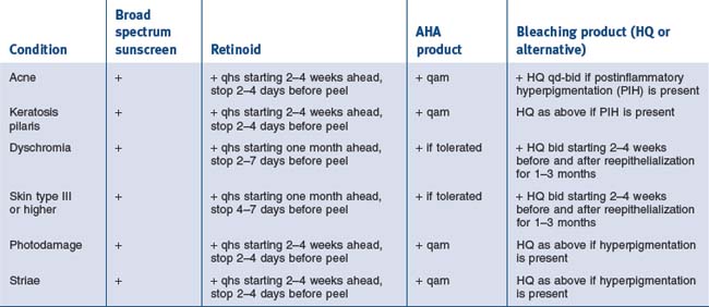
When discussing the approach to chemical peeling off the face, adjectives such as conservative and gradual should come to mind. Common indications are much the same as their facial counterparts and can be grouped into those that would be best treated with superficial peels and those that would benefit from medium-depth peels (see Table 12.1).

Table 12.1 Indications for superficial and medium-depth peels

Superficial peels Medium-depth peels

Patient selection

All skin types may undergo body peeling but extra caution should be employed in darker skin (III–VI) types since there is increased risk of unwanted pigmentation changes. Skin types I and II can easily undergo medium-depth peels with little risk of untoward effects. Appropriate patient expectations should be established from the start.

While medium-depth peels on the face can be considered one-stage procedures, this is often not the case on the body. Due to increased potential for adverse events such as scarring off the face, similar peels are done in a series and at lower concentrations. Results are seen after a few sessions and should be accompanied by a regular topical maintenance program to prolong the benefits. Additionally, for specific indications, body peels can be combined with other minimally invasive procedures including lasers, light sources, electrodessication, cryosurgery to expedite resolution while minimizing complications and downtime.

While all superficial peels are virtually pain-free, a few patients may opt for a sedative (like diazepam) prior to a medium-depth body peel. Overall, the skin on the body is much less sensitive than the face. Even our most anxious patients find the nonfacial medium-depth chemical peeling experience to be ‘easy and quick’ and prefer to avoid an anxiolytic so that they can drive themselves to and from the procedure.

Whether peeling in facial or nonfacial locations, similar background medical and surgical history should be collected. Waiting periods of greater than 6–12 months after isotretinoin and greater than 6 months after invasive surgery may be observed due to potential for delayed wound healing and abnormal scarring.

Past exposure to radiation in the neck, chest or other proposed site to be peeled is also relevant. Radiation diminishes the number of pilosebaceous units in treated areas and places these patients at an increased risk for development of postoperative scarring. Presence of adnexal structures can be assessed clinically by observation of vellus hairs or microscopically by performing a small punch biopsy as Brody describes. Similarly, a history of hypertrophic scar or keloid formation is also relevant especially if peeling will take place in high risk areas such as the chest, back shoulders and junction of the jaw and neck.

Common medications such as oral contraceptives, hormone replacement, and minocyclines can predispose to postinflammatory hyperpigmentation after a peel. Patients taking these medications should be warned of this possibility and advised on sun avoidance after the peel and regular usage of broad-spectrum sun protection.

Vasoconstriction due to smoking can create delayed wound healing and predispose to infections. Additionally, smoking also impairs existing collagen and elastic fiber. While chemical peeling can be performed in smokers, benefits may not be as pronounced.

Peeling in patients with active inflammatory conditions such as dermatitis and acne should be carefully undertaken. Likewise, if an individual has open excoriations, one may wish to delay a peeling until these areas are healed.

When peeling off the face, herpes simplex reactivation is less likely and antiviral prophylaxis is not required. Due to the fact that all of the peeling agents have some degree of bacteriostatic activity, postprocedural antibiotics are not routinely prescribed.

All patients undergoing medium or deeper peels should have preoperative and postoperative photographs to document the baseline extent of solar damage, scarring, and/or other conditions and to demonstrate the degree of improvement after the peeling process.

A signed informed consent form should outline the risks, benefits, alternatives and limitations associated with chemical peeling. Expected responses to peels include burning and stinging sensations, redness, and peeling. Less common effects following medium and deep peels include prolonged erythema, and abnormal pigmentation. Rarely, infection, delayed healing or scarring may result. Alternative resurfacing procedures such as microdermabrasion, dermabrasion, nonablative, fractional and ablative resurfacing lasers should also be mentioned.

Once a patient decides to pursue chemical peeling, we also like to give them a written information sheet which details what to expect (specific information regarding downtime limits future phone calls), what they will need to do or purchase for after care, and contact phone numbers if questions or problems arise in the postpeel period.

Preconditioning the skin

As might be expected, preparing the skin of the body is similar to what is done to the face prior to chemical peeling . For the sake of ease, only specific differences will be highlighted in the text while common preparations are listed in Table 12.2.

Daily broad spectrum sunscreen application is important in all conditions prior to peeling. It functions to reduce background pigmentation and potentially reduce the likelihood of postpeel hyperpigmentation.

In nonfacial acne, peels improve acne and mild scarring indirectly by improving the absorption of topical preparations and directly through the comedolytic and neo-collagenesis effects. While tretinoin is the most studied and commonly used retinoid for other indications such as photodamage and dyschromias, many acne patients may be on other products such as adapalene and tazorotene depending on tolerability. The purpose of starting topical retinoids ideally at least 14 days in advance of a peel is to harness their ability to promote an even, uniform peel and expedite time to healing. Patients may continue on their oral antibiotics throughout the process. Other topical therapies like benzoyl peroxide containing leave-on preparations should be discontinued a few days before the peel and restarted once the skin inflammation has subsided.

Of note, in patients who are being treated for abnormal pigment conditions or have a greater amount of background pigmentation in their skin, application of bleaching agents both before and after the peel can minimize the occurrence of postinflammatory hyperpigmentation. While hydroquinones (2–10%) are the most commonly used bleaching agents, other agents include, azelaic acid, aloesin, vitamin C, arbutin, licorice extract, or glabridin (licorice extract), mequinol (4-hydroxyanisol), melatonin, peptides (Lumixyl), niacinamide, and paper mulberry, soy, vitamin E, kojic acid, alpha and beta hydroxy acids, and retinoids. Alternating usage of hydroquinone in 3 to 4 month cycles with one of the natural depigmenting agents may prevent or reduce the possibility of side effects such as irritation or even exogenous ochronosis.

The Procedures on Location

Chest and neck

For treatment of photodamage and dyschromias alone or associated with Poikiloderma of Civatte, we find that the Jessner’s + TCA (20–25%) combination peel produces good improvement in a safe and reproducible manner. Common locations where this type of peel would be performed include the neck, chest, back, hands, arms and legs. Depending on the sebaceous quality of the skin and overall skin sensitivity, the skin may be degreased fully with alcohol and acetone (sebaceous, non-atopic skin) or minimally with alcohol or simply with a mild cleanser and water (translucent, paper-thin skin or dermatographic atopic skin). Specific locations such as the neck also warrant a more gentle approach (generally no need for degreasing with alcohol or acetone) since the neck has fewer adnexal structures and is more prone to scarring. Compared to the face, the degreasing process when performed on the body is much lighter, in order to reduce the chance of the peel penetrating more deeply than anticipated.

At this point hyperkeratotic actinic keratoses or thickened seborrheic keratoses can be spot-treated with liquid nitrogen, light curettage or hyfercation if desired.

Darker lentigines that may not fully clear with the overall peel alone can be treated with a stronger concentration of TCA (35%) after the two coats of Jessner’s solution have been applied. As expected, the frosting on these discrete lesions will be more intense than the background areas where lower concentrations of TCA are used. Patients should be advised that these lentigines may require a touch-up treatment afterwards if they are not fully resolved. In our practices, this can be achieved with TCA again, spot liquid nitrogen application, Intense pulse light or Q-switched laser.

Once the areas have been cleansed and/or degreased, two coats of Jessner’s solution will be applied. The purpose of the Jessner’s superficial peel is to encourage stratum corneum debridement and a subsequent even penetration of the TCA peel. This is especially important if patients have not been wholly compliant with the full duration of their daily retinoid application. As opposed to the face, larger surface areas will be treated so application is usually with large cotton tip applicator or 2″ × 2″ gauze pads. When a more intense reaction from the Jessner’s peel is desired, the solution can be applied with broad sable paint brushes. Only a mottled white speckling with a background of pink skin is expected. After letting the skin rest for 3–5 minutes, the TCA solution can be applied.

Alternately, a superficial glycolic acid peel can substituted for the Jessner’s peel as was developed by Coleman and Futrell for the face. Preparation of the skin is similar. The glycolic acid (GA) solution usually 50–70% can be applied using large cotton tip applicators, 2 × 2 gauze pads or with a prepackaged pledget. Since the glycolic acid peel will not be used by itself, a shortened application time of 1.5–2.5 minutes will be utilized. The GA peel is then neutralized with either copious amounts of water or a sodium bicarbonate solution. The skin is then patted dry and is ready for the TCA application.

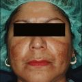
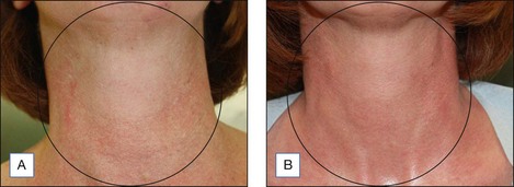
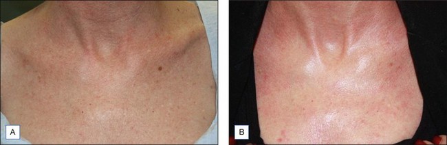
At this point, 25% TCA is applied. The resulting stinging sensation is brief (a few minutes) and patients can be consoled with the fact that cool compresses will soon be applied once the appropriate level of frosting appears. The TCA solution is typically applied using large, smooth cotton applicators so as not to introduce additional friction to the process which would unintentionally create a deeper peel. To promote patient comfort further the application is done in an expedited fashion. Frosting on nonfacial areas is usually slower, so it is important not to over treat a given areas because the frost has not uniformly appeared. At the maximum, a level 2 frost (even white enamel with erythematous background) should be the goal for medium-depth peels off the face while areas such as the neck should undergo less intense frosting. The major determinants for the degree of TCA penetration or frost are as follows: extent of degreasing, the amount of peeling agent and concentration used, type of applicator chosen, the duration of contact with skin, the degree of rubbing/pressure of application, sebaceous nature of the skin being treated and the number of coats applied. Figure 12.1 shows improvement of neck photodamage following Jessner’s/25% TCA peel.

Immediate relief of the peel’s stinging sensation in frosted areas is accomplished by application of iced saline/water compresses or ice packs. Alternately, a Zimmer cold air chiller can be employed during and after the peel to help soothe the skin. After 5–8 minutes the patient will state that the skin is feeling essentially normal again. They may state that treated areas feel ‘puffy’ and ‘tight’ at the same time. In almost all of our medium-depth peel patients, we pretreat them with oral loratidine (10 mg) to minimize the postpeel swelling. As the frost is receding, Aquaphor or petrolatum can be applied thickly. To create a short-term inexpensive occlusive dressing, plastic wrap from the grocery store can be placed on top of the ointment. Since fungal infections can result with prolonged (>48 hours continuous occlusion), we advise patients to utilize this plastic wrap if desired for the first two days then only when sleeping for 3–4 additional days. It has been shown that wounds occluded wounds reepithelialize faster than those that are desiccated from being left open to the air.

With respect to the neck, both the physician and patient should expect gradual improvement which will be evident after a series of peels. Even if it is technically possible to use more aggressive concentrations of TCA here, a sequential approach is advisable. In the case of poikiloderma of Civatte, as the dyschromia fades the vascular component may actually become more prominent. Patients should understand that 2–3 treatments with intense pulsed light or pulsed dye laser are needed to address the vascular aspect. Typical settings on the with the Candela VBEAM Perfecta: 7–10 mm spot size, 5–6.5 Joules, 10 msec pulse duration and cooling. Settings with the Cutera Limelight intense pulse light system are: Program A, 12–16 joules after application of thin layer of clear ultrasound gel.

In the late 1990s, Cook & Cook developed a technique which exfoliates nonfacial skin in a controlled manner. In their medium-depth peel, 70% glycolic acid gel is applied to the skin followed by TCA 40% solution to the same area. The gel vehicle for the glycolic acid is essential because it serves as a partial barrier to TCA penetration. Typically, the endpoint of white speckles appear with a background of pink skin which occurs within three minutes of application. Once this occurs, the treated area is neutralized with sodium bicarbonate 10% solution, and the peel is completed. Reported clinical results included smoother skin texture, decreased wrinkling and striae, fading of lentigines and other dyschromias.

Striae

Chemical peeling can also be a useful modality for improving the appearance of mature striae distensae. We often combine a light chemical peel with subcision for maximal clinical effect. Usually these treatments are performed as a series of 3–6 sessions at monthly intervals. The purpose of the peel is to improve atrophy within the epidermis and dermis, while the intent of subcision is to interrupt any dermal tethering as well as promotion of collagen remodeling. Striae are characterized histologically by epidermal flattening with loss of rete ridges, dermal atrophy as well as breakage and retraction of elastic fibers.

In a pilot study, we reported safe and equally effective treatment of mature abdominal striae in women with phototypes II–IV utilizing subcision, focal TCA (20%) peeling and the combination of both of these procedures. Clinically we found that there was a reduction in width of the striae with subcision while TCA application produced a reduction in overall length. Unlike the study of Luis-Montoya et al, we did not note necrosis or other significant adverse events following subcision.

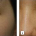
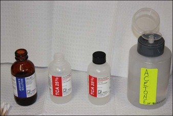
Priming of the skin as listed in Table 12.1 is encouraged. The skin is prepared by degreasing it with alcohol and acetone on a gauze pad. Proximal and distal limits of the striae are then marked. Local infiltration is with 2% lidocaine with epinephrine. After anesthesia is achieved, an 18G Nokor BD needle is inserted first at a 45° angle. Once the deep dermal plane is entered, the needle is adjusted to a more shallow angle. Back and forth movements along the length of striae are performed until no resistance is met and dissection of this plane is accomplished. A blush of purple ecchymosis is expected toward the end of the procedure and may be present for 2–5 days. Following subcision, the area is re-swabbed with alcohol and 3–4 coats of 20% TCA (using a cotton tipped applicator) is applied directly to the striae (Figure 12.2). A faint frost will be appreciated.

At the conclusion of the procedure, Aquaphor or petrolatum ointment is applied to treated areas. Patients are instructed to cleanse these areas with a gentle cleanser and may apply bland moisturizer twice daily to promote healing and hasten exfoliation. Sun avoidance in the immediate postprocedure period and broad spectrum photo protection thereafter are recommended. From the outset, patients are advised that multiple sessions (3–6 in most people) are needed to see substantive improvement.

Keratosis pilaris on the extremities

Keratosis Pilaris (KP) is characterized by epidermal hyperkeratosis, hypergranulosis, and plugging of individual hair follicles and predominantly occurs on the lateral upper arms and thighs. While there is no cure per se for the condition, the appearance and texture can be ameliorated with the addition of a series of superficial peels. Most patients with KP are initially started on a variety of topical medications including hydroxy acids (like lactic acid, salicyclic acid, or glycolic acid), urea preparations, retinoids (tretinoin, adapalene or tazarotene), topical steroids or immune modulators (pimecrolimus or tacrolimus). If excess pigmentation is present, bleaching creams containing hydroquinone or alternatives can be applied.

Since there is an inflammatory component to KP, the areas are only gently cleansed and lightly degreased with alcohol. Glycolic acid (70%) peels can be applied for 5–7 minutes initially and time can be titrated upwards as tolerated. Peeled areas are then neutralized with either water or sodium bicarbonate. If significant erythema develops afterwards, low to medium potency topical steroids can be applied twice daily for 1–2 days. Otherwise, areas will be cleansed with a gentle cleanser followed by a bland moisturizer for the first 48 hours. After this point, the regular topical KP program can be restarted. The goal of peeling in KP is twofold: exfoliation of keratotic follicular debris and enhancing the penetration of at home skin maintenance regimens.

Acne on the trunk

For acne occurring on the chest and back, superficial chemical peels can be a useful adjunct to standard topical and oral therapy. The main differences when treating body versus facial locations are that peeling solutions concentrations are generally higher and applications times are longer. While salicylic acid peels are more lipophyllic, glycolic acid peels may also be used effectively for this indication. Patients should be counseled that more than one peel (often 3–5 peels spaced at 2–4 weeks intervals) is required for improvement. On the back where sebaceous structures are numerous, a more vigorous degreasing with both alcohol and acetone can be performed. The chest is approached with a more mild degreasing with alcohol ± acetone. Typical peeling agents that are used to treat back and chest acne include salicylic acid (20–30%) and glycolic acid (70%). While salicylic acid is considered to be autoneutralizing, the precipitated white crystals which appear on the skin’s surface can be rinsed off the areas after 3–5 minutes. Application times for glycolic acid peels on sites such as the trunk and arms are 7–10 minutes or longer until mild erythema appears. These glycolic acid peels require neutralization with water or sodium bicarbonate. In patients with darker phototypes or where postinflammatory hyperpigmentation is already present, a topical steroid lotion or cream can be applied immediately after the peel and then twice daily for 24–48 hours to minimize the risk of developing further hyperpigmentation due to excess inflammation. Patients can continue with their oral medications while delaying application of potentially irritating topical acne preparations for 2–4 days.

Side Effects and Complication Management

The most common complication with chemical peeling is irregular pigmentation. When using lower concentrations of TCA (20–30%) hyperpigmentation is more likely; while hypopigmentation is more common when using higher concentrations. Fortunately, when postpeel hyperpigmentation occurs it is transient and can be resolved with topical bleaching products (hydroquinone or alternatives). In some cases, we actually perform superficial chemical peels (such as glycolic acid 30–40%) to expedite resolution. Patients in whom postpeel hyperpigmentation occurs should be advised regarding the desirability of temporary sun avoidance to the affected areas and daily usage and reapplication of physical sunscreens.

The expected duration of erythema after a chemical peel is dependent on the depth of injury of the peeling solution. With medium-depth peels, erythema is usually resolved by the second month. However, if ‘hot spots’ of redness are noted, we will generally have the patient apply a medium potency topical steroid two to three times daily for a few days. In a scenario where early postpeel erythema is accompanied by burning, stinging or itching, contact dermatitis (to retinoids, other components of the home regimen or aerosol chemical irritants), incipient infection or flaring of an underlying skin condition should be on the differential diagnosis. The underlying cause should be identified and possible irritants avoided. When the erythema is persistent or is accompanied by induration, impending scarring should be suspected and aggressively treated. Depending on the severity of the erythema, close monitoring along with a combination of topical steroids (medium to high potency bid to tid), antihistamines (such as loratidine 10 mg qd), low level vascular laser sessions (every 2–4 weeks), intralesional steroids for firm erythematous lesions (triamcinolone 10–40 mg/mL) or a tapering course of oral steroids can be employed. Patients should come in weekly for clinical and photographic assessment until resolution.

Infection with chemical peeling is very rare since most peeling agents are bactericidal. Proper wound care can minimize the risk of infection. Patients are given both verbal and written wound care instructions. For most of our medium-depth peels, patients will be cleansing the areas with dilute acetic acid 0.25% solution (1 pint warm water mixed with one tablespoon white vinegar) 2–4 times daily and rinsing with tap water. The mild acidity of this solution has antibacterial effects. After cleansing, liberal application of bland emollients like (Aquaphor, petrolatum and others) is suggested until re-epithelialization is complete. If infection is suspected, a culture should be taken and empiric antibiotics should be started. While gram positive bacterial causes (Streptococcal and Staphlococcal organisms) are more commonly documented, gram negative (Pseudomonas and Escherichia coli) and fungal (Candida) infections can also be seen. When peeling off the face, reactivation of herpes infection is much less common and prophylaxis is not routinely given. Nonetheless, if localized pain, erythema, ulcers and/or grouped vesicles appear after peeling, herpes infection should be suspected. Evaluation with viral cultures or direct fluorescent antibody testing should be performed and empiric anti-viral medication should be started.

Further Reading

Alam M, Omura N, Kaminer MS. Subcision for acne scarring: technique and outcomes in 40 patients. Dermatologic Surgery. 2005;31:310-317.

Baumann L. Depigmenting agents. In: Day DJ, editor. Understanding hyperpigmentation. What you need to know. Aurora, California: Intellyst medical communications, 2004.

Brody HJ. Chemical peeling and resurfacing, 2nd ed. Atlanta: Emory University Digital Library Publications; 2008.

Coleman WP, Futrell JM. The Glycolic acid trichloroacetic acid peel. Journal of Dermatologic Surgery and Oncology. 1994;20:76-80.

Cook KK, Cook WR. Chemical peel of nonfacial skin using glycolic acid gel augmented with TCA and neutralized based on visual staging. Dermatologic Surgery. 2000;26:994-999.

Grimes PE. Glycolic acid peels in blacks. In: Moy R, Luffman D, Kakita L, editors. Glycolic acid peels. Marcel Dekker, Inc; 2002:179-186.

Hevia O, Nemeth AJ, Taylor JR. Tretinoin accelerates healing after trichloroacetic acid chemical peel. Archives of Dermatology. 1991;127:678-682.

Hexsel DM, Mazzucco R. Subcision a treatment for cellulite. International Journal of Dermatology. 2000;39:539-544.

Kang WH, et al. Atrophic acne scar treatment using triple combination therapy: dot peeling, subcision and fractional laser. Journal of Cosmetic and Laser Therapy. 2009;11:212-215.

Luis-Montoya P, et al. Evaluation of subcision as a treatment for cutaneous striae. Journal of Drugs in Dermatology. 2005;4:346-350.

Maibach HF, Rovee DT. Epidermal Wounds Healing. St Louis: Mosby; 1972.

Monheit GD. The Jessner’s and TCA peel: A medium depth chemical peel. Journal of Dermatologic Surgery and Oncology. 1989;15:945-950.

Rendon MI, Gaviria JI. Review of skin-lightening agents. Dermatologic Surgery. 2005;31:886-889.

Rubin M. Manual of chemical peels. Philadelphia: Lippincott; 1995. pp. 120–121

Sato MS, et al. Clinical evaluation of the efficacy of trichloroacetic acid and subcision, combined or isolated, for abdominal striae. Sociedad Brasileira de Dermatologia-Surgical and Cosmetic Dermatology. Online access for English translation at. www.surgicalcosmetic.org.br, 2009. 1(4)