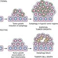
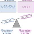
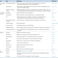
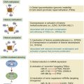
Figure 21-1 Biomarkers in cancer etiology Biomarkers in cancer etiologic studies can be classified into three broad categories: biomarkers of exposure, biomarkers of effect, and biomarkers of susceptibility. Biomarkers of susceptibility (cancer risk) can be derived from each of the steps along the continuum of the carcinogenic process, reflecting interindividual variations in absorption, distribution, metabolism (activating and detoxifying), and excretion of carcinogens; sensitivity to formation of macromolecule (DNA and protein) adducts; and ability to repair macromolecule damage, restore normal cellular functions, and eliminate premalignant cells.
Biomarkers of Internal Dose
Biomarkers of internal dose measure levels of a carcinogen or its metabolite in human tissues, bodily fluids, and excreta. 5 These biomarkers are not bound to cellular targets but provide a measure of exposure, absorption, metabolism, and excretion. There is generally a good correlation between external exposure and internal dose; however, the involvement of absorption and metabolism and interindividual variation in these processes suggest that the relationship may not always be simple. One of the classical examples of validated biomarkers of internal dose that contributed greatly to elucidate the environmental cause of human cancer is urinary aflatoxin and its metabolites. Aflatoxins have long been suspected to be human hepatic carcinogens, but the strongest evidence came from a prospective nested case-control study in which the authors measured urinary aflatoxin B1 (AFB1), its metabolites AFP1 and AFM1, and DNA adducts (AFB1-N7-Guanine) to assess the relation between aflatoxin exposure and liver cancer. Subjects with liver cancer were more likely to have detectable concentrations of any of the aflatoxin metabolites than controls, and the highest relative risk was for AFP1 (6.2-fold). Moreover, there was a strong interaction between chronic hepatitis B infection and aflatoxin exposure in liver cancer risk. 6 Tobacco-specific metabolites are the most studied biomarkers of internal dose. Cotinine is the main metabolite of nicotine, and the measurement of serum/plasma cotinine offers higher accuracy than self-reports in assessing tobacco smoking. 7 Two prospective studies have reported that higher levels of serum cotinine 8 and urinary cotinine 9 were associated with higher risk of lung cancer. The risk estimate of tobacco smoking and lung cancer from serum cotinine might be stronger than from questionnaire-based studies. The highest risk group had a 55-fold increased risk with no clear suggestion of a plateau in risk at high exposure levels, suggesting that analyzing the relationship between serum cotinine and lung cancer risk might contribute to a better quantitative assessment of tobacco-related lung carcinogenesis. 8 Additional prediagnostic urinary metabolites of tobacco carcinogens, such as NNAL (a metabolite of tobacco carcinogen NNK) and PheT (a metabolite of tobacco carcinogen polycyclic aromatic hydrocarbons [PAHs]), have also been associated with increased risk of lung cancer. 9,10 Other examples of biomarkers of internal dose include hormones or nutrients in body fluids and circulating antibodies to infectious agents (e.g., Helicobacter pylori in gastric cancer, hepatitis B virus [HBV] in liver cancer, and HPV in cervical cancer). 5
Biomarkers of Biologically Effective Dose
Many carcinogens are metabolically activated and bind to DNA and/or protein to form adducts. DNA adducts have been the most evaluated biomarker of biologically effective dose, and a broad range of different DNA adducts have been measured in human samples using various approaches, including 32P-postlabeling, immunoassays and immunohistochemistry, and mass spectrometry. 11,12 Because of the difficulty in measuring adducts in target tissues, almost all of the population studies have used easily accessible surrogate samples, typically plasma/serum, white blood cells, or urine. The first prospective study showing a significant association between carcinogen-DNA adducts and subsequent cancer development was the aforementioned aflatoxin study in liver cancer. 6,13 Men with detectable urinary aflatoxin-DNA adduct (AFB1-N7-Guanine) and no HBV infection exhibited a ninefold increased liver cancer risk compared to men without detectable AFB1-N7-Guanine and no HBV infection. Tobacco smoke contains many different carcinogens, including PAHs, aromatic amines, and nitrosamines. Three prospective studies have evaluated the association of PAH-DNA or related aromatic-DNA adducts with the risk of lung cancer. 14–16 In a pioneering nested case-control study within the prospective Physicians’ Health Study, the authors measured aromatic-DNA adducts in white blood cells (WBCs) at baseline and found current smokers who had higher levels of aromatic-DNA adducts in WBCs exhibited approximately threefold increased risk of lung cancer compared to current smokers with lower adduct concentrations. There were no associations in former and never smokers. 14 Two subsequent prospective studies, a pooled analysis of the three prospective studies, as well as a meta-analysis of nine studies (including retrospective case-control studies) recapitulated the main observation that bulky DNA adducts are associated with elevated lung cancer risk in current smokers after a follow-up of several years. 15–17 Protein adducts, such as hemoglobin (Hb) adducts of tobacco carcinogens and AFB1-albumin adducts, have also been used as biomarkers of biologically effective dose. 1,18,19

