Barrett Esophagus

Published on 18/07/2015 by admin

Filed under Radiology

Last modified 22/04/2025

Print this page

rate 1 star rate 2 star rate 3 star rate 4 star rate 5 star
Your rating: none, Average: 0 (0 votes)

This article have been viewed 2281 times

 Due to more severe reflux disease

image Hiatal hernia in almost all patients
image Mid esophageal mucosal irregularity, stricture, deep ulceration
image Risk of cancer > short-segment type
• Short segment: Columnar epithelium ≤ 3 cm above GE junction

image More common than long segment (reported in 2-12% of patients with chronic reflux at endoscopy)
image Due to less severe reflux disease
image Distal esophageal reticular mucosa, ± stricture, ± shallow ulceration

TOP DIFFERENTIAL DIAGNOSES

• Esophageal carcinoma
• Reflux esophagitis
• Candida esophagitis
• Viral esophagitis
• Radiation esophagitis
• Caustic esophagitis
• Drug-induced esophagitis
• Scleroderma

CLINICAL ISSUES

• Risk of adenocarcinoma based on morphology

image High risk: Midesophageal stricture, ulcer, reticular mucosa
image Moderate risk: Distal peptic stricture and reflux esophagitis
image Low risk: If none of above findings are present
• Diagnosis: Endoscopy with biopsy
image
(Left) Graphic shows a type 1 hiatal hernia, distal esophageal stricture, and nodular mucosal surface. Note the discrete ulcer image 
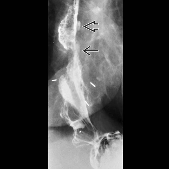
and an adenocarcinoma image represented by a raised sessile lesion with an irregular surface.

image
(Right) 2 views from an esophagram show a mid esophageal stricture image 
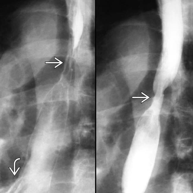
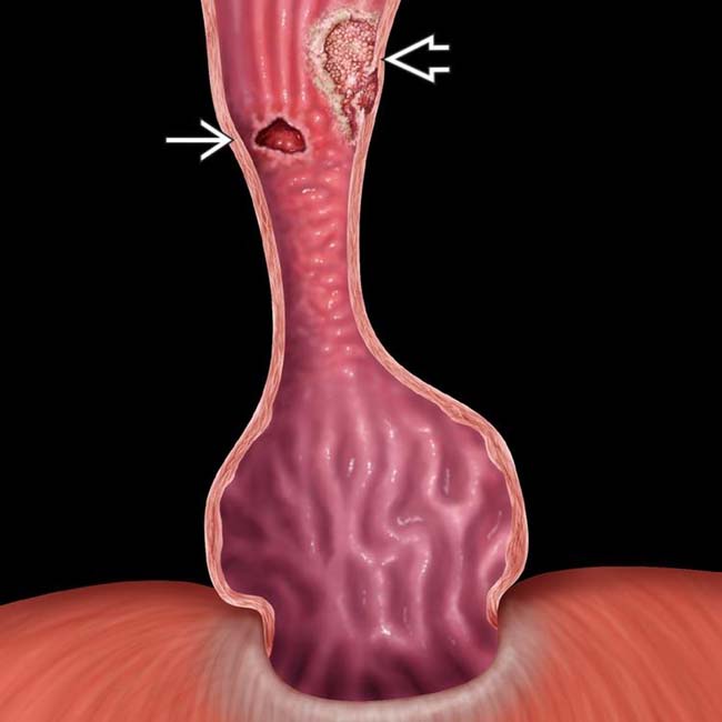
and ulcer in a patient with a small hernia image and reflux.
image
(Left) Endoscopic image shows a large ulcer image 
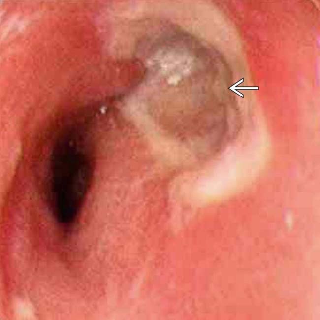
with the velvet texture of Barrett mucosa and stricture. Normal esophageal mucosa has a shiny, smooth, pink surface.

image
(Right) Two views from an esophagram show a polypoid mass image 
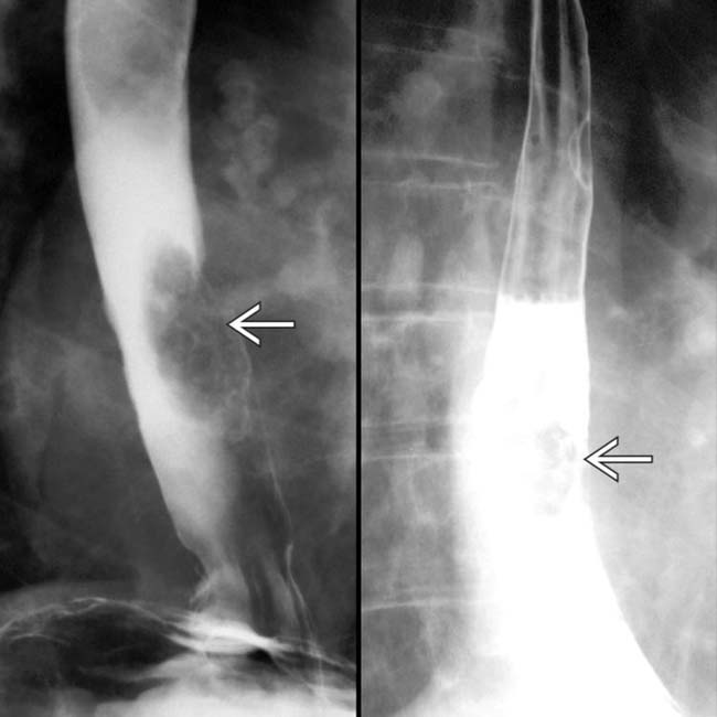
that represents an adenocarcinoma arising in Barrett mucosa.

TERMINOLOGY

Definitions

• Metaplasia of distal esophageal squamous epithelium to columnar epithelium

IMAGING

General Features

• Best diagnostic clue

image Mid esophageal stricture with hiatal hernia and reflux is essentially pathognomonic
• Other general features

image Acquired condition due to reflux esophagitis
image Premalignant condition associated with increased risk of esophageal adenocarcinoma

– Risk: 30-40x higher than in general population
– 90-100% of adenocarcinomas arise from Barrett mucosa

Radiographic Findings

• Double contrast esophagography is imaging of choice
• Classified into 2 types based on endoscopy and histopathologic findings

image Long segment: Columnar epithelium > 3 cm above gastroesophageal (GE) junction

– Due to more severe reflux disease
– Hiatal hernia in almost all patients
– Mid esophageal mucosal irregularity, stricture, ulceration
– Greater cancer risk in long- vs. short-segment type
image Short segment: Columnar epithelium ≤ 3 cm above GE junction

– Due to less severe reflux disease
– Hiatal hernia present in 72% of patients

DIFFERENTIAL DIAGNOSIS

Esophageal Carcinoma

• Asymmetric contour with abrupt proximal borders (“rat tail” appearance)

Esophagitis

• Reflux esophagitis

image May be impossible to distinguish from short-segment Barrett esophagus on imaging
• Candida esophagitis

image Multiple tiny, round lucencies ± ulcers
• Viral esophagitis

image Superficial ulcers on normal mucosa
image Usually in immunocompromised patients
• Caustic esophagitis

image Long ulceration and stricture
image Diagnosis: History and endoscopic biopsy
• Drug-induced esophagitis

image Acute onset of odynophagia with ulceration and spasm
image Diagnosis made by imaging and classic history

Scleroderma

• Aperistalsis, dilated esophagus, patulous or strictured GE junction

PATHOLOGY

General Features

• Etiology

image Chronic GE reflux of acid and pepsin with chronic esophagitis

– Re-epithelialization by pluripotent stem cells

image Differentiate into gastric or intestinal epithelium

Gross Pathologic & Surgical Features

• Velvety, pinkish-red columnar mucosa

Microscopic Features

• Proximally: Specialized columnar epithelium

image Villous architecture with goblet cells
• Distal to above: Junctional-type epithelium

image Cardiac mucous glands
• More distally: Fundic-type epithelium

image Parietal and chief cells

CLINICAL ISSUES

Presentation

• Most common signs/symptoms

image Reflux symptoms

– “Heartburn,” and angina-like pain
– 20-40% of patients are asymptomatic
• Diagnosis

image Endoscopy, biopsy, and histopathology

Demographics

• Age

image Mean: 55-65 years; prevalence increases with age
• Gender

image M:F = 2:1
• Ethnicity

image Caucasians > African Americans (10:1)
• Epidemiology

image Prevalence: 2-12 % of patients with reflux esophagitis
image Increased risk of adenocarcinoma in Barrett mucosa

– Estimated 30-40x higher risk than general population
– Accounts for almost all cases of adenocarcinoma

Treatment

• Medical (nonoperative): Antacids, cessation of irritants
• Surgical

image Fundoplication to prevent reflux
image Partial esophagectomy for long-segment Barrett esophagus with severe ulceration, stricture, or dysplasia

Clinical Features

• Risk of adenocarcinoma based on morphology

image High risk: Midesophageal stricture, ulcer, reticular mucosa
image Moderate risk: Distal peptic stricture and reflux esophagitis
image Low risk: If none of above findings are present
• Diagnosis: Endoscopy with biopsy

DIAGNOSTIC CHECKLIST

Consider

• Rule out other causes of esophageal stricture ± ulceration; consider carcinoma

Image Interpretation Pearls

• Long to mid esophageal or short distal esophageal stricture/ulcer associated with hiatal hernia/GE reflux
image
Esophagram shows a hiatal hernia and strictures at the GE junction image 
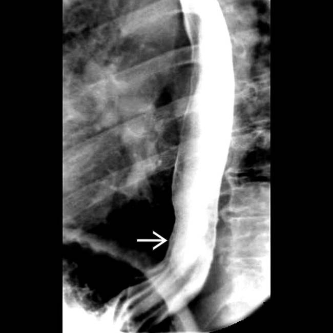
and higher. Note the discrete ulcer image.

image
Esophagram shows subtle raised plaque image in the distal esophagus in this patient with adenocarcinoma on Barrett mucosa.
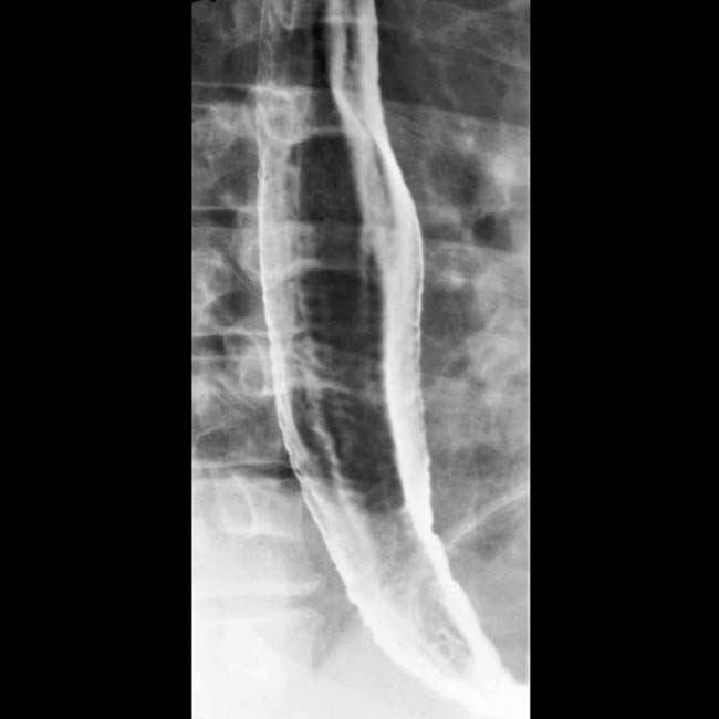
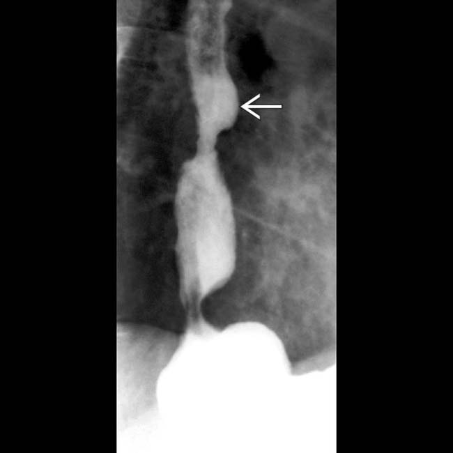
image
Esophagram shows diffuse nodularity of the distal esophagus.
image
Esophagram shows 2 strictures on an ulcer image and hiatal hernia.

Share this: