Bacterial Genetics, Metabolism, and Structure

Published on 08/02/2015 by admin

Filed under Basic Science

Last modified 22/04/2025

Print this page

rate 1 star rate 2 star rate 3 star rate 4 star rate 5 star
Your rating: none, Average: 3.8 (5 votes)

This article have been viewed 3853 times

Bacterial Genetics, Metabolism, and Structure

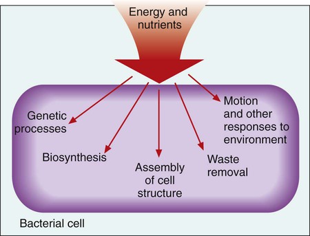
Microbial genetics, metabolism, and structure are the keys to microbial viability and survival. These processes involve numerous pathways that are widely varied, often complicated, and frequently interactive. Essentially, survival requires energy to fuel the synthesis of materials necessary to grow, propagate, and carry out all other metabolic processes (Figure 2-1). Although the goal of survival is the same for all organisms, the strategies microorganisms use to accomplish this vary substantially.

Knowledge regarding genetic, metabolic, and structural characteristics of microorganisms provides the basis for understanding almost every aspect of diagnostic microbiology, including:

Microorganisms vary significantly in many genetic and therefore physiologic aspects. A detailed consideration of these differences is beyond the scope of this textbook. Therefore, a generalized description of bacterial systems is used as a model to discuss microbial genetics, metabolism, and structure. Information regarding characteristics of fungi, parasites, and viruses can be found in subsequent chapters that discuss these specific taxonomic groups.

Bacterial Genetics

Genetics, the process of heredity and variation, is the starting point from which all other cellular pathways, functions, and structures originate. The ability of a microorganism to maintain viability, adapt, multiply, and cause disease is determined by the organism’s genetic composition. The three major aspects of microbial genetics that require discussion include:

Nucleic Acid Structure and Organization

For all living entities, hereditary information resides or is encoded in nucleic acids. The two major classes of nucleic acids are deoxyribonucleic acid (DNA), which is the most common macromolecule that encodes genetic information, and ribonucleic acid (RNA). In some forms, RNA encodes genetic information for various viruses; in other forms, RNA plays an essential role in several of the genetic processes in prokaryotic and eukaryotic cells, including the regulation and transfer of information. Prokaryotic or “prenuclear” organisms do not have membrane bound organelles and the cells’ genetic material is therefore not enclosed in a nucleus. Eukaryotic “true nucleus” are all of the organisms that have their genetic material enclosed in a nuclear envelope.

Nucleotide Structure and Sequence

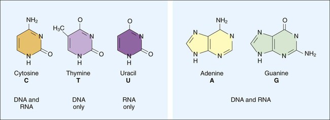
DNA consists of deoxyribose sugars connected by phosphodiester bonds (Figure 2-2, A). The bases that are covalently linked to each deoxyribose sugar are the key to the genetic code within the DNA molecule. The four bases include two purines, adenine (A) and guanine (G), and the two pyrimidines, cytosine (C) and thymine (T) (Figure 2-3). In RNA, uracil replaces thymine. The combined sugar, phosphate, and a base form a single unit referred to as a nucleotide (adenosine triphosphate [ATP], guanine triphosphate [GTP], cytosine triphosphate [CTP], and thymine triphosphate [TTP]). DNA and RNA are nucleotide polymers (i.e., chains or strands), and the order of bases along a DNA or RNA strand is known as the base sequence. This sequence provides the information that codes for the proteins that will be synthesized by microbial cells; that is, the sequence is the genetic code.

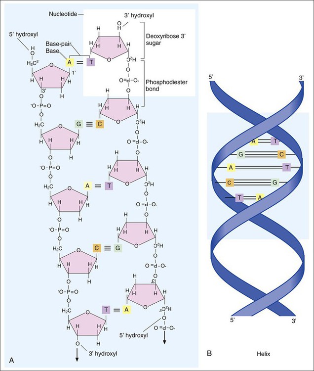
DNA Molecular Structure

The intact DNA molecule is composed of two nucleotide polymers. Each strand has a 5’ (prime) phosphate and a 3’ (prime) hydroxyl terminus (see Figure 2-2, A). The two strands run antiparallel, with the 5’ of one strand opposed to the 3’ terminal of the other. The strands are also complementary, because the adenine base of one strand always binds to the thymine base of the other strand by means of two hydrogen bonds. Similarly, the guanine base of one strand always binds to the cytosine base of the other strand by means of three hydrogen bonds. As a result of the molecular restrictions of these base pairings, along with the conformation of the sugar-phosphate backbones oriented in antiparallel fashion, DNA has the unique structural conformation often referred to as a “twisted ladder” or double helix (see Figure 2-2, B). Additionally, the dedicated base pairs provide the format essential for consistent replication and expression of the genetic code. In contrast to DNA, which carries the genetic code, RNA rarely exists as a double-stranded molecule. The three major types of RNA (messenger RNA [mRNA], transfer RNA [tRNA], and ribosomal RNA [rRNA]) play key roles in gene expression.

Genes and the Genetic Code

A DNA sequence that encodes for a specific product (RNA or protein) is defined as a gene. Thousands of genes in an organism encode messages or blueprints for the production of one or more proteins and RNA products that play essential metabolic roles in the cell. All the genes in an organism comprise the organism’s genome. The size of a gene and an entire genome is usually expressed in the number of base pairs (bp) present (e.g., kilobases [103 bases], megabases [106 bases]).

Certain genes are widely distributed among various organisms while others are limited to particular species. Also, the base pair sequence for individual genes may be highly conserved (i.e., show limited sequence differences among different organisms) or be widely variable. As discussed in Chapter 8, these similarities and differences in gene content and sequences are the basis for the development of molecular tests used to detect, identify, and characterize clinically relevant microorganisms.

Chromosomes

The genome is organized into discrete elements known as chromosomes. The set of genes within a given chromosome is arranged in a linear fashion, but the number of genes per chromosome is variable. Similarly, although the number of chromosomes per cell is consistent for a given species, this number varies considerably among species. For example, human cells contain 23 pairs (i.e., diploid) of chromosomes whereas bacteria contain a single, unpaired (i.e., haploid) chromosome.

Bacteria are classified as prokaryotes; therefore, the chromosome is not located in a membrane-bound organelle (i.e., nucleus). The bacterial chromosome contains the genes essential for viability and exists as a double-stranded, closed, circular macromolecule. The molecule is extensively folded and twisted (i.e., supercoiled) in order to fit within the confined space of the bacterial cell. The linearized, unsupercoiled chromosome of the bacterium Escherichia coli is about 1300 µm long, but it fits within a cell 1 × 3 µm; this attests to the extreme compact structure of the supercoiled bacterial chromosome. For genes in the compacted chromosome to be expressed and replicated, unwinding or relaxation of the molecule is required.

In contrast to the bacterial chromosome, the chromosomes of parasites and fungi number more than one per cell, are linear, and are housed within a membrane-bound organelle (the nucleus) of the cell. This difference is a major criterion for classifying bacteria as prokaryotic organisms and fungi and parasites as eukaryotes. The genome topology of a virus may consist of DNA or RNA contained within a protein coat rather than a cell.

Nonchromosomal Elements of the Genome

Although the bacterial chromosome represents the majority of the genome, not all genes in a given cell are confined to the chromosome. Many genes may also be located on plasmids and transposable elements. Both of these extrachromosomal elements are able to replicate and encode information for the production of various cellular products. Although considered part of the bacterial genome, they are not as stable as the chromosome and may be lost during cellular replication, often without any detrimental effects on the viability of the cell.

Plasmids exist as double-stranded, closed, circular, autonomously replicating extrachroosomal genetic elements ranging in size from 1 to 2 kilobases up to 1 megabase or more. The number of plasmids per bacterial cell varies extensively, and each plasmid is composed of several genes. Some genes encode products that mediate plasmid replication and transfer between bacterial cells, whereas others encode products that provide a specialized function, such as determinants of antimicrobial resistance or a unique metabolic process. Unlike most chromosomal genes, plasmid genes do not usually encode for products essential for viability. Plasmids, in whole or in part, may also become incorporated into the chromosome.

Transposable elements are pieces of DNA that move from one genetic element to another, from plasmid to chromosome or vice versa. Unlike plasmids, they are unable to replicate independently and do not exist as separate entities in the bacterial cell. The two types of transposable elements are the simple transposon or insertion sequence (IS) and the composite transposon. Insertion sequences are limited to containing the genes that encode information required for movement from one site in the genome to another. Composite transposons are a cassette (grouping of genes) flanked by insertion sequences. The internal gene imbedded in the insertion sequence encodes for an accessory function, such as antimicrobial resistance. Plasmids and transposable elements coexist with chromosomes in the cells of many bacterial species. These extrachromosomal elements play a key role in the exchange of genetic material throughout the bacterial microbiosphere, including genetic exchange among clinically relevant bacteria.

Replication and Expression of Genetic Information

Replication

Bacteria multiply by cell division, resulting in the production of two daughter cells from one parent cell. As part of this process, the genome must be replicated so that each daughter cell receives an identical copy of functional DNA. Replication is a complex process mediated by various enzymes, such as DNA polymerase and cofactors; replication must occur quickly and accurately. For descriptive purposes, replication may be considered in four stages (Figure 2-4):
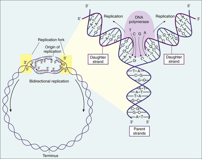
Relaxation of supercoiled chromosomal DNA is required so that enzymes and cofactors involved in replication can access the DNA molecule at the site where the replication process will originate (i.e., origin of replication). The origin of replication (a specific sequence of approximately 300 base pairs) is recognized by several initiation proteins, followed by the separation of the complementary strands of parental DNA. Each parental strand serves as a template for the synthesis of a new complementary daughter strand. The site of active replication is referred to as the replication fork; two bidirectional forks are involved in the replication process. Each replication fork moves through the parent DNA molecule in opposite directions so that replication is a bidirectional process. Activity at each replication fork involves different cofactors and enzymes, with DNA polymerases playing a central role. Using each parental strand as a template, DNA polymerases add nucleotide bases to each growing daughter strand in a sequence that is complementary to the base sequence of the template (parent) strand. The complementary bases of each strand are then held together by hydrogen bonding between nucleotides and the hydrophobic nature of the nitrogenous bases. The new nucleotides can be added only to the 3’ hydroxyl end of the growing strand so that synthesis for each daughter strand occurs only in a 5’ to 3’ direction.

Termination of replication occurs when the replication forks meet. The result is two complete chromosomes, each containing two complementary strands, one of parental origin and one newly synthesized daughter strand. Although the time required for replication can vary among bacteria, the process generally takes approximately 20 to 40 minutes in rapidly growing bacteria such as E. coli. The replication time for a particular bacterial strain can vary depending on environmental conditions, such as the availability of nutrients or the presence of toxic substances (e.g., antimicrobial agents).

Expression of Genetic Information

Gene expression is the processing of information encoded in genetic elements (i.e., chromosomes, plasmids, and transposons), which results in the production of biochemical molecules, including RNA molecules and proteins. The overall process of gene expression is composed of two complex steps, transcription and translation. Gene expression requires various components, including a DNA template representing a single gene or cluster of genes, various enzymes and cofactors, and RNA molecules of specific structure and function.

Transcription.

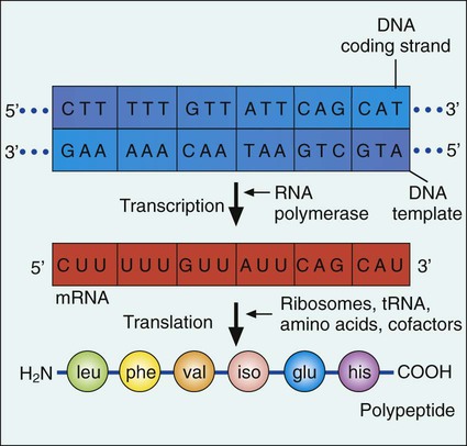
Gene expression begins with transcription. During transcription the DNA base sequence of the gene (i.e., the genetic code) is converted into an mRNA molecule that is complementary to the gene’s DNA sequence (Figure 2-5). Usually only one of the two DNA strands (the sense strand) encodes for a functional gene product. This same strand is the template for mRNA synthesis.

RNA polymerase is the enzyme central to the transcription process. The enzyme is composed of four protein subunits and a sigma factor. Sigma factors are required for the RNA polymerase to identify the appropriate site on the DNA template where transcription of mRNA is initiated. This initiation site is also known as the promoter sequence. The remainder of the enzyme functions to unwind the double-stranded DNA at the promoter sequence and use the DNA strand as a template to sequentially add ribonucleotides (ATP, GTP, uracil triphosphate [UTP], and CTP) to form the growing mRNA strand.

Transcription proceeds in a 5’ to 3’ direction. However, in mRNA, the TTP of DNA is replaced with UTP. TTP contains thymine, and UTP contains uracil. Both molecules contain a heterocyclic ring and are classified as pyrimidines. During synthesis and modification of these molecules, a portion of the molecules are dehydroxylated, forming a 2′-deoxy-nucleotide monophosphate. The dUMP (dehydroxylated uracil monophosphate) is then methylated, forming dTMP (dehydroxylated thymine monophosphate). Following phosphorylation, thymine is only found in the final state as deoxythymidine and therefore cannot be incorporated into an RNA molecule. Synthesis of the single-stranded mRNA product ends when specific nucleotide base sequences on the DNA template are encountered. Termination of transcription may be facilitated by a rho (a prokaryotic protein) cofactor or an intrinsic termination sequence. Both of these mechanisms disrupt the mRNA-RNA polymerase template DNA complex.

In bacteria, the mRNA molecules that result from the transcription process are polycistronic, that is, they encode for several gene products. Frequently, polycistronic mRNA may encode several genes whose products (proteins) are involved in a single or closely related cellular function. When a cluster of genes is under the control of a single promoter sequence, the gene group is referred to as an operon.

The transcription process not only produces mRNA but also tRNA and rRNA. All three types of RNA have key roles in protein synthesis.

Translation.

The next phase in gene expression, translation, involves protein synthesis. Through this process the genetic code in mRNA molecules is translated into specific amino acid sequences that are responsible for protein structure and function (see Figure 2-5).

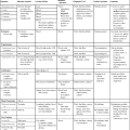
Before addressing the process of translation, a discussion of the genetic code that is originally transcribed from DNA to mRNA and then translated from mRNA to protein is warranted. The code consists of triplets of nucleotide bases, referred to as codons; each codon encodes for a specific amino acid. Because there are 64 different codons for 20 amino acids, an amino acid can be encoded by more than one codon (Table 2-1). Each codon is specific for a single amino acid. Therefore, through translation, the codon sequences in mRNA direct which amino acids are added and in what order. Translation ensures that proteins with proper structure and function are produced. Errors in the process can result in aberrant proteins that are nonfunctional, underscoring the need for translation to be well controlled and accurate.

TABLE 2-1

The Genetic Code as Expressed by Triplet-Base Sequences of mRNA*

Codon Amino Acid Codon Amino Acid Codon Amino Acid Codon Amino Acid
UUU Phenylalanine CUU Leucine GUU Valine AUU Isoleucine
UUC Phenylalanine CUC Leucine GUC Valine AUC Isoleucine
UUG Leucine CUG Leucine GUG Valine AUG (start) Methionine
UUA Leucine CUA Leucine GUA Valine AUA Isoleucine
UCU Serine CCU Proline GCU Alanine ACU Threonine
UCC Serine CCC Proline GCC Alanine ACC Threonine
UCG Serine CCG Proline GCG Alanine ACG Threonine
UCA Serine CCA Proline GCA Alanine ACA Threonine
UGU Cysteine CGU Arginine GGU Glycine AGU Serine
UGC Cysteine CGC Arginine GGC Glycine AGC Serine
UGG Tryptophan CGG Arginine GGG Glycine AGG Arginine
UGA None (stop signal) CGA Arginine GGA Glycine AGA Arginine
UAU Tyrosine CAU Histidine GAU Aspartic AAU Asparagine
UAC Tyrosine CAC Histidine GAC Aspartic AAC Asparagine
UAG None (stop signal) CAG Glutamine GAG Glutamic AAG Lysine
UAA None (stop signal) CAA Glutamine GAA Glutamic AAA Lysine

image

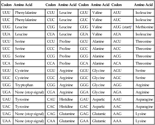
*The codons in DNA are complementary to those given here. Thus, U is complementary to the A in DNA, C is complementary to G, G to C, and A to T. The nucleotide on the left is at the 5’-end of the triplet.

AUG codes for N-formylmethionine at the beginning of messenger ribonucleic acid (mRNA) in bacteria.

Modified from Brock TD et al, editors: Biology of microorganisms, Upper Saddle River, NJ, 2009, Prentice Hall.

To accomplish the task of translation, intricate interactions between mRNA, tRNA, and rRNA are required. Sixty different types of tRNA molecules are responsible for transferring different amino acids from intracellular reservoirs to the site of protein synthesis. These molecules, which have a structure that resembles an inverted t, contain one sequence recognition site (anticodon) for binding to specific 3-base sequences (codons) on the mRNA molecule (Figure 2-6). A second site binds specific amino acids, the building blocks of proteins. Each amino acid is joined to a specific tRNA molecule through the enzymatic activity of aminoacyl-tRNA synthetases. Therefore, tRNA molecules have the primary function of using the codons of the mRNA molecule as the template for precisely delivering a specific amino acid for polymerization. Ribosomes, which are compact nucleoproteins, are composed of rRNA and proteins. They are central to translation, assisting with coupling of all required components and controlling the translational process.

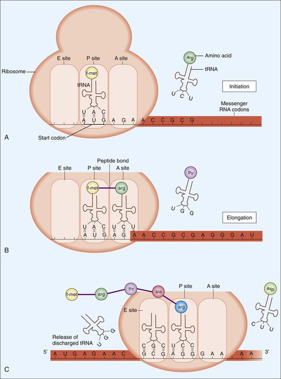
Translation, diagrammatically shown in Figure 2-6, involves three steps: initiation, elongation, and termination. Following termination, bacterial proteins often undergo posttranslational modifications as a final step in protein synthesis.

Initiation begins with the association of ribosomal subunits, mRNA, formylmethionine tRNA ([f-met] carrying the initial amino acid of the protein to be synthesized), and various initiation factors (see Figure 2-6, A). Assembly of the complex begins at a specific 3- to 9-base (Shine-Dalgarno sequence) on the mRNA about 10 bp upstream of the AUG start codon. After the initial complex has been formed, addition of individual amino acids begins.

Elongation involves tRNAs mediating the sequential addition of amino acids in a specific sequence that is dictated by the codon sequence of the mRNA molecule (see Figure 2-6, B and C, and Table 2-1). As the mRNA molecule threads through the ribosome in a 5’ to 3’ direction, peptide bonds are formed between adjacent amino acids, still bound by their respective tRNA molecules in the P (peptide) and A (acceptor) sites of the ribosome. During the process, the forming peptide is moved to the P site, and the most 5’ tRNA is released from the E (exit) site. This movement vacates the A site, which contains the codon specific for the next amino acid, so that the incoming tRNA−amino acid can join the complex (see Figure 2-6, C).

Because multiple proteins encoded on an mRNA strand can be translated at the same time, multiple ribosomes may be simultaneously associated with one mRNA molecule. Such an arrangement is referred to as a polysome; its appearance resembles a string of pearls.

Termination, the final step in translation, occurs when the ribosomal A site encounters a stop or nonsense codon that does not specify an amino acid (i.e., a “stop signal”; see Table 2-1). At this point, the protein synthesis complex disassociates and the ribosomes are available for another round of translation. After termination, most proteins must undergo modification, such as folding or enzymatic trimming, so that protein function, transportation, or incorporation into various cellular structures can be accomplished. This process is referred to as posttranslational modification.

Regulation and Control of Gene Expression

The vital role that gene expression and protein synthesis play in the survival of cells dictates that bacteria judiciously control these processes. The cell must regulate gene expression and control the activities of gene products so that a physiologic balance is maintained. Regulation and control are also key factors. These are highly complex mechanisms by which single-cell organisms are able to respond and adapt to environmental challenges, regardless of whether the challenges occur naturally or result from medical intervention (e.g., antibiotics).

Regulation occurs at one of three levels of information transfer from the gene expression and protein synthesis pathway: transcriptional, translational, or posttranslational. The most common is transcriptional regulation. Because direct interactions with genes and their ability to be transcribed to mRNA are involved, transcriptional regulation is also referred to as genetic control. Genes that encode enzymes involved in biosynthesis (anabolic enzymes) and genes that encode enzymes for biodegradation (catabolic enzymes) are used as examples of genetic control.

In general, genes that encode anabolic enzymes for the synthesis of particular products are repressed (i.e., are not transcribed and therefore are not expressed) in the presence of the gene end product. This strategy prevents waste and overproduction of products that are already present in sufficient supply. In this system, the product acts as a co-repressor that forms a complex with a repressor molecule. In the absence of co-repressor product (i.e., gene product), transcription occurs (Figure 2-7, A). When present in sufficient quantity, the product forms a complex with the repressor. The complex then binds to a specific base region of the gene sequence known as the operator region (Figure 2-7, B). This binding blocks RNA polymerase progression from the promoter sequence and inhibits transcription. As the supply of product (co-repressor) dwindles, an insufficient amount remains to form a complex with the repressor. The operator region is no longer bound to the repressor molecule. Transcription of the genes for the anabolic enzymes commences and continues until a sufficient supply of end product is again available.

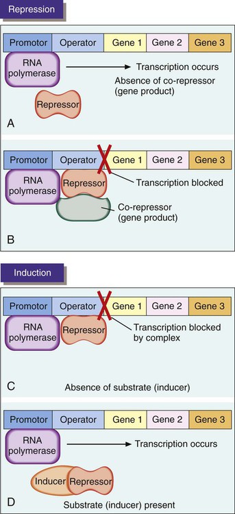
In contrast to repression, genes that encode catabolic enzymes are usually induced; that is, transcription occurs only when the substrate to be degraded by enzymatic action is present. Production of degradative enzymes in the absence of substrates would be a waste of cellular energy and resources. When the substrate is absent in an inducible system, a repressor binds to the operator sequence of the DNA and blocks transcription of the genes for the degradative enzymes (Figure 2-7, C). In the presence of an inducer, which often is the target substrate for degradation, a complex is formed between inducer and repressor and results in the release of the repressor from the operator site, allowing transcription of the genes encoding the specific catabolic enzymes (Figure 2-7, D).

Certain genes are not regulated; that is, they are not under the control of inducers or repressors. These genes are referred to as constitutive. Because they usually encode for products that are essential for viability under almost all growth and environmental conditions, these genes are continuously expressed. Also, not all regulation occurs at the genetic level (i.e., transcriptional regulation). For example, the production of some enzymes may be controlled at the protein synthesis (i.e., translational) level. The activities of other enzymes that have already been synthesized may be regulated at a posttranslational level; that is, certain catabolic or anabolic metabolites may directly interact with enzymes either to increase or to decrease their enzymatic activity.

Among different bacteria and even among different genes in the same bacterium, the mechanisms by which inducers and co-repressors are involved in gene regulation vary widely. Furthermore, bacterial cells have mechanisms to detect environmental changes. These changes can generate signals that interact with the gene expression mechanism, ensuring that appropriate products are made in response to the environmental change. In addition, several complex interactions between different regulatory systems are found within a single cell. Such diversity and interdependence are necessary components of metabolism that allow an organism to respond to environmental changes in a rapid, well-coordinated, and appropriate way.

Genetic Exchange and Diversity

In eukaryotic organisms, genetic diversity is achieved by sexual reproduction, which allows the mixing of genomes through genetic exchange. Bacteria multiply by simple binary cell division in which two daughter cells result by division of one parent cell. Each daughter cell receives the full and identical genetic complement contained in the original parent cell. This process does not allow for the mixing of genes from other cells and leaves no means of achieving genetic diversity among bacterial progeny. Without genetic diversity and change, the essential ingredients for evolution are lost. However, microorganisms have been on earth for billions of years, and microbiologists have witnessed their ability to change as a result of exposure to chemicals (i.e., antibiotics). It is evident that these organisms are fully capable of evolving and altering their genetic composition.

Genetic alterations and diversity in bacteria are accomplished by three basic mechanisms: mutation, genetic recombination, and exchange between bacteria, with or without recombination. Throughout diagnostic microbiology and infectious diseases, there are numerous examples of the impact these genetic alteration and exchange mechanisms have on clinically relevant bacteria and the management of the infections they cause.

Mutation

Mutation is defined as an alteration in the original nucleotide sequence of a gene or genes within an organism’s genome; that is, a change in the organism’s genotype. This alteration may involve a single DNA base in a gene, an entire gene, or several genes. Mutational changes in the sequence may arise spontaneously, perhaps by an error made during DNA replication. Alternatively, mutations may be induced by chemical or physical factors (i.e., mutagens) in the environment or by biologic factors, such as the introduction of foreign DNA into the cell. Alterations in the DNA base sequence can result in changes in the base sequence of mRNA during transcription. This, in turn, can affect the types and sequences of amino acids that will be incorporated into the protein during translation.

Depending on the site and extent of the mutation, various outcomes may affect the physiologic functions of the organism. For example, a mutation may be so devastating that it is lethal to the organism; the mutation, therefore, “dies” along with the organism. In other instances the mutation may be silent so that no changes are detected in the organism’s observable properties (i.e., the organism’s phenotype). Alternatively, the mutation may result in a noticeable alteration in the organism’s phenotype, and the change may provide the organism with a survival advantage. This outcome, in Darwinian terms, is the basis for prolonged survival and evolution. Nonlethal mutations are considered stable if they are passed on from one generation to another as an integral part of the cell’s genotype (i.e., genetic composition). Additionally, genes that have undergone stable mutations may also be transferred to other bacteria by one of the mechanisms of genetic exchange. In other instances, the mutation may be lost as a result of cellular repair mechanisms capable of restoring the original genotype and phenotype, or it may be lost spontaneously during subsequent cycles of DNA replication.

Genetic Recombination

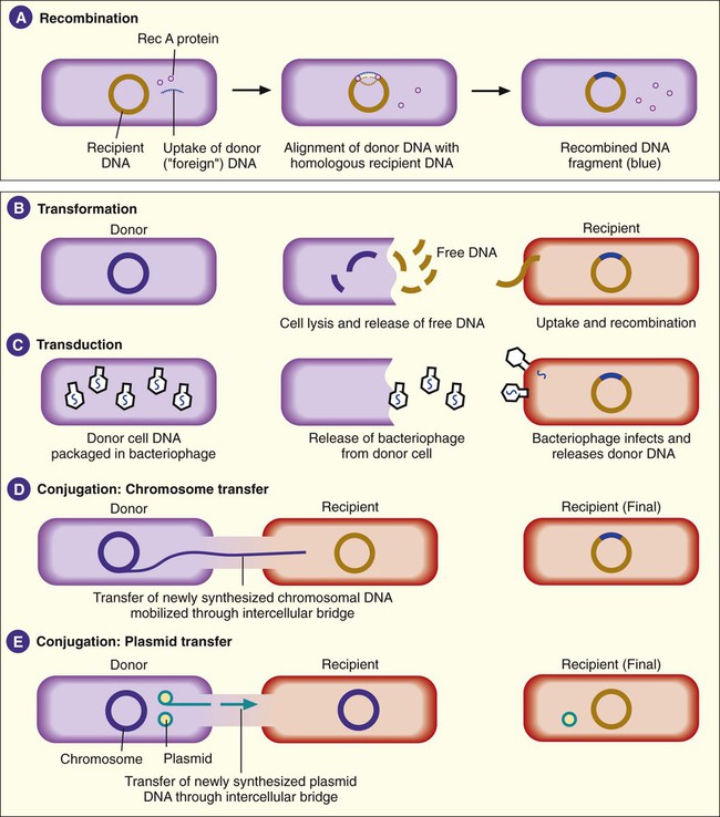
Besides mutations, bacterial genotypes can be altered through recombination. In this process, some segment of DNA originating from one bacterial cell (i.e., donor) enters a second bacterial cell (i.e., recipient) and is exchanged with a DNA segment of the recipient’s genome. This is also referred to as homologous recombination, because the pieces of DNA that are exchanged usually have extensive homology or similarities in their nucleotide sequences. Recombination involves a number of binding proteins, with the RecA protein playing a central role (Figure 2-8, A). After recombination, the recipient DNA consists of one original, unchanged strand and a second strand from the donor DNA fragment that has been recombined.

Recombination is a molecular event that occurs frequently in many varieties of bacteria, including most of the clinically relevant species, and it may involve any portion of the organism’s genome. However, the recombination event may go unnoticed unless the exchange of DNA results in a distinct alteration in the phenotype. Nonetheless, recombination is a major means by which bacteria may achieve genetic diversity and continue to evolve.

Genetic Exchange

An organism’s ability to undergo recombination depends on the acquisition of “foreign” DNA from a donor cell. The three mechanisms by which bacteria physically exchange DNA are transformation, transduction, and conjugation.

Transformation.

Transformation involves recipient cell uptake of naked (free) DNA released into the environment when another bacterial cell (i.e., donor) dies and undergoes lysis (see Figure 2-8, B). This genomic DNA exists as fragments in the environment. Certain bacteria are able to take up naked DNA from their surroundings; that is, they are able to undergo transformation. Such bacteria are said to be competent. Among the bacteria that cause human infections, competence is a characteristic commonly associated with members of the genera Haemophilus, Streptococcus, and Neisseria.

Once the donor DNA, usually as a singular strand, gains access to the interior of the recipient cell, recombination with the recipient’s homologous DNA can occur. The mixing of DNA between bacteria via transformation and recombination plays a major role in the development of antibiotic resistance and in the dissemination of genes that encode factors essential to an organism’s ability to cause disease. Additionally, gene exchange by transformation is not limited to organisms of the same species, thus allowing important characteristics to be disseminated to a greater variety of medically important bacteria.

Transduction.

Transduction is a second mechanism by which DNA from two bacteria may come together in one cell, thus allowing for recombination (see Figure 2-8, C). This process is mediated through viruses capable of infecting bacteria (i.e., bacteriophages). In their “life cycle,” these viruses integrate their DNA into the bacterial cell’s chromosome, where viral DNA replication and expression occur. When the production of viral products is complete, viral DNA is excised (cut) from the bacterial chromosome and packaged within a protein coat. This virion contains bacterial and viral DNA. The newly formed recombinant virion, along with the additional multiple virions (virus particles), is released when the infected bacterial cell lyses. In transduction, the recombinant virion incorporates its own DNA but may also pick up a portion of the donor bacterium’s DNA.

The bacterial DNA may be randomly incorporated with viral DNA (generalized transduction), or it may be incorporated along with adjacent viral DNA (specialized transduction). In either case, when the viruses infect another bacterial cell, they release their DNA contents, which includes the previously incorporated bacterial donor DNA. Therefore, the newly infected cell is the recipient of donor DNA introduced by the bacteriophage, and recombination between DNA from two different cells occurs.

Conjugation.

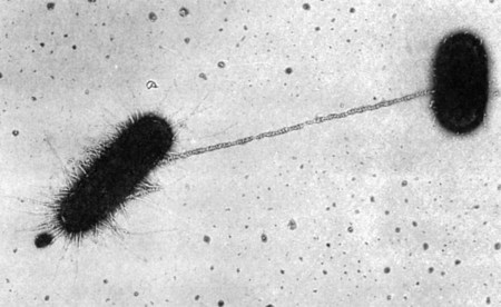
The third mechanism of DNA exchange between bacteria is conjugation. This process occurs between two living cells, involves cell-to-cell contact, and requires mobilization of the donor bacterium’s chromosome. The nature of intercellular contact is not well characterized in all bacterial species capable of conjugation. However, in E. coli, contact is mediated by a sex pilus (Figure 2-9). The sex pilus originates from the donor and establishes a conjugative bridge that serves as the conduit for DNA transfer from donor to recipient cell. With intercellular contact established, chromosomal mobilization is undertaken and involves DNA synthesis. One new DNA strand is produced by the donor and is passed to the recipient (see Figure 2-8, D). The amount of DNA transferred depends on how long the cells are able to maintain contact, but usually only portions of the donor molecule are transferred. In any case, the newly introduced DNA is then available to recombine with the recipient’s genome.

In addition to chromosomal DNA, genes encoded in extrachromosomal genetic elements, such as plasmids and transposons, may be transferred by conjugation (see Figure 2-8, E). Not all plasmids are capable of conjugative transfer, but for those that are, the donor plasmid usually is replicated so that the donor retains a copy of the plasmid transferred to the recipient. (See the discussion of the F plasmid in the section Cellular Appendages, later in the chapter.) Plasmid DNA may also become incorporated into the host cell’s chromosome.

In contrast to plasmids, transposons do not exist independently in the cell. Except when they are moving from one location to another, transposons must be incorporated into the chromosome or plasmids or both. These elements are often referred to as “jumping genes” because of their ability to change location within and even between the genomes of bacterial cells. Transposition is the process by which these genetic elements excise from one genomic location and insert into another. Transposons carry genes that have products that help mediate the transposition process, in addition to genes that encode for other accessory characteristics, such as antimicrobial resistance. Homologous recombination between the genes of plasmids or transposons and the host bacterium’s chromosomal DNA may occur.

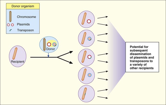
Plasmids and transposons play a key role in genetic diversity and dissemination of genetic information among bacteria. Many characteristics that significantly alter the activities of clinically relevant bacteria are encoded and disseminated on these elements. Furthermore, as shown in Figure 2-10, the variety of strategies that bacteria can use to mix and match genetic elements provides them with a tremendous capacity to genetically adapt to environmental changes, including those imposed by human medical practices. A good example of this is the emergence and widespread dissemination of resistance to antimicrobial agents among clinically important bacteria. Bacteria have used their capacity for disseminating genetic information to establish resistance to many of the commonly prescribed antibiotics. (See Chapter 11 for more information about antimicrobial resistance mechanisms.)

Bacterial Metabolism

Fundamentally, bacterial metabolism involves all the cellular processes required for the organism’s survival and replication. Familiarity with bacterial metabolism is essential for understanding bacterial interactions with human host cells, the mechanisms bacteria use to cause disease, and the basis of diagnostic microbiology; that is, the tests and strategies used for laboratory identification of infectious etiologies. Because metabolism is an extensive and complicated topic, this section focuses on processes typical of medically relevant bacteria.

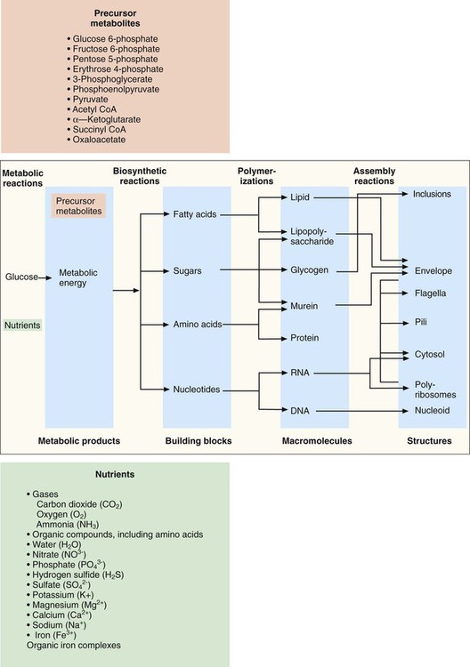
For the sake of clarity, metabolism is discussed in terms of four primary, but interdependent, processes: fueling, biosynthesis, polymerization, and assembly (Figure 2-11).

Fueling

Fueling is considered the utilization of metabolic pathways involved in the acquisition of nutrients from the environment, production of precursor metabolites, and energy production.

Acquisition of Nutrients

Bacteria use various strategies for obtaining essential nutrients from the external environment and transporting these substances into the cell’s interior. For nutrients to be internalized, they must cross the bacterial cell wall and membrane. These complex structures help protect the cell from environmental insults, maintain intracellular equilibrium, and transport substances into and out of the cell. Although some key nutrients (e.g., water, oxygen, and carbon dioxide) enter the cell by simple diffusion across the cell membrane, the uptake of other substances is controlled by membrane-selective permeability; still other substances use specific transport mechanisms.

Active transport is among the most common methods used for the uptake of nutrients such as certain sugars, most amino acids, organic acids, and many inorganic ions. The mechanism, driven by an energy-dependent pump, involves carrier molecules embedded in the membrane portion of the cell structure. These carriers combine with the nutrients, transport them across the membrane, and release them inside the cell. Group translocation is another transport mechanism that requires energy but differs from active transport in that the nutrient being transported undergoes chemical modification. Many sugars, purines, pyrimidines, and fatty acids are transported by this mechanism.

Production of Precursor Metabolites

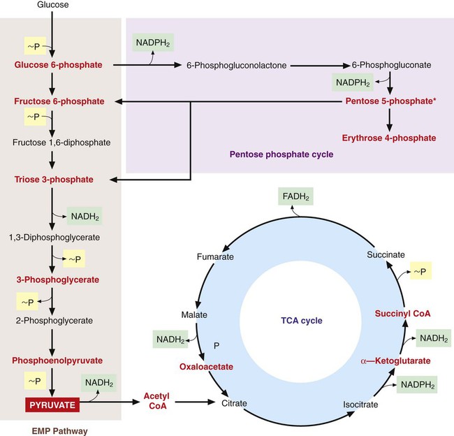
Once inside the cell, many nutrients serve as the raw materials from which precursor metabolites for subsequent biosynthetic processes are produced. These metabolites, listed in Figure 2-11, are produced through three central pathways; the Embden-Meyerhof-Parnas (EMP) pathway, the tricarboxylic acid (TCA) cycle, and the pentose phosphate shunt. These pathways and their relationship to one another are shown in Figure 2-12; not shown are the several alternative pathways (e.g., the Entner-Douder off pathway) that play key roles in redirecting and replenishing the precursors as they are used in subsequent processes.

image
Figure 2-12 Overview diagram of the central metabolic pathways (Embden-Meyerhof-Parnas [EMP], the tricarboxylic acid [TCA] cycle, and the pentose phosphate shunt). Precursor metabolites (see also Figure 2-11) that are produced are highlighted in red; production of energy in the form of ATP (~P) by substrate-level phosphorylation is highlighted in yellow; and reduced carrier molecules for transport of electrons used in oxidative phosphorylation are highlighted in green. (Modified from Niedhardt FC, Ingraham JL, Schaechter M, editors: Physiology of the bacterial cell: a molecular approach, Sunderland, Mass, 1990, Sinauer Associates.)

The production efficiency of a bacterial cell resulting from these precursor-producing pathways can vary substantially, depending on the growth conditions and availability of nutrients. This is an important consideration because the accurate identification of medically important bacteria often depends heavily on methods that measure the presence of products and byproducts of these metabolic pathways.

Energy Production

The third type of fueling pathway is one that produces energy required for nearly all cellular processes, including nutrient uptake and precursor production. Energy production is accomplished by the breakdown of chemical substrates (i.e., chemical energy) through the degradative process of catabolism coupled with oxidation-reduction reactions. In this process, the energy source molecule (i.e., substrate) is oxidized as it donates electrons to an electron-acceptor molecule, which is then reduced. The transfer of electrons is mediated through carrier molecules, such as nicotinamide-adenine-dinucleotide (NAD+) and nicotinamide-adenine-dinucleotide-phosphate (NADP+). The energy released by the oxidation-reduction reaction is transferred to phosphate-containing compounds, where high-energy phosphate bonds are formed. ATP is the most common of such molecules. The energy contained in this compound is eventually released by the hydrolysis of ATP under controlled conditions. The release of this chemical energy, coupled with enzymatic activities, specifically catalyzes each biochemical reaction in the cell and drives nearly all cellular reactions.

The two general mechanisms for ATP production in bacterial cells are substrate-level phosphorylation and electron transport, also referred to as oxidative phosphorylation. In substrate-level phosphorylation, high-energy phosphate bonds produced by the central pathways are donated to adenosine diphosphate (ADP) to form ATP (see Figure 2-12). Additionally, pyruvate, a primary intermediate in the central pathways, serves as the initial substrate for several other pathways to generate ATP by substrate level phosphorylation. These other pathways constitute fermentative metabolism, which does not require oxygen and produces various end products, including alcohols, acids, carbon dioxide, and hydrogen. The specific fermentative pathways and the end products produced vary with different bacterial species. Detection of these products is an important basis for laboratory identification of bacteria. (See Chapter 7 for more information on the biochemical basis for bacterial identification.)

Oxidative Phosphorylation.

Oxidative phosphorylation involves an electron transport system that conducts a series of electron transfers from reduced carrier molecules such as NADH2 and NADPH2, produced in the central pathways (see Figure 2-12), to a terminal electron acceptor. The energy produced by the series of oxidation-reduction reactions is used to generate ATP from ADP. When oxidative phosphorylation uses oxygen as the terminal electron acceptor, the process is known as aerobic respiration. Anaerobic respiration refers to processes that use final electron acceptors other than oxygen.

A knowledge of which mechanisms bacteria use to generate ATP is important for designing laboratory protocols for cultivating and identifying these organisms. For example, some bacteria depend solely on aerobic respiration and are unable to grow in the absence of oxygen (strictly aerobic bacteria). Others can use either aerobic respiration or fermentation, depending on the availability of oxygen (facultative anaerobic bacteria). For still others, oxygen is absolutely toxic (strictly anaerobic bacteria).

Biosynthesis

The fueling reactions essentially bring together all the raw materials needed to initiate and maintain all other cellular processes. The production of precursors and energy is accomplished through catabolic processes and the degradation of substrate molecules. The three remaining pathways for biosynthesis, polymerization, and assembly depend on anabolic metabolism. In anabolic metabolism, precursor compounds are joined for the creation of larger molecules (polymers) required for assembly of cellular structures (see Figure 2-11).

Biosynthetic processes use the precursor products in dozens of pathways to produce a variety of building blocks, such as amino acids, fatty acids, sugars, and nucleotides (see Figure 2-11). Many of these pathways are highly complex and interdependent, whereas other pathway are completely independent. In many cases, the enzymes that drive the individual pathways are encoded on a single mRNA molecule that has been transcribed from contiguous genes in the bacterial chromosome (i.e., an operon).

As previously mentioned, bacterial genera and species vary extensively in their biosynthetic capabilities. Knowledge of these variations is necessary to use optimal conditions for growing organisms under laboratory conditions. For example, some organisms may not be capable of synthesizing an essential amino acid necessary as a building block for proteins. Without the ability to synthesize the amino acid, the bacterium must obtain the building block from the environment. Similarly, if the organism is cultivated in the microbiology laboratory, the amino acid must be provided in the culture medium.

Structure and Function of the Bacterial Cell

Based on key characteristics, all cells are classified into two basic types: prokaryotic and eukaryotic. Although these two cell types share many common features, they have many important differences in terms of structure, metabolism, and genetics.

Eukaryotic and Prokaryotic Cells

Among clinically relevant organisms, bacteria are single-cell prokaryotic microorganisms. Fungi and parasites are single-cell or multicellular eukaryotic organisms, as are plants and all higher animals. Viruses are dependent on host cells for survival and therefore are not considered cellular organisms but rather infectious agents.

A notable characteristic of eukaryotic cells, such as those of parasites and fungi, is the presence of membrane-enclosed organelles that have specific cellular functions. Examples of these organelles and their respective functions include:

Additionally, eukaryotic cells have an infrastructure, or cytoskeleton, that provides support for cellular structure, organization, and movement.

Prokaryotic cells, such as bacteria, do not contain organelles. All functions take place in the cytoplasm or cytoplasmic membrane of the cell. Prokaryotic and eukaryotic cell types differ considerably at the macromolecular level, including protein synthesis machinery, chromosomal organization, and gene expression. One notable structure present only in prokaryotic bacterial cells is a cell wall composed of peptidoglycan. This structure has an immeasurable impact on the practice of diagnostic bacteriology and the management of bacterial diseases.

Bacterial Morphology

Most clinically relevant bacterial species range in size from 0.25 to 1 µm in width and 1 to 3 µm in length, thus requiring microscopy for visualization (see Chapter 6 for more information on microscopy). Just as bacterial species and genera vary in their metabolic processes, their cells also vary in size, morphology, and cell-to-cell arrangements and in the chemical composition and structure of the cell wall. The bacterial cell wall differences provide the basis for the Gram stain, a fundamental staining technique used in bacterial identification schemes. This staining procedure separates almost all medically relevant bacteria into two general types: gram-positive bacteria, which stain a deep blue or purple, and gram-negative bacteria, which stain a pink to red (see Figure 6-3). This simple but important color distinction is due to differences in the constituents of bacterial cell walls that influence the cell’s ability to retain differential dyes following treatment with a decolorizing agent.

Common bacterial cellular morphologies include cocci (circular), coccobacilli (ovoid), and bacillus (rod shaped), as well as fusiform (pointed end), curved, or spiral shapes. Cellular arrangements are also noteworthy. Cells may characteristically occur singly, in pairs, or grouped as tetrads, clusters, or in chains (see Figure 6-4 for examples of bacterial staining and morphologies). The determination of the Gram stain reaction and the cell size, morphology, and arrangement are essential aspects of bacterial identification.

Bacterial Cell Components

Bacterial cell components can be divided into those that make up the outer cell structure and its appendages (cell envelope) and those associated with the cell’s interior. It is important to note that the cellular structures work together to function as a complex and integrated unit.

Cell Envelope

As shown in Figure 2-13, the outermost structure, the cell envelope, comprises:
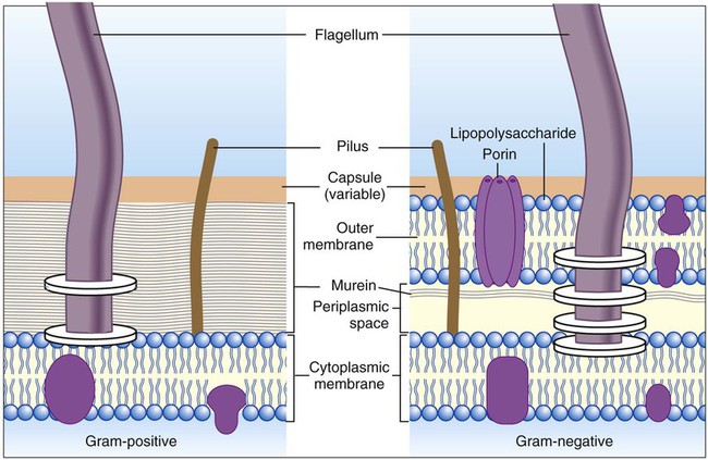
Outer Membrane.

Outer membranes, which are found only in gram-negative bacteria, function as the cell’s initial barrier to the environment. These membranes serve as primary permeability barriers to hydrophilic and hydrophobic compounds and contain essential enzymes and other proteins located in the periplasmic space. The membrane is a bilayered structure composed of lipopolysaccharide, which gives the surface of gram-negative bacteria a net negative charge. The outer membrane also plays a significant role in the ability of certain bacteria to cause disease.

Scattered throughout the lipopolysaccharide macromolecules are protein structures called porins. These water-filled structures control the passage of nutrients and other solutes, including antibiotics, through the outer membrane. The number and types of porins vary with bacterial species. These differences can substantially influence the extent to which various substances pass through the outer membranes of different bacteria. In addition to porins, other proteins (murein lipoproteins) facilitate the attachment of the outer membrane to the next internal layer in the cell envelope, the cell wall.

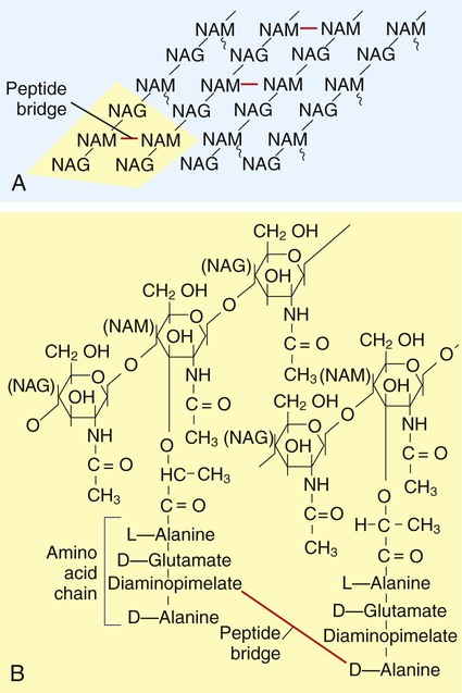
Cell Wall (Murein Layer).

The cell wall, also referred to as the peptidoglycan, or murein layer, is an essential structure found in nearly all clinically relevant bacteria. This structure gives the bacterial cell shape and strength to withstand changes in environmental osmotic pressures that would otherwise result in cell lysis. The murein layer protects against mechanical disruption of the cell and offers some barrier to the passage of larger substances. Because this structure is essential for the survival of bacteria, its synthesis and structure are often the primary targets for the development and design of several antimicrobial agents.

The structure of the cell wall is unique and is composed of disaccharide-pentapeptide subunits. The disaccharides N-acetylglucosamine and N-acetylmuramic acid are the alternating sugar components (moieties), with the amino acid chain linked to N-acetylmuramic acid molecules (Figure 2-14). Polymers of these subunits cross-link to one another by means of peptide bridges to form peptidoglycan sheets. In turn, layers of these sheets are cross-linked with one another, forming a multilayered, cross-linked structure of considerable strength. Referred to as the murein sacculus, or sack, this peptidoglycan structure surrounds the entire cell.

A notable difference between the cell walls of gram-positive and gram-negative bacteria is the substantially thicker peptidoglycan layer in gram-positive bacteria (see Figure 2-13). Additionally, the cell wall of gram-positive bacteria contains teichoic acids (i.e., glycerol or ribitol phosphate polymers combined with various sugars, amino acids, and amino sugars). Some teichoic acids are linked to N-acetylmuramic acid, and others (e.g., lipoteichoic acids) are linked to the next underlying layer, the cellular membrane. Other gram-positive bacteria (e.g., mycobacteria) have waxy substances within the murein layer, such as mycolic acids. Mycolic acids make the cells more refractory to toxic substances, including acids. Bacteria with mycolic acid in the cell walls require unique staining procedures and growth media in the diagnostic laboratory.

Cytoplasmic (Inner) Membrane.

The cytoplasmic (inner) membrane is present in both gram-positive and gram-negative bacteria and is the deepest layer of the cell envelope. The cytoplasmic membrane is heavily laced with various proteins, including a number of enzymes vital to cellular metabolism. The cell membrane serves as an additional osmotic barrier and is functionally similar to the membranes of several of eukaryotic cellular organelles (e.g., mitochondria, Golgi complexes, lysosomes). The cytoplasmic membrane functions include:

Cellular Appendages.

In addition to the components of the cell envelope proper, cellular appendages (i.e., capsules, fimbriae, and flagella) are associated with or proximal to this portion of the cell. The presence of these appendages, which can play a role in the mediation of infection and in laboratory identification, varies among bacterial species and even among strains within the same species.

The capsule is immediately exterior to the murein layer of gram-positive bacteria and the outer membrane of gram-negative bacteria. Often referred to as the “slime layer,” the capsule is composed of high-molecular-weight polysaccharides, the production of which may depend on the environment and growth conditions surrounding the bacterial cell. The capsule does not function as an effective permeability barrier or add strength to the cell envelope, but it does protect bacteria from attack by components of the human immune system. The capsule also facilitates and maintains bacterial colonization of biologic (e.g., teeth) and inanimate (e.g., prosthetic heart valves) surfaces through the formation of biofilms. A biofilm consists of a monomicrobic or polymicrobic group of bacteria housed in a complex polysaccharide matrix. (See Chapter 3 for further discussion of microbial biofilms.)

Fimbriae, or pili, are hairlike, proteinaceous structures that extend from the cell membrane into the external environment; some may be up to 2 µm long. Fimbriae may serve as adhesins that help bacteria attach to animal host cell surfaces, often as the first step in establishing infection. In addition, a pilus may be referred to as a sex pilus; this structure, which is well characterized in the gram-negative bacillus E. coli, serves as the conduit for the passage of DNA from donor to recipient during conjugation. The sex pilus is present only in cells that produce a protein referred to as the F factor. F-positive cells initiate mating or conjugation only with F-negative cells, thereby limiting the conjugative process to cells capable of transporting genetic material through the hollow sex pilus.

Flagella are complex structures, mostly composed of the protein flagellin, intricately embedded in the cell envelope. These structures are responsible for bacterial motility. Although not all bacteria are motile, motility plays an important role in survival and the ability of certain bacteria to cause disease. Depending on the bacterial species, flagella may be located at one end of the cell (monotrichous flagella) or at both ends of the cell (lophotrichous flagella), or the entire cell surface may be covered with flagella (peritrichous flagella).

Cell Interior

Those structures and substances that are bounded internally by the cytoplasmic membrane compose the cell interior and include the cytosol, polysomes, inclusions, the nucleoid, plasmids, and endospores.

The cytosol, where nearly all other functions not conducted by the cell membrane occur, contains thousands of enzymes and is the site of protein synthesis. The cytosol has a granular appearance caused by the presence of many polysomes (mRNA complexed with several ribosomes during translation and protein synthesis) and inclusions (i.e., storage reserve granules). The number and nature of the inclusions vary depending on the bacterial species and the nutritional state of the organism’s environment. Two common types of granules include glycogen, a storage form of glucose, and polyphosphate granules, a storage form for inorganic phosphates that are microscopically visible in certain bacteria stained with specific dyes.

Unlike eukaryotic chromosomes, the bacterial chromosome is not enclosed within a membrane-bound nucleus. Instead the bacterial chromosome exists as a nucleoid in which the highly coiled DNA is intermixed with RNA, polyamines, and various proteins that lend structural support. At times, depending on the stage of cell division, more than one chromosome may be present per bacterial cell. Plasmids are the other genetic elements that exist independently in the cytosol, and their numbers may vary from none to several hundred per bacterial cell.

The final bacterial structure to be considered is the endospore. Under adverse physical and chemical conditions or when nutrients are scarce, some bacterial genera are able to form spores (i.e., sporulate). Sporulation involves substantial metabolic and structural changes in the bacterial cell. Essentially, the cell transforms from an actively metabolic and growing state to a dormant state, with a decrease in cytosol and a concomitant increase in the thickness and strength of the cell envelope. The spore remains in a dormant state until favorable conditions for growth are again encountered. This survival tactic is demonstrated by a number of clinically relevant bacteria and frequently challenges our ability to thoroughly sterilize materials and food for human use.

Share this: