[level-membership-for-radiology-category] Absent spleen in virtually all patients


• Polysplenia (PSP) syndrome: Left isomerism or bilateral left-sidedness


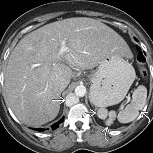
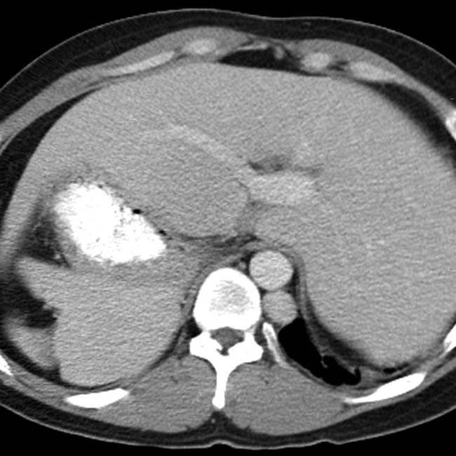
 in the left upper quadrant. The multiple spleens in PSP are typically in the left abdomen, but can rarely be on the right.
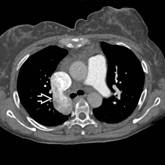
 .
.
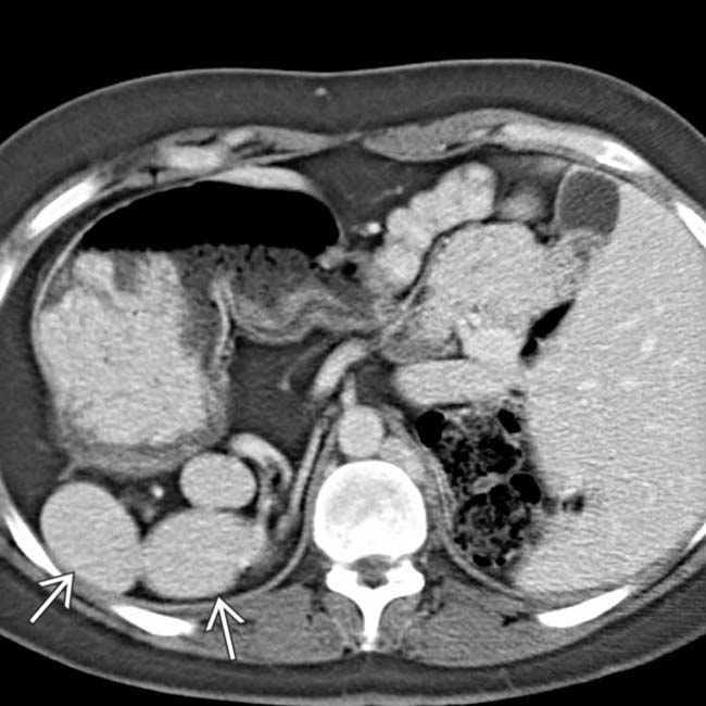
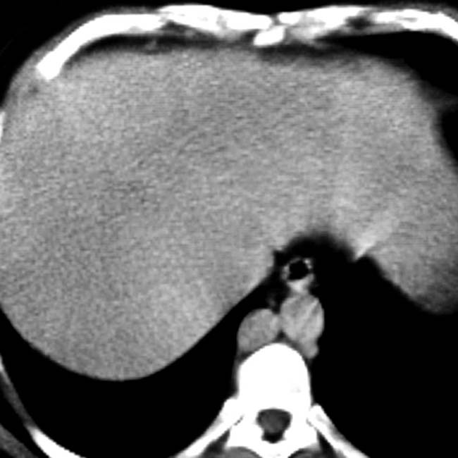
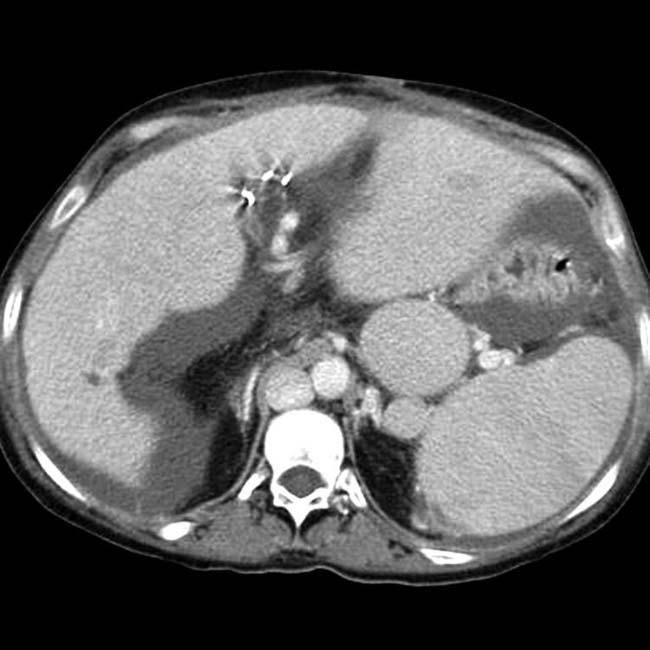
 and a dilated azygous vein
  to the right of the aorta. Azygous continuation of the inferior vena cava (IVC) is a very common abnormality in PSP syndrome.
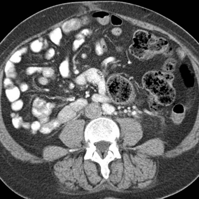


IMAGING
General Features
• Best diagnostic clue


• Key concepts
 ASP syndrome: Right isomerism or bilateral right-sidedness
 PSP syndrome: Left isomerism or bilateral left-sidedness
ASP syndrome: Right isomerism or bilateral right-sidedness
– Situs ambiguus and bilateral right-sidedness; no fixed set of findings, abnormalities exist across a spectrum


– Cardiovascular
 Total anomalous pulmonary venous return (almost 100%), endocardial cushion defect (85%), single ventricle (51%), transposition of great vessels (58%), pulmonary stenosis or atresia (70%), dextrocardia (42%), mesocardia, ventricular septal defect, single atrioventricular valve, bilateral superior vena cava (SVC)
 Total anomalous pulmonary venous return (almost 100%), endocardial cushion defect (85%), single ventricle (51%), transposition of great vessels (58%), pulmonary stenosis or atresia (70%), dextrocardia (42%), mesocardia, ventricular septal defect, single atrioventricular valve, bilateral superior vena cava (SVC)


 PSP syndrome: Left isomerism or bilateral left-sidedness
– Situs ambiguus and bilateral left-sidedness: No fixed set of findings and abnormalities exist across a spectrum


– Cardiovascular
 Increased risk of complex cardiac anomalies, although less common with PSP than with ASP, accounting for better long-term survival
 Transposition of great vessels (13%), double outlet right ventricle (13%), pulmonary valvular stenosis (23%), subaortic stenosis, or atresia
Increased risk of complex cardiac anomalies, although less common with PSP than with ASP, accounting for better long-term survival Transposition of great vessels (13%), double outlet right ventricle (13%), pulmonary valvular stenosis (23%), subaortic stenosis, or atresia


CLINICAL ISSUES
Presentation


 located in the right upper quadrant and situs inversus. Note the reversed positions of the liver and stomach.
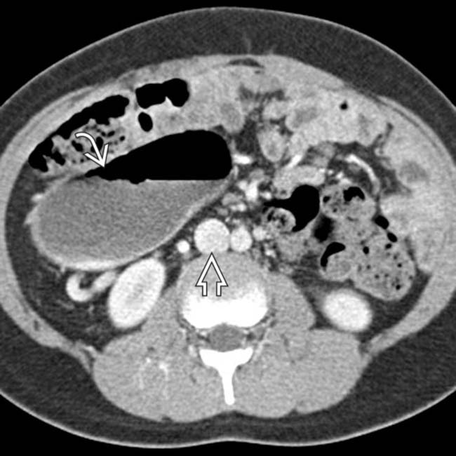
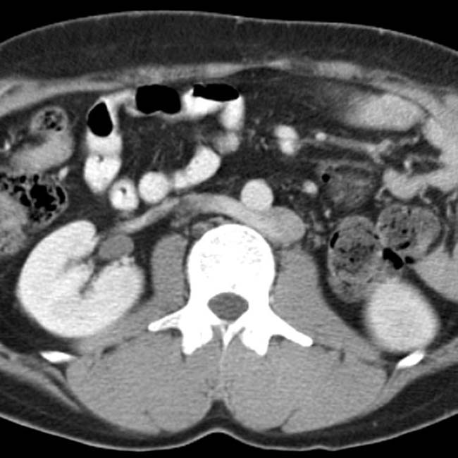
 . Note that the IVC
  is normally located on the right. The majority of the small bowel is on one side of the abdomen, in keeping with malrotation.
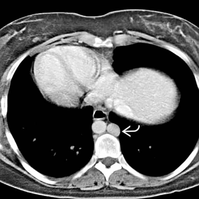
 .
.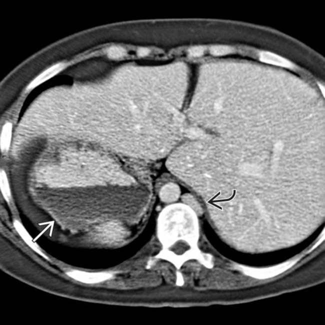
 and liver on the right-hand side. Note the left-sided IVC
  .
.
 in the right upper quadrant along with abdominal situs inversus.
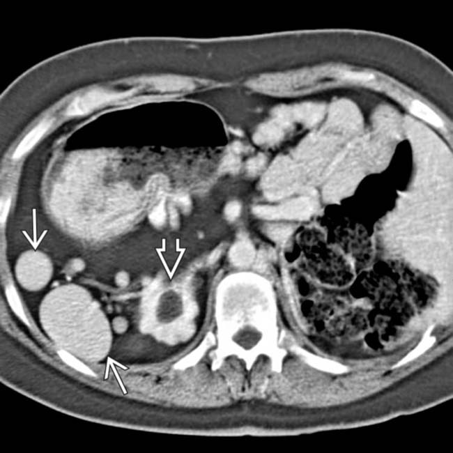
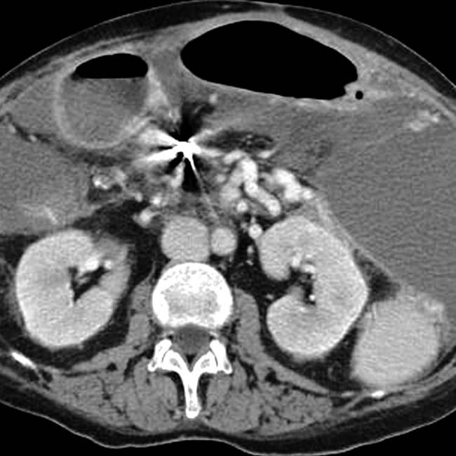
 , situs inversus, and cystic disease of the kidneys
  . This patient was 35 years old (patients with PSP are much more likely to reach adulthood than ASP).
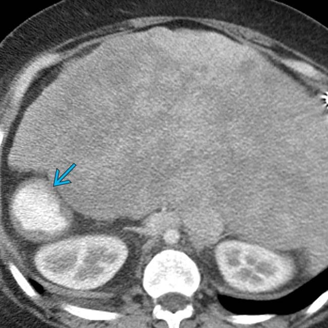
 . Extensive cardiac anomalies were also noted (not shown).
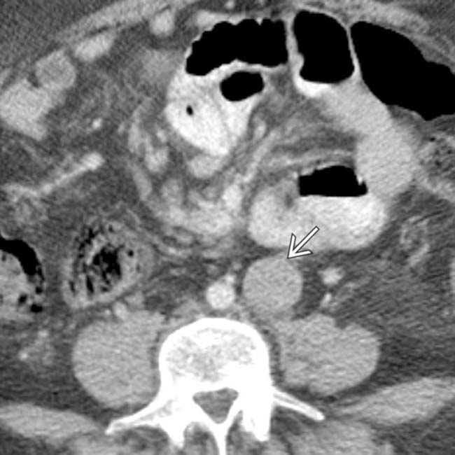
 .
.
 .
.
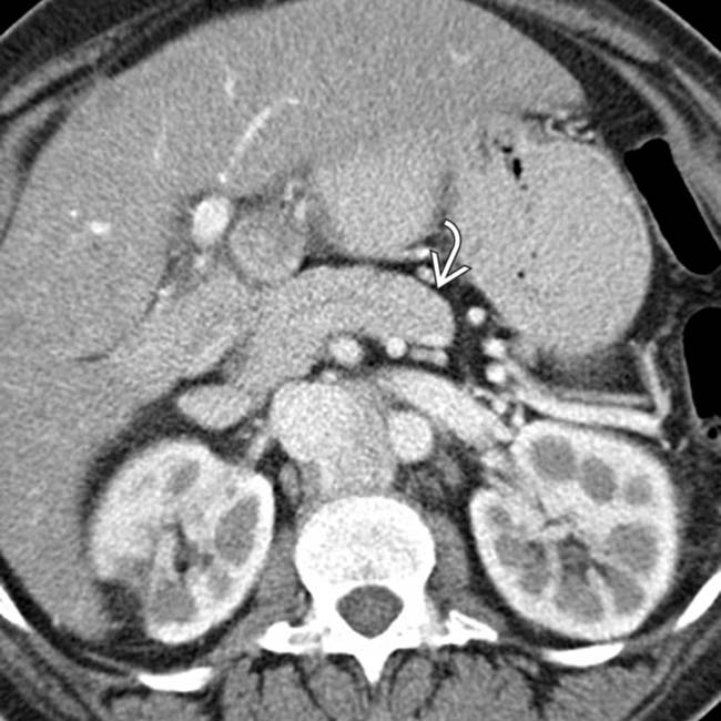
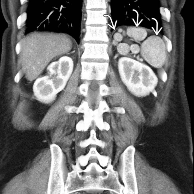
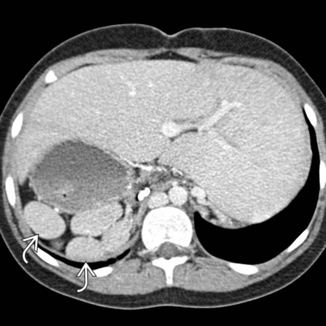
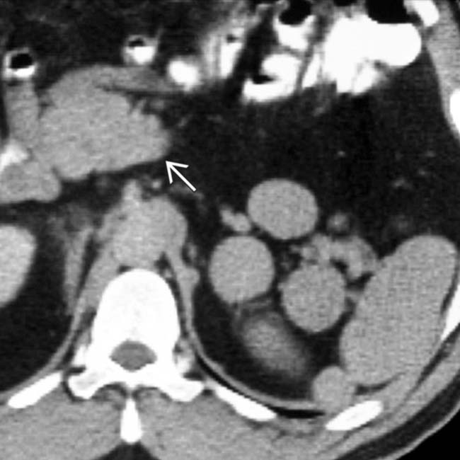
 , a commonly associated abnormality in PSP.
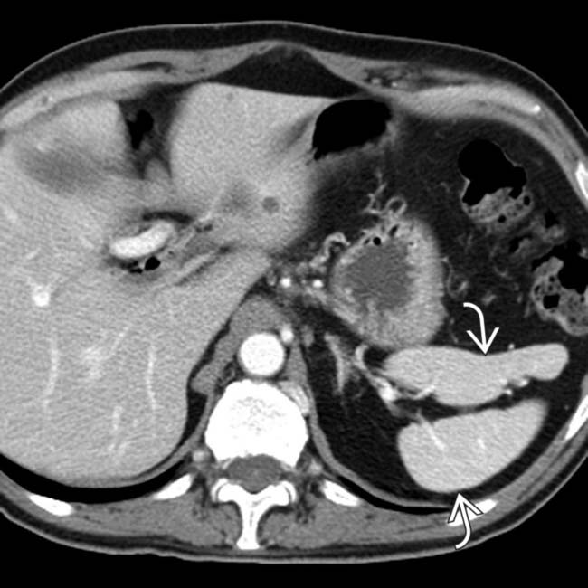
 in the left upper quadrant.
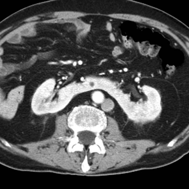
[/level-membership-for-radiology-category][not-level-membership-for-radiology-category] Absent spleen in virtually all patients
• Polysplenia (PSP) syndrome: Left isomerism or bilateral left-sidedness
in the left upper quadrant. The multiple spleens in PSP are typically in the left abdomen, but can rarely be on the right. and a dilated azygous vein to the right of the aorta. Azygous continuation of the inferior vena cava (IVC) is a very common abnormality in PSP syndrome.IMAGING
General Features
• Best diagnostic clue
• Key concepts
ASP syndrome: Right isomerism or bilateral right-sidedness
PSP syndrome: Left isomerism or bilateral left-sidedness
ASP syndrome: Right isomerism or bilateral right-sidedness
– Situs ambiguus and bilateral right-sidedness; no fixed set of findings, abnormalities exist across a spectrum
– Cardiovascular
Total anomalous pulmonary venous return (almost 100%), endocardial cushion defect (85%), single ventricle (51%), transposition of great vessels (58%), pulmonary stenosis or atresia (70%), dextrocardia (42%), mesocardia, ventricular septal defect, single atrioventricular valve, bilateral superior vena cava (SVC)
Total anomalous pulmonary venous return (almost 100%), endocardial cushion defect (85%), single ventricle (51%), transposition of great vessels (58%), pulmonary stenosis or atresia (70%), dextrocardia (42%), mesocardia, ventricular septal defect, single atrioventricular valve, bilateral superior vena cava (SVC) PSP syndrome: Left isomerism or bilateral left-sidedness
– Situs ambiguus and bilateral left-sidedness: No fixed set of findings and abnormalities exist across a spectrum
– Cardiovascular
Buy Membership for Radiology Category to continue reading. Learn more here
[/not-level-membership-for-radiology-category]
