Aspiration Syndromes

Published on 22/03/2015 by admin

Filed under Pediatrics

Last modified 22/04/2025

Print this page

rate 1 star rate 2 star rate 3 star rate 4 star rate 5 star
Your rating: none, Average: 0 (0 votes)

This article have been viewed 1086 times

Chapter 389 Aspiration Syndromes

Aspiration includes a wide clinical spectrum from an asymptomatic condition to acute life-threatening events, such as occur with massive aspiration of gastric contents or hydrocarbon products. Other chapters discuss mechanical obstruction of large or intermediate-sized airways (as occurs with foreign bodies; Chapter 379) and infectious complications of aspiration and recurrent microaspiration (Chapter 390), such as may occur with gastroesophageal reflux (Chapter 315.1) or dysphagia (Chapter 298). Occult aspiration of nasopharyngeal secretions into the lower respiratory tract is a normal event in healthy people, usually without apparent clinical significance.

Gastric Contents

Large-volume aspiration of gastric contents usually occurs in the context of vomiting. It is an infrequent complication of general anesthesia, gastroenteritis, and altered level of consciousness. Among 63,180 pediatric patients undergoing general anesthesia, 24 cases of aspiration occurred, but symptoms developed in only 9. Pathophysiologic consequences can vary, depending primarily on the pH and volume of the aspirate and the amount of particulate material. Increased clinical severity is noted with volumes greater than approximately 0.8 mL/kg and/or pH <2.5. Hypoxemia, hemorrhagic pneumonitis, atelectasis, intravascular fluid shifts, and pulmonary edema all occur rapidly after massive aspiration. These occur earlier, become more severe, and last longer with acid aspiration. Most clinical changes are present within minutes to 1-2 hr after the aspiration event. In the next 24-48 hr, there is a marked increase in lung parenchymal neutrophil infiltrations, mucosal sloughing, and alveolar consolidation that often correlates with increasing infiltrates on chest radiographs. These changes tend to occur significantly later and are more prolonged after aspiration of particulate material. Infection usually does not have a role in initial lung injury after aspiration of gastric contents; aspiration may impair pulmonary defenses, predisposing the patient to secondary bacterial pneumonia. In the patient who has shown clinical improvement but then demonstrates clinical worsening, especially with fever and leukocytosis, secondary bacterial pneumonia should be suspected.

Treatment

If large-volume or highly toxic substance aspiration occurs in a patient who already has an artificial airway in place, it is important to perform immediate suctioning of the airway. If immediate suctioning cannot be performed, later suctioning or bronchoscopy is usually of limited therapeutic value. An exception to this policy is suspicion of significant particulate aspiration. Attempts at acid neutralization are not warranted because acid is rapidly neutralized by the respiratory epithelium. Patients in whom large-volume or toxic aspiration is suspected should be observed, should undergo oxygenation measurement by oximetry or blood gas analysis, and should undergo a chest radiograph, even if they are asymptomatic. If the chest radiograph findings and oxygen saturation are normal, and the patient remains asymptomatic, home observation, after a period of observation in the hospital or office, is adequate. No treatment is indicated at that time, but the caregivers should be instructed to bring the child back in for medical attention should respiratory symptoms or fever develop. For patients who present with abnormal findings or in whom such findings develop during observation, oxygen therapy is given to correct hypoxemia. Endotracheal intubation and mechanical ventilation are often necessary for more severe cases. Bronchodilators may be tried, although they are usually of limited benefit. Animal studies indicate that treatment with corticosteroids does not appear to have any benefit, unless given nearly simultaneously with the aspiration event; use of these agents may increase the risk of secondary infection. Prophylactic antibiotics are not indicated, although in the patient with very limited reserve, early antibiotic coverage may be appropriate. If used, antibiotics should be selected that cover for anaerobic microbes. If the aspiration event occurs in a hospitalized or chronically ill patient, coverage of Pseudomonas and enteric gram-negative organisms should also be considered. A mortality rate of ≤5% is seen if 3 or fewer lobes are involved. Unless complications develop, such as infection or barotrauma, most patients recover in 2-3 wk, although prolonged lung damage may persist, with scarring, bronchiolitis obliterans, and bronchiectasis.

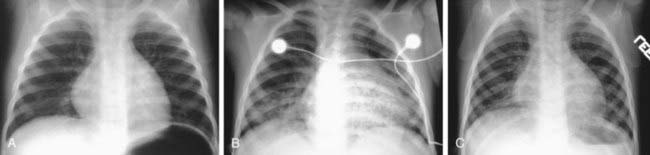
Hydrocarbon Aspiration

The most dangerous consequence of acute hydrocarbon ingestion is usually aspiration and resulting pneumonitis (Chapter 58). Although significant pneumonitis occurs in <2% of all hydrocarbon ingestions, an estimated 20 deaths occur annually from hydrocarbon aspiration in both children and adults. Some of these deaths represent suicides. Hydrocarbons with lower surface tensions (gasoline, turpentine, naphthalene) have more potential for aspiration toxicity than heavier mineral or fuel oils. Ingestion of >30 mL (approximate volume of an adult swallow) of hydrocarbon is associated with an increased risk of severe pneumonitis. Clinical findings such as chest retractions, grunting, cough, and fever may occur as soon as 30 min after aspiration or may be delayed for several hours. Lung radiographic changes usually occur within 2-8 hr, peaking in 48-72 hr (Fig. 389-1). Pneumatoceles and pleural effusions may occur. Patients presenting with cough, shortness of breath, or hypoxemia are at high risk for pneumonitis. Persistent pulmonary function abnormalities can be present many years after hydrocarbon aspiration. Other organ systems, especially the liver, central nervous system, and heart, may suffer serious injury. Cardiac dysrhythmias may occur and may be exacerbated by hypoxia and acid-base or electrolyte disturbances.

Treatment

Gastric emptying is nearly always contraindicated because the risk of aspiration is greater than any systemic toxicity. Treatment is generally supportive, consisting of oxygen, fluids, and ventilatory support as necessary. The child who has no symptoms and normal chest radiograph findings should be observed for 6-8 hr to ensure safe discharge. Certain hydrocarbons have more inherent systemic toxicity. The pneumonic CHAMP refers collectively to the following hydrocarbons: camphor, halogenated carbons, aromatic hydrocarbons, and those associated with metals and pesticides. Patients who ingest these compounds in volumes >30 mL, such as might occur with intentional overdose, may benefit from gastric emptying. This is still a high-risk procedure that can result in further aspiration. If a cuffed endotracheal tube can be placed without inducing vomiting, this procedure should be considered, especially in the presence of altered mental status. Treatment of each case should be considered individually, with guidance from a poison control center.

Other substances that are particularly toxic and cause significant lung injury when aspirated or inhaled include baby powder, chlorine, shellac, beryllium, and mercury vapors. Repeated exposure to low concentrations of these agents can lead to chronic lung disease, such as interstitial pneumonitis and granuloma formation. Corticosteroids may help reduce fibrosis development and improve pulmonary function, although the evidence for this benefit is limited.