Ascending Cholangitis

Published on 19/07/2015 by admin

Filed under Radiology

Last modified 22/04/2025

Print this page

rate 1 star rate 2 star rate 3 star rate 4 star rate 5 star
Your rating: none, Average: 0 (0 votes)

This article have been viewed 4526 times

 Dilation of intrahepatic ± extrahepatic ducts with abrupt cut off at site of obstruction

– Biliary dilatation may be central, diffuse, or segmental
image Bile duct wall thickening with hyperenhancement
image Intraductal purulent bile or pus: High density on CT, intermediate to low signal on T1 and T2WI MR
image Heterogeneous liver enhancement, which can be wedge-shaped, peribiliary, patchy, or diffuse
image Can be associated with liver abscesses (1/4 of cases) or portal vein thrombosis
• Ultrasound findings

image Dilatation, stenosis, and thickening of bile duct walls with intraluminal echogenic debris (purulent bile)

TOP DIFFERENTIAL DIAGNOSES

• Primary sclerosing cholangitis
• Recurrent pyogenic, AIDS-related, or chemotherapy-related cholangitis

PATHOLOGY

• Pathogenesis: Stone/stricture → obstruction → bile stasis → ↑ biliary pressure → infection
• Source of infection is usually bacteria from duodenum, which ascend biliary tree
• Obstruction due to gallstones accounts for 80% of cases
• Other common causes include biliary strictures (benign or malignant), recent intervention, and hepatobiliary surgery

CLINICAL ISSUES

• Treatment with broad spectrum parenteral antibiotics and biliary drainage (usually via ERCP)
• Patients classically present with Charcot triad (pain, fever, jaundice)
image
(Left) Coronal CECT in a patient with fever and leukocytosis after Whipple procedure shows the common bile duct anastomosed to the Roux limb with thickening and hyperenhancement of the bile duct wall image and adjacent free fluid image, characteristic of ascending cholangitis.
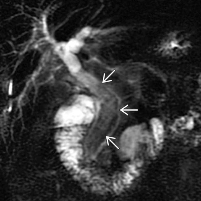
image
(Right) Coronal MRCP in a patient with markedly elevated liver function tests and leukocytosis shows intra- and extrahepatic biliary dilatation. Note the relatively low signal pus and infectious debris within the mid and distal common duct image.
image
(Left) Coronal CECT in a patient presenting with fever and hypotension demonstrates thickening and hyperenhancement of a dilated CBD image 
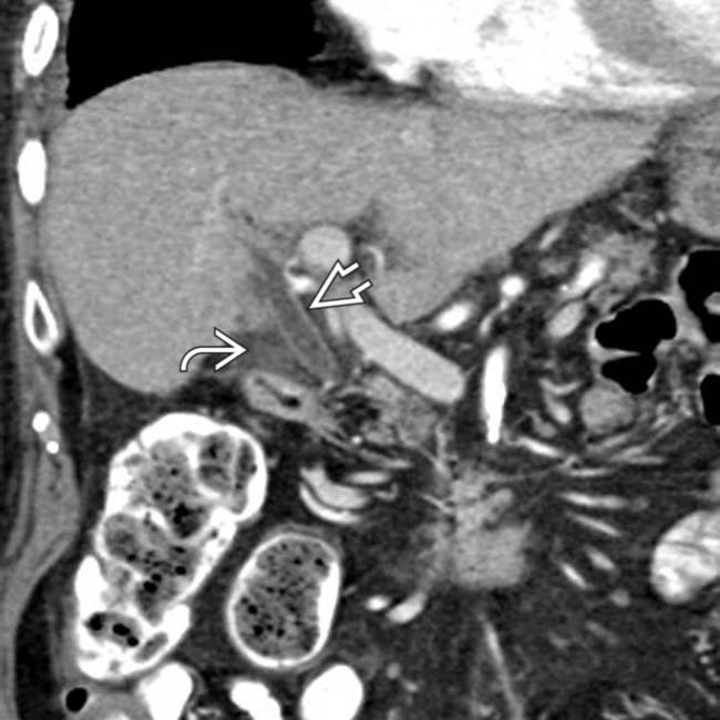
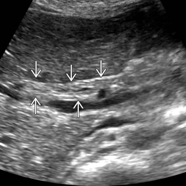
with a small amount of adjacent fluid image, suggesting ascending cholangitis.

image
(Right) Coronal NECT in the same patient demonstrates an obstructing stone image 
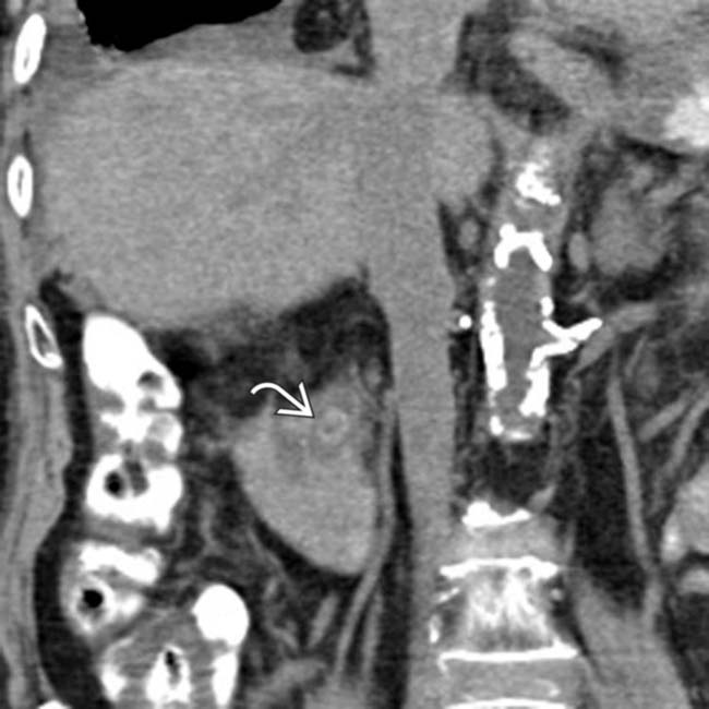
as the cause of the patient’s cholangitis.

TERMINOLOGY

Synonyms

• Bacterial cholangitis, acute cholangitis, suppurative cholangitis, biliary infection

Definitions

• Pyogenic infection of biliary tree as a result of biliary obstruction

IMAGING

General Features

• Best diagnostic clue

image Biliary ductal dilatation, often with an irregular branching pattern and bile duct wall thickening with hyperenhancement
• Location

image Dilation of intrahepatic ± extrahepatic ducts

CT Findings

• Dilation of intrahepatic ± extrahepatic ducts (depending on level of obstruction) with abrupt “cut-off” at site of obstruction

image Obstructing stone of variable density on CT depending on stone type

– Sensitivity of CT for stones is variable (25-90%) with roughly 1/4 of stones isoattenuating to bile
– Bull’s-eye sign: Rim of bile surrounding stone
– Meniscus sign at distal common bile duct (CBD) may suggest occult stone (even if stone is low density and not visible)
• High-density intraductal debris (purulent bile or pus)
• Concentric and diffuse bile duct wall thickening with hyperenhancement
• Heterogeneous hepatic parenchymal enhancement: Wedge-shaped, peribiliary, patchy, or diffuse

image May be more apparent on arterial phase imaging
• Can be associated with liver abscesses (1/4 of cases) or portal vein thrombosis

image Small abscesses may arise adjacent to biliary tree and may communicate with bile ducts

MR Findings

• Intrahepatic biliary dilatation almost always present ± extrahepatic ductal dilatation

image MRCP nicely demonstrates level of obstruction with abrupt cut-off of duct, as well as abnormal arborization and tapering of intrahepatic ducts
image Biliary dilatation may be central, diffuse, or segmental
• Presence of intermediate to low T2WI and intermediate T1WI signal purulent material (pus) within bile ducts

image Juxtaposed against normal T2 hyperintense and T1 hypointense bile
• Obstructing stones are low signal on T1WI and T2WI
• Bile duct walls appear thickened on T1WI and T2WI with progressive hyperenhancement of duct walls on T1WI C+

image Often outlined by high T2 signal periportal edema
• Diffusely heterogeneous hepatic parenchymal enhancement, which can be wedge-shaped, peribiliary, or patchy in distribution

image Abnormal enhancement most apparent in arterial and delayed phases
• Liver abscesses (1/4 of cases) and portal vein thrombosis are common complications

Fluoroscopic Findings

• Cholangiography

image Irregular, thickened bile duct wall with luminal narrowing
image Ductal stricture or radiolucent filling defect (stone) with obstruction and proximal ductal dilatation
image Abnormal arborization and tapering of intrahepatic ducts
image Intrahepatic bile ducts may show communication with hepatic abscesses

Ultrasonographic Findings

• Dilatation, stenosis, and thickening of bile duct walls
• Intraluminal echogenic material (purulent bile or pus)
• Thickened gallbladder wall ± calculi
• Relatively insensitive for distal CBD stones due to bowel gas

Nonvascular Interventions

• ERCP

image Can be performed (for stone clearance) on an elective basis (after treatment with antibiotics) in patients with mild cholangitis
image Performed urgently with stent placement in patients with severe cholangitis (hypotension, renal dysfunction, confusion)
• Transhepatic cholangiography

image Option for percutaneous drainage after failed initial ERCP or due to altered biliary anatomy or hilar obstruction

Imaging Recommendations

• Best imaging tool

image US is best screening tool to assess for presence of biliary dilatation/obstruction
image MRCP or CT helpful in patients with only mild symptoms to assess for cause of obstructions (stone, tumor, etc.)

DIFFERENTIAL DIAGNOSIS

Primary Sclerosing Cholangitis

• Segmental strictures with beading and pruning of ducts
• Can involve intrahepatic or extrahepatic ducts with thickening/hyperenhancement of duct wall in acute phase
• End stage: Cirrhotic-appearing liver with marked hypertrophy of caudate lobe/central liver and preferential atrophy of liver periphery

Other Forms of Cholangitis

• Recurrent pyogenic, AIDS-related, or chemotherapy-related cholangitis
• Patient history is key to correct diagnosis

PATHOLOGY

General Features

• Etiology

image Pathogenesis

– Stone/stricture → obstruction → bile stasis → ↑ biliary pressure → infection

image Biliary tree normally sterile due to bacteriostatic bile salts, mechanical barrier of sphincter of Oddi, and hepatocyte tight junctions
image Source of infection is usually bacteria from duodenum, which ascend biliary tree, although rarely may be hematogenous
image In cases with hematogenous spread, ↑ pressures in biliary tree due to obstruction may disrupt tight hepatocellular junctions and translocate bacteria from blood
– ↑ biliary pressure adversely affects host defense mechanism (bile flow, tight junctions, IgA production)
– ↑ biliary pressure results in hepatovenous reflux and bacteremia
image Risk factors

– Choledocholithiasis or hepatolithiasis in 80% of cases

image Most common cause of cholangitis in Western world
– Biliary stricture (e.g., primary sclerosing cholangitis or malignancy)

image Malignant obstruction is a very rare cause of cholangitis (except in setting of biliary intervention)
– Biliary stents (can act as nidus for infection)
– Hepatobiliary surgery
– Recent manipulation (i.e., ERCP, sphincterotomy, or percutaneous transhepatic cholangiography [PTC])
– Sphincter of Oddi dysfunction or stenosis
image Bacteriology

– Escherichia coli (25-50%), Klebsiella (15-20%), Enterococcus species (10-20%), Enterobacter species (5-10%)
– Anaerobes (Bacteroides  and  Clostridia) seen in mixed infections
– Parasitic infections (Ascaris, Clonorchis, etc.) common in developing world
• Associated abnormalities

image Liver abscess
image Portal vein thrombosis due to septic thrombophlebitis

Staging, Grading, & Classification

• Tokyo guidelines for initial diagnosis

image Clinical manifestations (fever, abdominal pain, jaundice)
image Inflammatory response (leukocytosis, C-reactive protein)
image Abnormal liver function tests
image Imaging evidence of etiology (stone, stricture, stent)
• Severity of disease

image Mild (responsive to antibiotic and supportive therapy)
image Moderate (not responsive to medical therapy, but no organ dysfunction)
image Severe (organ dysfunction)

Microscopic Features

• Acute inflammation of wall of biliary ducts
• Entry of neutrophils into luminal space

CLINICAL ISSUES

Presentation

• Most common signs/symptoms

image May be a life-threatening condition, although its severity can range from mild to severe
image Charcot triad (seen in 15-75%): Pain, fever, jaundice

– Distinction between cholangitis and cholecystitis often difficult based on clinical presentation and laboratory markers
• Other signs/symptoms

image Reynold pentad: Charcot triad + confusion and hypotension

– Present in only a small minority of patients (4-8%)
image Elderly patients are more likely to have atypical presentations

– Hypotension may be only symptom in elderly and patients on steroids
image Lab findings

– ↑ WBC, ↑ direct bilirubin, ↑ alkaline phosphatase, ↑ GGT, ↑ amylase (concomitant pancreatitis)
– ↑ transaminases
– Positive blood cultures

Demographics

• Age

image 20-50 years (but can occur at any age)
• Gender

image M = F
• Epidemiology

image Most common type of cholangitis in Western countries
image Usually due to poor nutrition and parasitic infestation in developing countries

Natural History & Prognosis

• Complications: Liver abscesses, sepsis, portal vein thrombosis
• Prognosis: 20-30% mortality in severe cases (including patients with Reynold pentad)

Treatment

• Broad spectrum parenteral antibiotics + decompression/drainage of biliary system

image 80% respond to conservative therapy and drainage can be delayed and performed on elective basis
image 20% deteriorate and need urgent drainage
• Drainage methods

image ERCP (preferred): Sphincterotomy with stone extraction ± stenting
image PTC: When ERCP unavailable, unsuccessful, or not technically feasible

– May be necessary with high biliary obstruction, intrahepatic stone, or prior biliary-enteric surgery
image Cholecystostomy
image Open surgical decompression
• Interventional management of stones/strictures

DIAGNOSTIC CHECKLIST

Consider

• Correlate with clinical and laboratory data to differentiate from other causes of cholangitis

Image Interpretation Pearls

• Cholangiography: Strictures, dilatations, intraluminal filling defects
image
(Left) Longitudinal oblique ultrasound in a patient with RUQ pain, leukocytosis, and jaundice shows a markedly thickened CD image, suggestive of cholangitis.

image
(Right) Longitudinal oblique ultrasound in a patient with mildly elevated white blood count (WBC) and alkaline phosphatase shows high-level echoes within a markedly dilated CD image
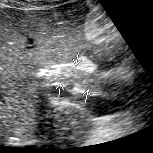
. An obstructing distal stone and abundant pus were identified at ERCP, compatible with cholangitis.
image
(Left) Transhepatic cholangiography in a patient S/P biliary-enteric anastomosis shows the anastomosis image 
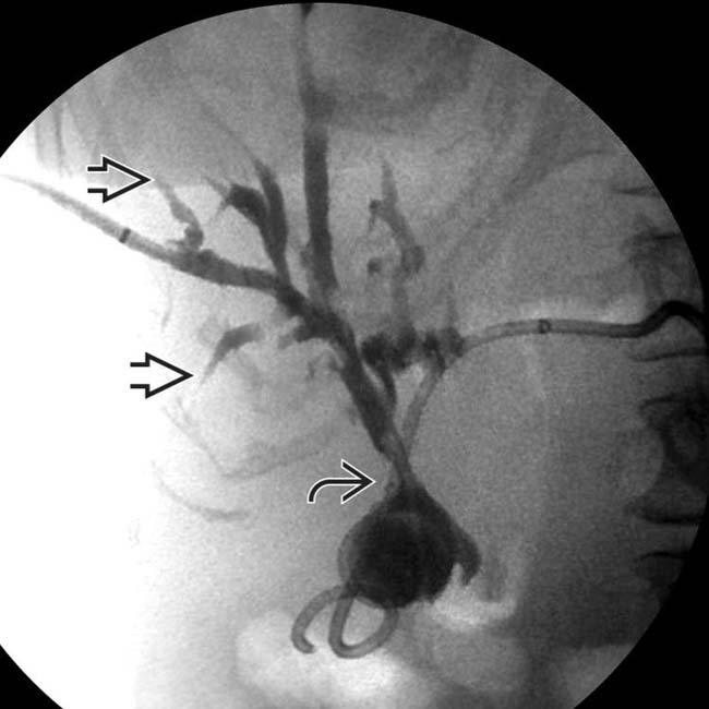
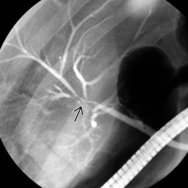
and proximal ductal dilation. Note the irregular arborization of the ducts and the abrupt “arrowhead” terminations image, characteristic of cholangitis.

image
(Right) Axial CECT in a 75-year-old woman with fever, RUQ pain, & large complex right lobe hepatic abscess shows that this complication of documented ascending cholangitis was treated successfully with biliary stenting & percutaneous drainage.
image
(Left) Cholangiogram in a patient with ascending cholangitis shows focal narrowing of the distal right and left hepatic ducts and proximal common hepatic duct image
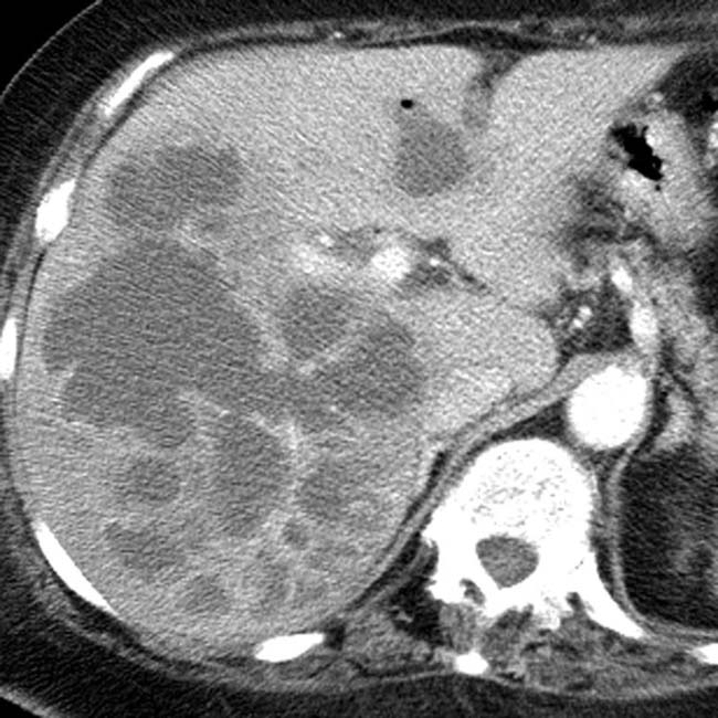
. This narrowing is likely due to inflammatory wall thickening associated with bacterial infection.

image
(Right) Cholangiogram in the same patient 2 weeks later following antibiotics shows near complete resolution of bile duct narrowing image
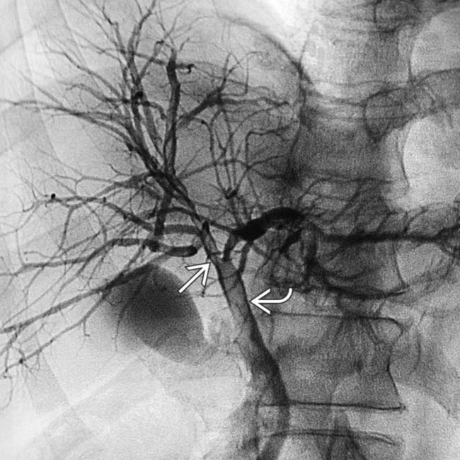
. The filling defect image within the CBD is a blood clot attributable to the imaging procedure.
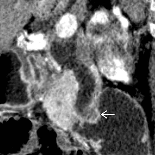
image
Coronal CECT minimum intensity image shows an ascending cholangitis related to an obstructing common duct stone. Note the dilatation of the common duct from an impacted stone image.

image
Coronal CECT in a patient presenting with sepsis demonstrates diffuse dilatation of the extrahepatic bile duct image 
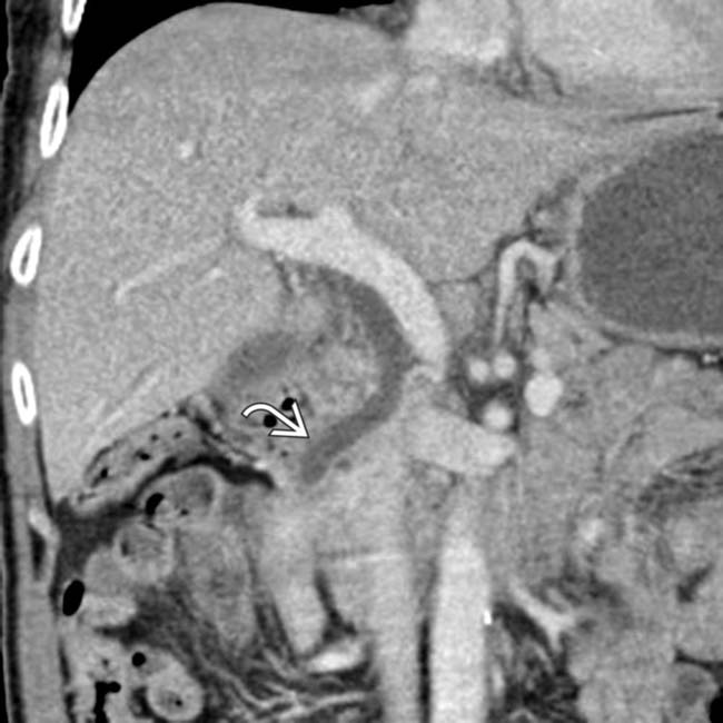
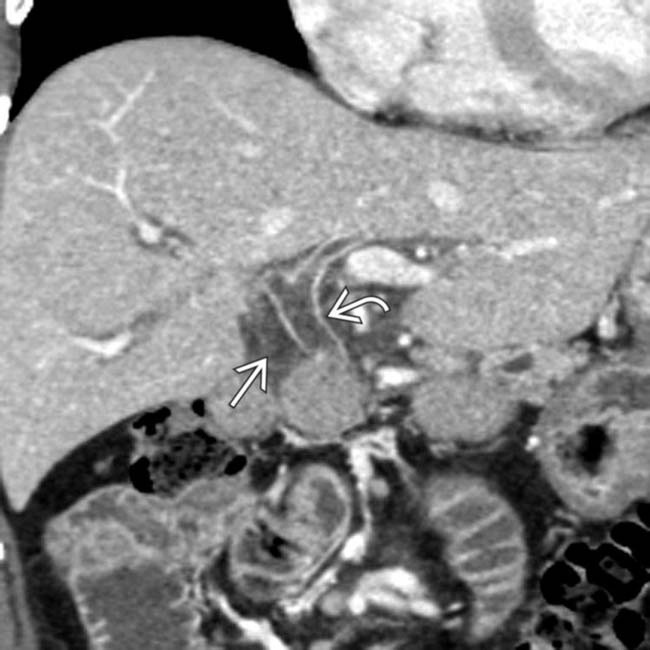
with subtle thickening and enhancement of the bile duct wall, suggesting ascending cholangitis. While not visible on CT, multiple stones were found at ERCP.
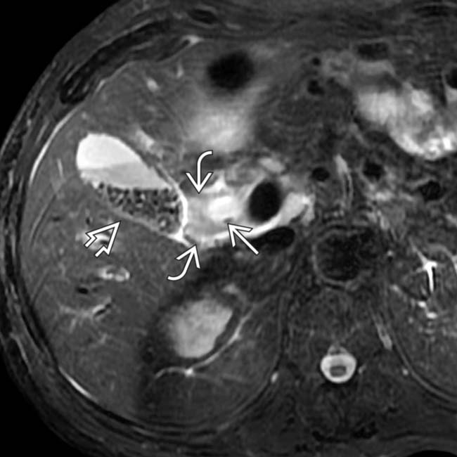
image
Axial T2WI FSE MR in an 80-year-old man with documented ascending cholangitis shows a dilated extrahepatic duct that contains a small dependent stone image, periductal edema image, and multiple gallstones image.

SELECTED REFERENCES

1. Costello, JR, et al. MR imaging of benign and malignant biliary conditions. Magn Reson Imaging Clin N Am. 2014; 22(3):467–488.

2. Kochar, R, et al. Infections of the biliary tract. Gastrointest Endosc Clin N Am. 2013; 23(2):199–218.

Patel, NB, et al. Multidetector CT of emergent biliary pathologic conditions. Radiographics. 2013; 33(7):1867–1888.

Lee, NK, et al. MR appearance of normal and abnormal bile: correlation with imaging and endoscopic finding. Eur J Radiol. 2010; 76(2):211–221.

Catalano, OA, et al. Biliary infections: spectrum of imaging findings and management. Radiographics. 2009; 29(7):2059–2080.

Kim, SW, et al. Transient arterial enhancement of the hepatic parenchyma in patients with acute cholangitis. J Comput Assist Tomogr. 2009; 33(3):398–404.

Lee, JG. Diagnosis and management of acute cholangitis. Nat Rev Gastroenterol Hepatol. 2009; 6(9):533–541.

Lee, NK, et al. Discrimination of suppurative cholangitis from nonsuppurative cholangitis with computed tomography (CT). Eur J Radiol. 2009; 69(3):528–535.

Salek, J, et al. Analysis of risk factors predictive of early mortality and urgent ERCP in acute cholangitis. J Clin Gastroenterol. 2009; 43(2):171–175.

Yeh, BM, et al. MR imaging and CT of the biliary tract. Radiographics. 2009; 29(6):1669–1688.

Attasaranya, S, et al. Choledocholithiasis, ascending cholangitis, and gallstone pancreatitis. Med Clin North Am. 2008; 92(4):925–960. [x].

Kinney, TP. Management of ascending cholangitis. Gastrointest Endosc Clin N Am. 2007; 17(2):289–306. [vi].

Watanabe, Y, et al. MR imaging of acute biliary disorders. Radiographics. 2007; 27(2):477–495.

Hanbidge, AE, et al. From the RSNA refresher courses: imaging evaluation for acute pain in the right upper quadrant. Radiographics. 2004; 24(4):1117–1135.

Arai, K, et al. Dynamic CT of acute cholangitis: early inhomogeneous enhancement of the liver. AJR Am J Roentgenol. 2003; 181(1):115–118.

Browning, JD, et al. Gallstone disease and its complications. Semin Gastrointest Dis. 2003; 14(4):165–177.

Håkansson, K, et al. MR characteristics of acute cholangitis. Acta Radiol. 2002; 43(2):175–179.

Bader, TR, et al. MR imaging findings of infectious cholangitis. Magn Reson Imaging. 2001; 19(6):781–788.

Hanau, LH, et al. Acute (ascending) cholangitis. Infect Dis Clin North Am. 2000; 14(3):521–546.

Song, HH, et al. Eosinophilic cholangitis: US, CT, and cholangiography findings. J Comput Assist Tomogr. 1997; 21(2):251–253.