Arthroscopic Management of the Trauma Patient

Published on 11/04/2015 by admin

Filed under Orthopaedics

Last modified 22/04/2025

Print this page

rate 1 star rate 2 star rate 3 star rate 4 star rate 5 star
Your rating: none, Average: 0 (0 votes)

This article have been viewed 1145 times

CHAPTER 24 Arthroscopic Management of the Trauma Patient

Introduction

Hip arthroscopy has been described as part of treatment regimens for hip trauma and related sequelae for decades, but the role of hip arthroscopy for this application has expanded rapidly during recent years. As early as 1931, Burman noted a role for a form of hip arthroscopy for loose body or fragment removal. The role for surgical debridement of the hip expanded substantially with the observation that loose bodies were ubiquitous with hip fracture dislocations. Epstein advised that all hip fracture–dislocations should be treated with debridement in an attempt to delay the appearance of traumatic arthritis and to minimize its severity. He advocated open debridement rather than arthroscopic debridement. The prevalence of loose bodies after injury to the hip and their impact on the development of hip joint arthrosis certainly contributed to the imperfect long-term results of dislocations and fractures around the hip joint (Figures 24-1 and 24-2). Until recently, loose bone and cartilage fragments were almost always retrieved with open arthrotomy. Advances in arthroscopic tools and techniques have made arthroscopic loose-body removal highly efficient. Arthroscopy advantages include diminished blood loss, smaller incisions, decreased recovery time, reduced potential for neurovascular damage, and decreased disruption of capsuloligamentous structures. Indications for arthroscopic debridement after trauma have been extended to include the extraction of bullets, the removal of broken hardware from the joint, and joint lavage for the treatment of infection or contamination in association with bullet fragments passing through the bowel and communicating with the hip joint.

image

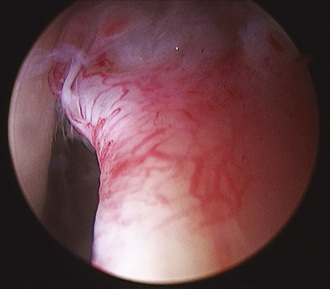
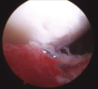
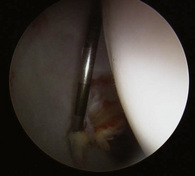
Figure 24–2 Another view of the same patient shown in Figure 24-1. Femoral head chondrolysis is evident. Intervention was limited to labral debridement, chondroplasty of the femoral head, and lavage.

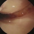
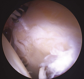
The arthroscopic treatment of acetabular labral pathology most typically involves atraumatic tears or labral disease associated with impingement or hip dysplasia. Isolated cases of traumatic labral pathology have been reported since 1959, when Dameron described a bucket-handle tear of the acetabular labrum that prohibited the reduction of a posterior dislocation of the hip that subsequently required open repair. Labral injury has more recently been described as a relatively common but previously poorly recognized phenomenon in association with acetabular fractures. Ganz described reproducible labral pathology in 14 patients with displaced transverse acetabular fractures who had been treated with open reduction and internal fixation. The labrum was partially or completely detached from the superior acetabular rim in all cases. In this series, an avulsed portion of the labrum was left if it was stable and undamaged, resected if it was unstable and damaged, and repaired if it was unstable but intact or attached to a bony fragment. Ganz proposed arthrotomy at the time of acetabular fracture fixation to search for associated intracapsular injuries in displaced transverse acetabular fractures and to treat injuries accordingly. In the case of acetabular fractures, multiple authors have identified reduction as the most important factor for avoiding the development of arthrosis and for obtaining a good clinical outcome, but they have noted that even anatomic fracture reduction fails to guarantee excellent outcomes. It is likely that additional factors such as chondral damage at the time of injury, loose fragments, and labral injuries all contribute to the patient’s final long-term outcome (Figures 24-3 and 24-4).
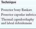
image

Figure 24–4 Another view of the same patient shown in Figure 24-3. There is a small anterior labral tear. The labral tear and the femoral head were debrided.

Brief history and physical examination

Patients with high-energy trauma require more urgent and comprehensive treatment than patients who have experienced lower-energy or focal trauma to the hip. The initial evaluation of a patient with high-energy trauma is based on the Advanced Trauma Life Support protocol and includes the “ABCs”—airway, breathing, and circulation—of the primary survey. The treatment of high-energy trauma patients is directed by general surgical colleagues with prompt cooperation from orthopedic surgeons. Standard radiographic trauma series include an anteroposterior pelvic radiograph, an anteroposterior chest radiograph, and a lateral cervical spine radiograph. The orthopedic examination includes the palpation of the spine and all extremities to look for crepitance, deformity, open injuries, and dislocation. A high index of suspicion for a posterior hip dislocation is maintained when a patient presents with a shortened extremity with the affected hip held in flexion, abduction, and internal rotation. Alternatively, an anterior dislocation leaves the hip in extension and neutral or slight abduction. The examiner must complete a thorough trauma evaluation, because 95% of patients with a hip dislocation have at least one other organ-system injury. Of the patients who have high-energy hip dislocations, 15% have abdominal injuries, 21% have thoracic injuries, 21% have craniofacial injuries, 24% have closed head injuries, and 33% have other orthopedic injuries.

Imaging and diagnostic studies

An anteroposterior pelvic radiograph is a routine part of the evaluation of a traumatically injured patient. If there is a disruption of the anterior or posterior pelvic ring, then the evaluation routinely includes pelvic inlet and outlet views. Similarly, if an associated acetabular fracture is present or suspected, radiographic evaluation should include the 45-degree oblique views described by Judet and Letournel. The closed reduction of a dislocated hip must be confirmed by a repeat anteroposterior pelvic radiograph. Follow up studies should also include pelvic computed tomography (CT) scans with 1.5-mm cuts through the acetabulum to search for loose bodies, to assess acetabular fractures, and to evaluate the reduction status of associated femoral head fractures. Final complete radiographs should include a dedicated hip series as well as full-length views of the ipsilateral femur to evaluate for associated fractures. In the case of gunshot wounds, metallic markers should be placed at all identifiable entrance and exit wounds to facilitate an understanding of the bullet’s trajectory. Radiographs and CT scans may not reliably demonstrate loose bodies within the hip joint. In a series of 36 patients who were treated with arthroscopy, Mullis and Dahners found loose bodies in 33 patients, including 7 out of 9 patients who had no loose bodies seen on preoperative radiographs or CT scans with 3-mm cuts.

Labral tears are best evaluated with magnetic resonance arthrography. Typically, labral tears associated with high-energy trauma have not been routinely addressed or even recognized acutely unless they are specifically sought intraoperatively during open fracture fixation. Thus, magnetic resonance arthrograms may be most frequently indicated for traumatically injured patients who continue to have unexplained pain during convalescence. Labral injuries have been demonstrated in association with acetabular fractures, and traumatic labral pathologies should be addressed. The extent to which minor labral injuries associated with high-energy trauma will become symptomatic or contribute to arthrosis remains unknown.

Surgical technique

The patient can be placed supine or in a lateral position, depending on surgeon preference. Because one must take into consideration other injuries of the traumatized patient (e.g., spine injury) when positioning him or her, a supine position is often required. By contrast, an obese patient’s pannus may interfere with the maneuverability of arthroscopic equipment if he or she is in the supine position, so a lateral position should be considered for these individuals. A standard fracture table or a custom distraction device is necessary to distract the joint space. In our practice, we prefer to use a commercially available distraction device system that accommodates hip arthroscopy. Distractor systems must possess a stable distraction mechanism and a well-padded perineal post; most described complications encountered with hip arthroscopy are neuropraxias caused by compression against an underpadded post or by distraction, especially if it is prolonged. In addition, the perineal post must be offset laterally against the medial thigh of the operative leg to achieve a sufficient vector to distract the hip joint. The hip is slightly abducted and flexed to relax the anterior hip capsule. Both feet are generously padded and securely placed into the foot holders. Fluoroscopy is introduced from the nonoperative side of the patient before the sterile preparation of the injured extremity to confirm the ability to distract the joint. Approximately 50 lb of traction is needed to distract the hip joint; however, less force may be needed for the distraction of recently injured hips with traumatic capsular disruptions.

Three standard portals have been described for hip arthroscopy: anterior, anterolateral, and posterolateral. All of these may be necessary for the retrieval of intra-articular fragments, particularly if the fragments have been impacted and are relatively immobile. The anterolateral portal lies most centrally in the safe zone for arthroscopy and therefore should be established first for the introduction of the arthroscope and direct visualization while introducing the remaining portals. The anterolateral portal is placed 1 cm proximally and 1 cm anterior to the tip of the greater trochanter. The posterolateral portal is made at the superoposterior margin of the greater trochanter. The anterior portal is placed at the intersection of a line drawn distally from the anterosuperior iliac spine and a transverse line from the tip of the greater trochanter. Alternative arthroscopic portals have been described, including an inferomedial approach to remove an intra-articular bullet. With this approach, the hip is placed in extension and in approximately 30 degrees of abduction. A 3-cm incision is made posterior to the adductor longus tendon to allow for the blunt dissection and identification of the psoas tendon. The capsule is then penetrated medial to the tendon and distended with saline.

The hip is distracted a minimum of 8 mm to avoid chondral and labral injuries during the insertion of instruments. Portals are made with the use of specially designed extra-long arthroscopic instruments, including a long spinal needle and a flexible guidewire. A long 18-gauge spinal needle is placed first under fluoroscopic imaging at the anterolateral portal. Special attention must be paid to avoid the penetration of the labrum. If the labrum is penetrated with the spinal needle, it will be subsequently damaged with the passage of the trocar. As the needle penetrates the capsule, a decrease in resistance is noted, whereas if the needle contacts the labrum, the resistance increases. The joint is then distended with saline, and intracapsular positioning is confirmed by the backflow of fluid. The guidewire is passed, and the spinal needle is removed. A sharp cannulated trocar is then introduced over the guidewire that is penetrating the joint capsule, and this is followed by a blunt cannulated trocar to avoid damage to the articular cartilage. The anterior portal can then be made by direct intra-articular visualization of the anterior triangle, which is comprised of the capsule, the labrum, and the femoral head. A long 18-gauge spinal needle is inserted within this triangle under direct visualization, and the steps are then repeated to introduce a trocar.

The inspection of the hip joint is accomplished systematically. One may switch among the three established portals with the use of a combination of 70- and 30-degree arthroscopic cameras. The 70-degree arthroscope is most commonly used, and it affords the best view of the labrum, the femoral head, and the acetabulum, including the most inferior portion of the acetabular fossa, which contains the ligamentum teres. The 30-degree arthroscope provides the best view of the central portion of the acetabulum, the femoral head, and the superior part of the fossa. Joint hematoma is addressed first to increase visualization. Subsequently, peripheral pathology is inspected and dealt with accordingly, with labral repair or excision, loose-body removal, or chondroplasty. Debridement within the acetabular fossa is performed last, because bleeding will obscure visualization. Electrocautery, epinephrine added to the pump fluid, and hypotensive anesthesia may be employed to optimize visualization. In addition, a high-flow pump may be used to allow for sufficient flow without requiring increased fluid pressure, which may be maintained at 60 mm Hg. Loose bodies can often be flushed from the joint with simple fluid lavage, whereas larger bodies require removal with a grasper.

Labral tears associated with hip trauma can be repaired with suture anchors if detached from the bone or with suture lassos if an intrasubstance tear is present. The suture anchor is positioned on the acetabular rim away from the articular surface. The anchor may be inserted with fluoroscopic assistance, but typically this is not necessary. The anchor is gently tapped into place while aiming away from the articular surface and simultaneously visualizing the joint surface to identify improper placement into the articular cartilage. With the anchor solidly in position, a curved spectrum is used to capture capsular tissue, and a suture passer is delivered through the spectrum. The passing suture and one limb of the anchor suture are retrieved with a grasping instrument through the anterior portal, and these are tied tightly together. The other end of the passing suture is then pulled out through the anterior portal, carrying with it the anchor suture limb through the capsular tissue and back out through the anterior portal. The passing suture is then untied from the suture anchor limb. The second suture anchor limb is retrieved from the joint through the anterior portal with a grasping instrument. The authors prefer a Weston knot, which results in a vertical mattress and the maintenance of the knot on the capsular side of the labrum. The suture is cut with a boxed suture cutter, and the technique is repeated as needed to reattach the labrum to the acetabular rim. Alternatively, intrasubstance tears may be repairable if a stable outer rim is present. The cleavage plane is initially debrided of nonviable tissue to healthy bleeding edges to facilitate healing. A curved spectrum is passed through the portion of the labrum that is attached to the acetabular rim. A passing suture is passed through the spectrum, delivered through the portal, and tied to a suture, which is pulled through the joint back out through the working cannula. A bird’s beak is then used to pierce the labrum peripheral to the tear. The suture is grasped, and the free end is brought out through the working cannula. A Weston knot is placed on the capsular side, and the free sutures are cut with a boxed suture cutter. Additional sutures are placed as needed to stabilize the tear.

To address a ligamentum teres tear, the arthroscope is first placed in the anterolateral portal. The ligament is most readily accessed from the anterior portal. External rotation of the hip will help to deliver a portion of the ligament anteriorly to allow for debridement with an arthroscopic shaver. To completely debride the ligament, one must address the acetabular attachment via the posterolateral portal. The released ligament is then drawn to the mechanical debrider with suction.

Bullet extraction is accomplished with an appropriate portal that depends on the location of the missile. Cory and Ruch described removing a bullet from the femoral head with a pituitary rongeur through the anterior portal. Goldman used a mini open posterior approach and freed the missile with osteotomes to lever the bullet from the femoral head. In that particular case, the bullet fragmented on the first attempt to remove it, and a second hip arthroscopy was undertaken to remove all fragments. Singleton described the removal of a bullet from the acetabulum via the anterior portal by seating a pin with a threaded tip into the bullet and then extracting the bullet with the pin.

Broken fracture implants may rarely be considered for arthroscopic removal. A case study by Lu described the use of an arthroscope after the removal of a dynamic hip screw to examine the hip joint cavity. Hip arthroscopy in this case revealed synovitis, which was debrided, and an osteochondral defect that was caused by lag screw penetration. The new screw length was measured with the assistance of the arthoscopic camera.

A traumatically contaminated hip joint can be irrigated with arthroscopy. Irrigation is generally accomplished with an arthroscopic camera with associated inflow in the anterolateral portal, whereas the outflow trocar is placed in the anterior portal position. Synovial or hematoma debridement can be accomplished with a standard arthroscopic shaver in the anterior portal and with a high-flow pump that delivers at least 9 L of saline. Antibiotics can be added to the saline bags at the surgeon’s discretion. The joint should be inspected for loose bodies as well, especially when hip contamination is associated with a gunshot to the hip.

Hip arthroscopy has reportedly been used to visualize the reduction of femoral head fractures. We have no experience with this technique, and we do not anticipate using the technique for significantly displaced or highly comminuted fractures. However, the technique may be useful to verify the minimal displacement of femoral head fractures and to confirm that fracture debris is absent from the joint.

Results and outcomes

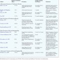
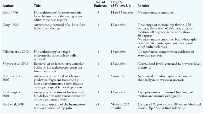
The majority of results for hip arthroscopy performed for traumatic injuries are extracted from case reports, which typically have minimal follow up (Table 24-1). Keene and Villar described two cases of posterior hip dislocation with the successful removal of retained loose bodies with hip arthroscopy. Byrd described three cases of hip arthroscopy for the management of posttraumatic loose fragments. At 14 to 33 months of follow up, each case had a reportedly successful outcome with no mechanical symptoms. Svoboda discussed a case of loose-body removal from a posterior hip dislocation in a 23-year-old military recruit that was confirmed with a postoperative CT scan. Mullis reported about 36 patients with either a simple dislocation, a fracture dislocation, or a wall fracture. When arthroscopy was performed, 92% of these patients were found to have loose bodies. Interestingly, loose bodies were found even in 7 of the 9 patients in which standard radiographic studies (anteroposterior pelvic imaging and CT scanning) failed to demonstrate them.

The use of hip arthroscopy for bullet extraction has been documented mostly with case reports that have minimal follow up times. Goldman described a mini open posterior arthroscopic approach for bullet extraction from the hip, whereas Meyer described an intra-articular bullet removal via a lateral approach. Their case reports did not include patient follow up or outcome. In 1998, Ruch removed a .44-caliber bullet and associated loose fragments from the femoral head of a 45-year-old male. Clinical examination at 1 year of follow up revealed a full range of motion, negative heel impaction, and no mechanical symptoms. Radiographic evaluation revealed joint space narrowing with subchondral sclerosis and no evidence of avascular necrosis. Teloken described an inferomedial approach for bullet removal. In this case report, the bullet was removed, and this resulted in a full range of motion without crepitus or pain at 18 months of follow up. Singleton detailed a case of a bullet lodged in a patient’s acetabulum after traversing through the rectum. The patient underwent hip arthroscopy, irrigation, debridement, and missile removal, with no evidence of subsequent hip infection.

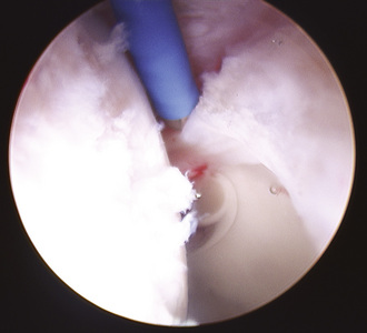
Violent trauma to the hip, including dislocations, is frequently associated with injury or rupture of the ligamentum teres (Figure 24-5). Isolated injuries may present a diagnostic challenge. In 2001, Kashiwagi published a case report of a 10-year-old girl who fell awkwardly from a swing onto her leg, which resulted in an avulsion of the ligamentum teres from its acetabular attachment. The patient presented with pain and the inability to bear weight on the affected leg. On physical examination, her hip lay in an abducted position, with limited range of motion. Radiographs demonstrated a slightly widened joint space, and a CT scan confirmed a bone fragment that was associated with the avulsed ligamentum teres. The patient was treated with hip arthroscopy, and the osteochondral fragment was removed from the anterior portal using forceps. The girl was asymptomatic with a full range of hip motion and no abnormal radiographic findings at 1 year postoperatively. Byrd treated 23 patients with traumatic injuries to the ligamentum teres (15 violent injuries, including 6 dislocations and 8 twisting injuries). The duration of symptoms averaged 28.5 months and included deep anterior groin pain in all patients; 19 patients had mechanical symptoms, and 4 patients had pain during activity. On examination, 15 patients had pain during logrolling of the hip, whereas 23 had pain with maximal flexion and internal rotation. Upon intervention, it was found that 12 patients had complete ruptures and that 11 had partial tears. Most commonly, there was associated pathology (9 labral tears, 5 loose bodies, 5 chondral injuries). The average preoperative Modified Harris Hip Score of 47 improved to 90 postoperatively.

Complications

Complication rates associated with hip arthroscopy range from 0.5% to 6.4%. The majority of complications encountered during hip arthroscopy are neuropraxias. Funke performed hip arthroscopy with the patient in the lateral decubitus position on 19 patients and noted three complications: neuropraxia of the pudendal nerve, hematoma of the labia majora, and a postoperative acute onset of abdominal pain that was surmised to result from fluid extravasation into the retroperitoneum that caused peritoneal irritation. Griffin reported an overall complication rate of 1.6% in 640 patients. Complications in this series included neuropraxias of the sciatic and femoral nerves, a small vaginal tear, a portal hematoma, and the breakage of two arthroscopic instruments in the joint. Sampson reported 34 complications in 530 cases performed since 1977. Complications included 20 transient nerve injuries (10 peroneal, 4 pudendal, 4 sciatic, 1 that was both femoral and sciatic); 9 cases of fluid extravasation, which was felt to be related to prolonged surgery with a pump; and 1 case of avascular necrosis of the femoral head. Clarke reported about 1054 consecutive hip arthroscopies with an overall complication rate of 1.4%. The complications included neuropraxia, portal wound bleeding, portal hematoma, trochanteric bursitis, and instrument breakage.

The prospect of using hip arthroscopy for acute traumatic injuries of the hip or the acetabulum can be complicated by fluid extravasation. Bartlett and colleagues reported about a patient who underwent open reduction and internal fixation for a 3-week-old acetabular fracture. Hip arthroscopy was performed 12 days after the open reduction and internal fixation of the acetabular fracture for persistent intra-articular fragments and slight hip subluxation. In this report, an abdominal compartment syndrome developed as a result of intra-abdominal extravasated arthroscopy fluid that occurred during the procedure, which led to cardiopulmonary arrest, presumably as a result of paradoxic bradycardia from compression of the inferior vena cava. This report highlights the importance of strict fluid management and the maintenance of low intra-articular pressure to safely perform hip arthroscopy in the presence of acute acetabular trauma. This is especially important for intra-articular fractures or those that involve the tectal region, which can allow for extravasation into the retroperitoneum. Signs of fluid extravasation include the inability to distend the joint, increased fluid requirements to maintain distention, the frequent cutoff of pump irrigation systems, and abdominal distention or thigh swelling.

Annotated references and suggested readings

Bartlett C.S., DiFelice G.S., Buly R.L., Quinn T.J., Green D.S., Helfet D.L. Cardiac arrest as a result of intraabdominal extravasation of fluid during arthroscopic removal of a loose body from the hip joint of a patient with an acetabular fracture. J Orthop Trauma. 1998;12(4):294-299.

Burman M.S. Arthroscopy or the direct visualization of joints: an experimental cadaver study. J Bone Joint Surg. 1931;13:669-695.

Byrd J.W. Hip arthroscopy for post-traumatic loose fragments in the young adult: three case reports. Clin J Sport Med. 1996;6:129-134.

Byrd J.W., Jones K.S. Traumatic rupture of the ligamentum teres as a source of hip pain. Arthroscopy. 2003;20(4):385-391.

Clarke M.T., Arora A., Villar R.N. Hip arthroscopy: complications in 1054 cases. Clin Orthop Relat Res (406); 2003:84-88.

Cory J.W., Ruch D.S. Arthroscopic removal of a .44 caliber bullet from the hip. Arthroscopy. 1998;14(6):624-626.

Dameron T.B.Jr. Bucket-handle tear of acetabular labrum accompanying posterior dislocation of the hip. J Bone Joint Surg. 1959;41-A:131-134.

Epstein H.C. Posterior fracture-dislocations of the hip; long-term follow-up. J Bone Joint Surg Am. 1974;56:1103-1127.

Funke E.L., Munzinger U. Complications in hip arthroscopy. Arthroscopy. 1996;12(2):156-159.

Griffin D.R., Villar R.N. Complications of arthroscopy of the hip. J Bone Joint Surg Br. 1999;81(4):604-606.

Goldman A., Minkoff J., Price A., Krinick R. A posterior arthroscopic approach to bullet extraction from the hip. J Trauma. 1987;27(11):1294-1300.

Ilizaliturri V.M.Jr, Zarate-Kalfopulos B., Martinez-Escalante F.A., Cuevas-Olivo R., Camacho-Galindo J. Arthroscopic retrieval of a broken guidewire fragment from the hip joint after cannulated screw fixation of slipped capital femoral epiphysis. Arthroscopy. 2007;23(2):227.e1-227.e4. Epub 2006 Sep 11

Judet R., Judet J., Letournel E. Fractures of the acetabulum: classification and surgical approaches for open reduction—preliminary report. J Bone Joint Surg Am. 1964;46:1615-1646.

Kashiwagi N., Suzuki S., Seto Y. Arthroscopic treatment for traumatic hip dislocation with avulsion fracture of the ligamentum teres. Arthroscopy. 2001;17(1):67-69.

Keene G.S., Villar R.N. Arthroscopic loose body retrieval following traumatic hip dislocation. Injury. 1994;25:507-510.

Lee G.H., Virkus W.W., Kapotas J.S. Arthroscopically assisted minimally invasive intraarticular bullet extraction: technique, indications, and results. J Orthop Trauma. 2008;56:513-516.

During a 5-year period, 3 out of 11 patients with retained intra-articular missiles in the hip underwent arthroscopically assisted minimally invasive bullet extraction. The four associated bullets were successfully extracted without complication..

Leunig M., Sledge J.B., Gill T.J., Ganz R. Traumatic labral avulsion from the stable rim: a constant pathology in displaced transverse acetabular fractures. Arch Orthop Trauma Surg. 2003;123(8):392-395.

Lu K.H. Arthroscopically assisted replacement of dynamic hip screw for unrecognized joint penetration of lag screw through a new portal. Arthroscopy. 2004;20(2):201-205.

Meyer N.J., Thiel B., Ninomiya J.T. Retrieval of an intact, intraarticular bullet by hip arthroscopy using the lateral approach. J Orthop Trauma. 2002;16(1):51-53.

Mullis B.H., Dahners L.E. Hip arthroscopy to remove loose bodies after traumatic dislocation. J Orthop Trauma. 2006;20(1):22-26.

Philippon M.J., Schenker M.L., Briggs K.K., Maxwell R.B. Can microfracture produce repair tissue in acetabular chondral defects? Arthroscopy. 2008;24:46-50.

The authors wrote a case series about second-look arthroscopy for acetabular defects that had been treated with microfracture. Eight out of 9 patients had 95% to 100% coverage with grade 1 or 2 appearance of the cartilage at an average follow up of 20 months..

Sampson T.G. Complications of hip arthroscopy. Clin Sports Med. 2001;20(4):831-835.

Singleton S.B., Joshi A., Schwartz M.A., Collinge C.A. Arthroscopic bullet removal from the acetabulum. Arthroscopy. 2005;21(3):360-364.

Svoboda S.J., Williams D.M., Murphy K.P. Hip arthroscopy for osteochondral loose body removal after a posterior hip dislocation. Arthroscopy. 2003;19:777-781.

Teloken M.A., Schmietd I., Tomlinson D.P. Hip arthroscopy: a unique inferomedial approach to bullet removal. Arthroscopy. 2002;18(4):E21.

Upadhyay S.S., Moulton A. The long-term results of traumatic posterior dislocation of the hip. J Bone Joint Surg Br. 1981;63B:548-1541.

The authors reviewed 81 cases of patients with traumatic posterior dislocation of the hip who were treated between 1936 and 1974. The average follow-up period was 12.5 years. The overall results were surprisingly poor despite the early reduction of the dislocation in the majority of cases. The authors found that, at 15 years after simple dislocation, 24% of cases had a poor result by both clinical and radiologic criteria. The results deteriorated with more severe injuries, with 73.3% of the patients being graded as fair or poor..

Watson D., Walcott-Sapp S., Westrich G. Symptomatic labral tear post femoral shaft fracture: case report. J Orthop Trauma. 2007;21:731-733.

A 33-year-old male fell 4.5 m and sustained a Winquist type III left femoral shaft fracture, which was treated with a standard reamed statically locked antegrade femoral nail. The patient presented 22 months after his fall with significant groin pain that was worse with activity and an occasional sharp pain associated with a catching sensation in the hip. The patient underwent hip arthroscopy, with which a large labral tear was confirmed and treated with debridement back to a stable edge. At his 2-month postoperative visit, the patient had no complaints of groin pain, and he was walking better than he had for the previous 2 years. The incidence of hip pain with antegrade femoral nailing is reported to be as high as 26% with an incomplete resolution of symptoms after nail removal, whereas even those patients treated with retrograde femoral nails have a 4% reported incidence of residual hip pain. The authors propose that labral tears may be an underinvestigated and underestimated cause of hip pain after femoral shaft fracture that can be confirmed and treated safely with arthroscopy..

Yamamoto Y., Ide T., Ono T., Hamada Y. Usefulness of arthroscopic surgery in hip trauma cases. Arthroscopy. 2003;19:269-273.

Eleven joints from 10 hip trauma cases with hip fracture dislocations, including 5 femoral head fractures, were studied. All of the patients underwent successful hip arthroscopy with lavage, debridement, loose-body removal, and arthroscopically assisted fracture fixation in 2 patients. Their results at final follow up more than 5 years postoperatively showed that none of the arthroscopically treated hip joints with or without fractures had developed osteoarthritis..