Arthroscopic and Open Anatomy of the Hip

Published on 11/04/2015 by admin

Filed under Orthopaedics

Last modified 22/04/2025

Print this page

rate 1 star rate 2 star rate 3 star rate 4 star rate 5 star
Your rating: none, Average: 0 (0 votes)

This article have been viewed 3644 times

CHAPTER 2 Arthroscopic and Open Anatomy of the Hip

Introduction

The hip joint is defined by the articulation between the head of the femur and the acetabulum of the pelvis. It is covered by a large soft-tissue envelope and a complex array of neurovascular and musculotendinous structures. The joint’s morphology and orientation are complex, and there are wide anatomic variations seen among individuals. The joint’s deep location makes both arthroscopic and open access challenging. To avoid iatrogenic injury while establishing functional and efficient access, the hip surgeon should possess a sound anatomic knowledge of the hip.

The human “hip” can be subdivided into three categories: 1) the superficial surface anatomy; 2) the deep femoroacetabular joint and capsule; and 3) the associated structures, including the muscles, nerves, and vasculature, all of which directly affect its function.

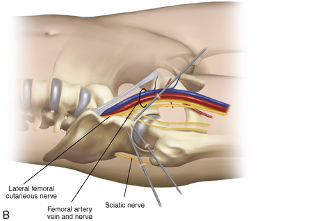
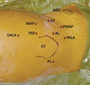
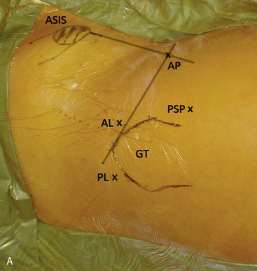
Several bony landmarks define the surface anatomy of the hip. The anterosuperior iliac spine (ASIS) and anteroinferior iliac spine (AIIS) are located anteriorly, with the former being palpable. These structures serve as the insertion points for the sartorius and the direct head of the rectus femoris, respectively. Posterolaterally, two bony landmarks are palpable: the greater trochanter and the posterosuperior iliac spine. The greater trochanter serves as the insertion point of the tendon of the gluteus medius, the gluteus minimus, the obturator externus, the obturator internus, the gemelli, and piriformis. The posterosuperior iliac spine serves as the attachment point of the oblique portion of the posterior sacroiliac ligaments and the multifidus. During arthroscopic and open access to the joint, both of these landmarks are useful tools for incision planning, and, in combination with the anterior bony prominences, for initial orientation (Figure 2-1, A).

The hip is a synovial, diarthrodial, ball-and-socket joint that is comprised of the bony articulation between the proximal femur and the acetabulum. The acetabulum is formed at the cartilaginous confluence of the three bones of the pelvis: the ilium, the ischium, and the pubis. When aligned with the anterior pelvis plane, the acetabulum is inclined at approximately 55 degrees and anteverted at approximately 20 degrees, although there are gender variations. The acetabular cup is roughly hemispheric in shape. It covers 70% of the femoral head, and it has a wave-like profile of three peaks and three troughs, including the acetabular notch. The proximal femur consists of a femoral head, which articulates with the acetabulum, and a tapered neck, which is angled onto the femoral shaft. A normal femur has a neck–shaft angle of approximately 130 degrees.

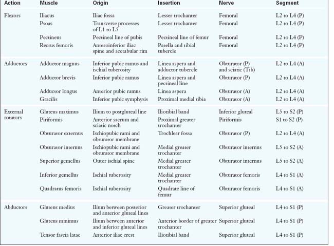
There are a total of 27 muscles that cross the hip joint. They can be categorized into six groups according to the functional movements that they induce at the joint: 1) flexors; 2) extensors; 3) abductors; 4) adductors; 5) external rotators; and 6) internal rotators. Although some muscles have dual roles, their primary functions define their group placement, and they all have unique neurovascular supplies (Table 2-1).

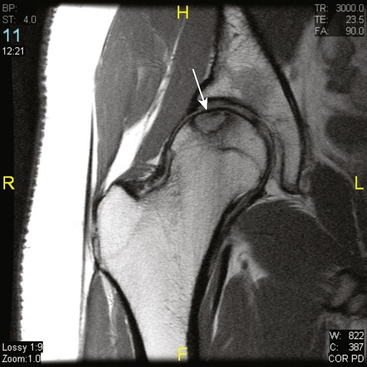
The vascular supply of the hip stems from the external and internal iliac arteries. An understanding of the course of these vessels is critical for avoiding catastrophic vascular injury. In addition, the blood supply to the femoral head is vulnerable to both traumatic and iatrogenic injury; the disruption of this supply can result in avascular necrosis (Figure 2-2).

Hip musculature

Hip Flexors

The primary hip flexors are the rectus femoris, iliacus, psoas, iliocapsularis, and sartorius muscles. The rectus femoris muscle has two distinct origins proximally: the direct head and the reflected head. They originate at the AIIS and the anterior acetabular rim (in close proximity to the anterior hip capsule), respectively. The tendinous fibers of the rectus femoris coalesce distally and become confluent with the other quadriceps musculature in the thigh. The quadriceps consists of four distinct muscles: 1) the vastus intermedius; 2) the vastus lateralis; 3) the vastus medialis; and 4) the rectus femoris. The rectus femoris is the only quadriceps muscle that traverses both the hip and the knee joint. The rectus femoris is a powerful hip flexor, but it is largely dependent on the position of the knee and hip to assert its influence. It is most powerful when the knee is flexed, whereas significant power is lost when the knee is extended. The rectus femoris is innervated by the femoral nerve (i.e., the posterior division of L2 to L4).

The iliopsoas is another powerful hip flexor that begins in two distinct regions proximally. The iliacus has a broad origin, arising from the inner table of the iliac wing, the sacral alae, and the iliolumbar and sacroiliac ligaments. The psoas originates at the lumbar transverse processes, the intervertebral discs, and the adjacent bodies from T12 to L5, in addition to the tendinous arches between these points. Distally, the two large muscular bodies converge to become one distinct structure—the iliopsoas—and subsequently jointly insert at the lesser trochanter of the proximal femur. The nerve to the iliopsoas (i.e., the anterior division of L1 to L3) supplies the iliopsoas muscle.

The sartorius originates at the ASIS and proceeds to traverse obliquely and laterally down the thigh to eventually insert at the anterior surface of the tibia, just inferomedial to the tibial tuberosity, as part of the pes anserinus. In addition to flexing the hip and knee, the sartorius aids in the abduction of the hip. It is innervated by the femoral nerve (i.e., the posterior division of L2 and L3).

Other muscles that can be recruited to assist with hip flexion include the tensor fascia latae (TFL), the pectineus, the adductors, the gracilis, and the anterior aspects of the gluteus medius and the gluteus minimus. The contribution of these secondary hip flexors largely depends on the position of the hip at the time at which movement is initiated.

Hip Extensors

The major contributors to hip extension are the gluteus maximus, the ischiocondylar part of the adductor magnus, the semimembranosus, the semitendinosus, and the biceps femoris (i.e., the long and the short heads). The most powerful muscle of this group is the gluteus maximus, which is responsible for more than 75% of the total power output of the extensor group. The gluteus maximus originates at the posteromedial outer rim of the iliac wing (i.e., behind the posterior gluteal line), the sacrococcygeal junction, the sacrotuberous ligaments, and the aponeurosis of the gluteus medius origin. Distally, the majority of the muscle inserts at the posterior aspect of the iliotibial tract of fascia lata, whereas the remainder inserts at the gluteal tuberosity of the proximal femur. In addition to extending the flexed femur, the gluteus maximus assists with lateral rotation and abduction of the thigh, and it stabilizes both the hip and knee joints through its influence on the iliotibial tract. It is innervated by the inferior gluteal nerve (i.e., the posterior divisions of L5 to S2).

The hamstring muscles also collaboratively assist with the extension of the hip. The long head of the biceps femoris, the semitendinosus, and the semimembranosus originate at the ischial tuberosity and insert below the knee. The combined hip extensor strength of these three hamstring muscles is still significantly lower than that of the gluteus maximus. However, in maximal hip flexion, the gluteus maximus loses its mechanical advantage, and the hamstrings become the dominant hip extensors. Because the hamstrings cross the knee joint, they are also able to flex and rotate the leg at the knee. They are innervated by the sciatic nerve (i.e., the posterior divisions of L5 to S2).

Abductors

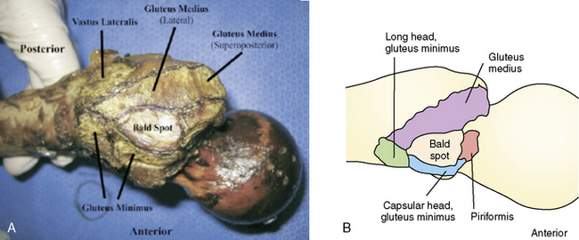
The abductors consist of the gluteus medius and gluteus minimus muscles. Both of these muscles are innervated by the superior gluteal nerve (i.e., the posterior division of L5 to S2). The TFL and the iliotibial band also contribute to hip abduction. This action is only apparent with the hip in a flexed position, and the TFL and the iliotibial band are therefore considered secondary abductors. The gluteus medius, which is the primary hip abductor, originates at the posterior external table of the iliac wing. It is completely covered by the overlying gluteus maximus as it travels distally toward its insertion at the lateral and superoposterior facet of the greater trochanter (Figure 2-3, A). The gluteus minimus fibers run in close approximation to the lateral hip capsule, onto which some of the muscle may also insert. These fibers are often the first to be encountered during hip arthroscopy procedures when establishing the anterolateral portal. The gluteus minimus, which is responsible for 25% of abduction power, runs in the same plane deep to the gluteus medius. It inserts more anteriorly on the greater trochanter, and it has a separate long head component. Recent evidence suggests that a common cause of lateral hip pain may be tears of the hip abductor insertion and not simply trochanteric bursitis. These tears are referred to as rotator cuff tears of the hip. Anatomic restoration of the insertion of the torn gluteus medius can be achieved with standard arthroscopic technique in the recently described peritrochanteric compartment. When trochanteric bursitis does exist in isolation, it is most likely located at the posterior facet or the bald spot of the trochanter (see Figure 2-3, B).

Functionally, the gluteus medius and gluteus minimus are critical to the gait cycle, and they assert the major stabilizing force during the end of the terminal swing phase. This force provides tension among the pelvis, the iliotibial band, and the greater trochanter; it peaks during the initial part of the stance phase, and it persists through the middle of the stance. Injury to the gluteus medius or indeed to the superior gluteal nerve can be clinically recognized by the presence of a Trendelenburg sign, which is classically described as the dropping of the pelvis on the opposite side of the pathology.

Neurovascular supply of the hip

Vasculature of the Hip

The common iliac arteries provide the primary blood supply to the lower limbs. Each artery divides into the external and internal iliac arteries. These vessels run parallel with their venous counterparts, the internal and external iliac veins, which join to form the inferior vena cava. The external iliac artery, which travels obliquely over the psoas muscle, is particularly vulnerable to injury. Damage can occur during hip arthroplasty when accessing the acetabulum, during the placement of screws in the anterior quadrant, or, more commonly, from the aberrant placement of anterior acetabular retractors. Excessive medial reaming can also put the external iliac vessels at risk, especially during revision acetabular surgery. If iatrogenic injury does occur, the external iliac artery and vein can be accessed most easily through the ilioinguinal approach.

The obturator vessels arise from the internal iliac vessels. They pass over the quadrilateral surface of the pelvis to the upper part of the obturator foramen to emerge from the obturator canal. The obturator artery divides into anterior branch, which supplies the obturator externus and the adjacent bone, and the posterior branch, which supplies the soft tissue of the acetabular fossa. The obturator nerve mimics the course and divisions of the obturator vessels. It is responsible for the sensory cutaneous innervation of the medial thigh and the motor innervation of the adductor muscles. Overlying these neurovascular structures is a reflected portion of the parietal peritoneum and the obturator internus muscle. These structures are fairly consistent, and they are anchored firmly by the obturator membrane as they pass through the obturator foramen. Occasionally, an aberrant vessel may traverse the pelvic brim that connects the external iliac vessels and the obturator vessels. Although the obturator vessels are usually safe during arthroscopic approaches to the hip, errant passes of an inferomedially directed arthroscopic cannula can be potentially injurious. Similarly, there is little risk to the obturator vessels and nerve during open approaches for primary hip arthroplasty, but one still has to be cautious around the transverse acetabular ligament, because distal branches of the obturator vessels can be injured here. In addition, anteroinferior screw placement or excess traction on the proximal femur during an anterior approach to the hip can also be potentially harmful.

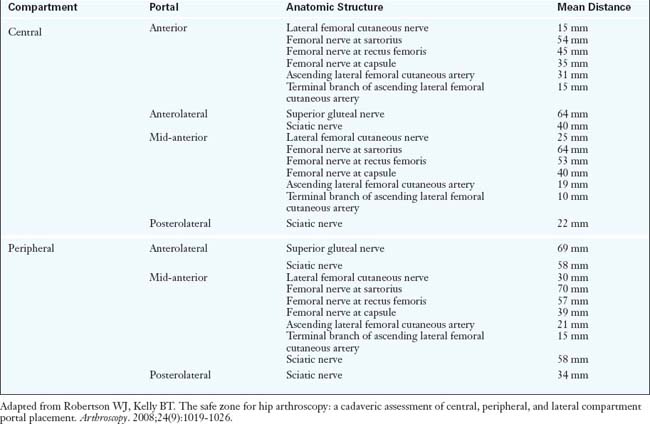
The common femoral artery is the first branch of the external iliac artery, and it traverses just anteromedial to the hip capsule as it travels distally. It is at high risk for damage during both arthroscopic and open anterior approaches to the hip. In fact, the traditional anterior arthroscopic portal is approximately 3.5 cm from the femoral neurovascular bundle (Table 2-2). During total hip arthroplasty (THA), femoral vessel injury and femoral nerve palsy have been described as arising from the incorrect placement of retractors, which can occur with all approaches to the hip. However, because the artery is a large, fairly superficial, and therefore readily palpable vascular structure, its exact location should be routinely identified and thus easily avoided.

The profundus femoris artery, which is also known as the deep femoral artery, is the first branch of the common femoral artery. It penetrates posteriorly between the pectineus, the adductor longus, and the adductor brevis, lying behind the femoral artery and vein on the medial side of the femur. The profundus femoris artery gives rise to the lateral circumflex femoral artery 90% of the time and the medial circumflex femoral artery only 30% of the time. Injuries to the profundus femoris and its branches have been described during arthroplasty approaches to the hip, but they are fairly unusual. When they do occur, it is usually as a result of anteriorly placed deep retractors or during cement extrusion in this region.

The superior gluteal vessels are branches of the internal iliac artery (i.e., the posterior branches). The vessels, along with the gluteal nerve, traverse the posterior column of the acetabulum as they exit through the sciatic notch. They emerge superior to the piriformis and then terminate in the gluteus medius and gluteus minimus muscles. The inferior gluteal and internal pudendal vessels are also branches of the internal iliac artery (i.e., the anterior branches). They exit inferior and medial to the piriformis. The inferior gluteal vessels pass through the lower part of the greater sciatic foramen. The internal pudendal vessels exit the greater sciatic notch and then reenter the pelvis via the lesser sciatic notch. Erroneous posterior screw placement can cause the disruption of these structures. Palpation of the sciatic notch and the posterior column can help to prevent the placement of proud screws and further decrease the risk of injury. However, arthroscopic approaches in the safe zones as described by Byrd and colleagues pose very little risk to these neurovascular structures.

Four sets of arteries are responsible for the arterial blood supply to the femoral head: 1) the medial circumflex artery; 2) the lateral circumflex artery; 3) the medullary artery from the shaft of the femur; and 4) the artery of the ligamentum teres. The last one provides minimal if any contribution to the vascular integrity to the femoral head, although the vessel remains patent in approximately 20% of the adult population. The exact contribution of the medullary artery to the femoral head is unknown, but it is believed that this also plays a relatively minor role in vascularization.

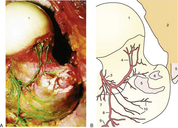
Therefore, the vessel that supplies the majority of the arterial supply to the head is the medial circumflex femoral artery, with varying contributions from the lateral circumflex femoral artery. These vessels branch off at the base of the femoral neck and then ascend toward the femoral head via the posterolateral and posteroinferior synovial retinacular folds (Figure 2-4, A and B). It is believed that disruption at this level (e.g., by a femoral neck fracture) poses the greatest risk for avascular necrosis. The lateral synovial folds, which contain the terminal branches of the medial circumflex femoral artery, can also be injured as a result of aggressive arthroscopic dissection (Figure 2-5) or open approaches. Therefore, they should be routinely identified and protected during peripheral compartment arthroscopy and during open joint-preserving hip surgery.

Neural Supply of the Hip

The sciatic nerve is the largest nerve in the body, and it is the main branch of the sacral plexus. It contains nerve branches primarily from L4 to S3. It consists of two main peripheral nerves contained within a single sheath: the tibial nerve (anterior division) and the common fibular or peroneal nerve (posterior division). These branches enter the gluteal region just inferior to the piriformis via the greater sciatic foramen. The sciatic nerve then descends in the plane between the superficial and deep group of gluteal region muscles, crossing the posterior surfaces first of the obturator internus and associated gemellus muscles and then of the quadratus femoris muscle. It lies just deep to the gluteus maximus at the midpoint between the ischial tuberosity and the greater trochanter. At the lower margin of the quadratus femoris muscle, the sciatic nerve enters the posterior thigh. The hip surgeon should be aware that, in a small subset of the population (i.e., 10% to 12%), the sciatic nerve can bifurcate proximal to the piriformis and pass through the piriformis.

The sciatic nerve is one of the most commonly injured structures during THA. The incidence ranges from 0.4% to 2.0%. The inadvertent lengthening of the operative limb is the most common cause for sciatic and peroneal palsies. Other risk factors include revision THA, increased intraoperative blood loss, the aberrant placement of retractors, and congenital hip deformities. As with THA, prolonged distraction time of the operative limb during arthroscopy is associated with sciatic nerve injury. Some studies have suggested that sciatic and femoral nerve distress is seen on intraoperative monitoring during hip arthroscopy. Fortunately, the clinical manifestation of sciatic nerve stretch palsy is very rare during hip arthroscopy. Despite this, it is still recommended that traction on the operative limb be let down within 2 hours in an effort to further reduce the risk of neurovascular distress.

The pudendal nerve, which supplies structures within the perineum, and the nerve to the obturator internus, which supplies the obturator internus muscle on its pelvic surface, are branches of the sacral plexus. They leave the pelvis via the greater sciatic foramen and below the piriformis before crossing the ischial spine and entering the pelvis via the lesser sciatic foramen. Neuropraxia of the pudendal nerve and its branches (including the perineal nerve) is a more common complication of hip arthroscopy. This is usually a compression phenomenon seen during limb traction and caused by the direct abutment of the perineal post on the groin. The genitofemoral nerve is also at risk here. These traction injuries manifest as a loss of cutaneous sensation around the labia or scrotum and the inner thigh; they are minimized intraoperatively by abundantly padding the perineal post.

The femoral nerve (L2 to L4) arises from the lumbar plexus and descends between the psoas and the iliacus. It enters the thigh posterior to the inguinal ligament, lateral to the femoral artery, and outside of the femoral sheath. The femoral nerve supplies muscles in the anterior thigh compartment, including the iliacus, psoas major, pectineus, and quadriceps muscles. In addition, it has several cutaneous branches that supply the skin over the medial and anterior thigh. The distal branches supply sections of skin that overlie the knee, leg, and foot. The lateral femoral cutaneous nerve (LFCN), however, provides the lateral cutaneous innervation of the thigh and knee. Finally, the superior gluteal nerve is also of particular importance to the hip arthroscopist. It leaves the pelvis through the greater sciatic foramen and above the piriformis to supply the gluteus medius, the gluteus minimus, and the TFL.

Arthroscopic anatomy of the hip

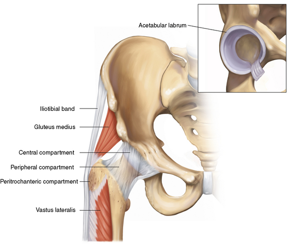
Hip arthroscopists divide the femoroacetabular joint and its surrounding areas into three compartments. The central compartment refers to the confines of the hip joint proper; the peripheral compartment provides access to the femoral neck and the outer acetabular rim. More recently, Kelly and colleagues described the peritrochanteric compartment, which lies between the iliotibial band and the proximal femur. Each compartment can be accessed by a number of arthroscopic portals and has pathologies that are individual to the specific area.

Hip arthroscopy has evolved with improvements in surgical equipment and technical skill. In addition, advances in magnetic resonance imaging have heralded the diagnosis of previously unrecognized intra-articular pathology. Arthroscopy now has numerous indications, including the treatment of labral lesions, degenerative disease, articular injuries, synovial abnormalities, femoroacetabular impingement, loose bodies in the central and peripheral compartments, gluteus medius tendon tears, iliotibial band problems, and trochanteric bursitis.

The biggest challenge in hip arthroscopy is gaining safe access to the various compartments of the hip. A detailed understanding of local anatomy is therefore intrinsic to establishing a safe and effective portal to the hip. In addition to negotiating neurovascular structures, the arthroscopist is confronted by the large soft-tissue envelope that encases the femoroacetabular joint. In obese patients, this envelope may make an arthroscopic approach to the hip an impossible endeavor, even with specialized long instrumentation. Even in the thinner patient, the tough, fibrous hip capsule can still make access problematic. Precise arthroscopic technique and adherence to established principles for the management of the soft-tissue envelope can make hip arthroscopy a reliable and successful procedure for the relief of hip symptoms.

Surface Anatomy

An accurate understanding of the surface anatomy is crucial for anatomic orientation and initial trocar insertion. Typically, the outlines of the greater trochanter and the ASIS are drawn, followed by the intersecting horizontal and vertical lines from each point, respectively. With the use of these basic landmarks, the three most commonly used portals as described by Thomas Byrd—the anterior, anterolateral, and posterolateral portals—can be accurately placed (see Figure 2-1, B).

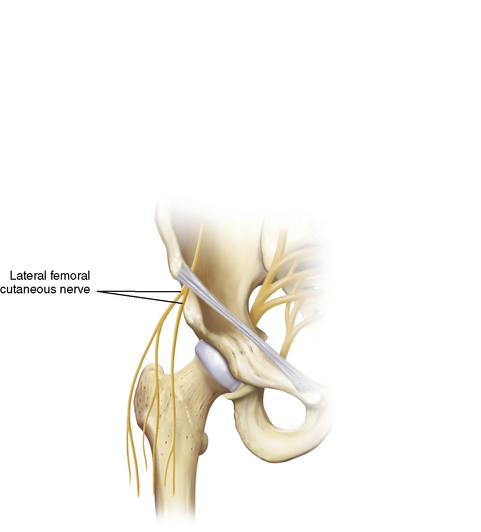
Anterolaterally, the arthroscopist should understand the course of the LFCN, which travels approximately 1 cm medial to the ASIS just below the epidermal layer and subsequently branches out in a fan-like distribution (Figure 2-6). Although the incidence of complications during hip arthroscopy is low, the LFCN branches are in close proximity to the anterior portals, and localized paresthesias of the thigh as a result of damage of this nerve are not uncommon. Although blunt trauma during portal placement can cause damage to the LFCN branches, the more likely culprit is inadvertent scalpel injury, which reiterates the importance of a careful skin incision.

It is also important to palpate and mark the main neurovascular bundle that traverses just anteromedial to the hip joint, which contains the femoral nerve, artery, and vein and the associated lymphatics. These structures are only a few centimeters medial to the anterior portal, so the arthroscopist should be keenly aware of their location when establishing anterior or medial portals. One should rarely if ever be medial to the imaginary line that extends distally from the ASIS. In addition, in the peripheral compartment, the distal part of the iliopsoas is within 3 cm of the femoral vessels, and care must be taken when performing an iliopsoas release at this level. Fortunately, these complications are extremely rare during arthroscopy of the hip. Finally, the arthroscopist should be familiar with the sciatic nerve and the gluteal vessels posteriorly and proximally. Localization of the portal pathway is then typically achieved with the use of a long 18-gauge spinal needle and image intensification. One measure that helps to avoid injury is to make skin incisions only after proper intra-articular placement has been achieved.

The Arthroscopic Portals

A keen appreciation of hip anatomy in combination with proper positioning and distraction is fundamental to safe and successful portal placement. Portals should be established in zones that minimize the risk of soft-tissue damage and that maximize arthroscope maneuverability and the visualization of anatomic structures. This is of particular importance in the hip joint, because it is deeply recessed and enveloped by the thick, fibrous capsule.

The anterolateral portal is the workhorse of all of the arthroscopic portals around the hip. It is considered to be the safest portal, and it is therefore established first and “blindly” (i.e., with the assistance of fluoroscopy alone). It is useful for the visualization of the central, peripheral, and peritrochanteric compartments. To create an anterolateral portal, an initial superficial incision no deeper than the subcutaneous adipose tissue is made over the anterosuperior tip of the greater trochanter. A fascial band just posterior to the skin incision is commonly encountered, and it can be used as a reference mark. A sheathed blunt trocar or snap is used to pass through the adipose tissue, fascia, and muscle tissue to the hip capsule. The superior gluteal nerve lays approximately 4.5 cm superiorly to this portal (see Table 2-2). A spinal needle is then used to enter the hip joint, and this is followed by a guidewire and a trocar. After the surgeon has entered the joint space, the superolateral labrum, the acetabular articular cartilage, and the femoral head are vulnerable to iatrogenic damage. Therefore, blunt trocars are preferred. Finally, image intensification and the injection of saline can confirm the proper positioning of the intra-articular spinal needle. Subsequent portals are established with intra-articular arthroscopic guidance.

After the anterolateral portal is established, the anterior portal is typically the second and most difficult portal to establish. It allows for the observation of the anterior femoral neck, the superior retinacular fold, the lateral acetabular labrum, portions of the transverse acetabular ligament, and the ligamentum teres. Traditionally, this portal is placed directly at the intersection of the lines drawn vertically from the ASIS and horizontally from the greater trochanter. However, this approach involves the direct penetration of the origin of the rectus femoris tendon. It has been suggested that a resultant tendinopathy causes increased soreness in the anterior groin region. Therefore, a recent a trend has emerged with the anterior portal placed just distal and lateral to the traditional portal site: the mid-anterior portal (Figure 2-7). To establish any anterior portal, one must be careful to superficially avoid the LFCN and its branches. On deeper exploration, the surgeon should be familiar with the ascending branch of the lateral circumflex femoral artery, which can be only 1.9 cm away; a small terminal branch of this artery can be as close as 1 cm away. As stated previously, one must also be aware of the femoral neurovascular bundle, which is located approximately 3.5 cm to 4 cm medially (see Table 2-2).

When creating an anterior or mid-anterior portal, it is important to penetrate the hip capsule with firm and controlled pressure so that the joint space is accessed without damage to the intra-articular anterior femoral head and the anterior labrum. Indeed, inadvertent plunging and sudden penetration of the obturator and cannula can have devastating results.

The final central compartment portal is the posterolateral portal. It is placed under direct arthroscopic visualization, and it is considered the easiest to establish because of a relative “soft spot” in the posterior soft-tissue envelope. The hip capsule is thinnest in this zone. The portal is placed on the same transverse line as the anterolateral portal but just posterior to the greater trochanter. It traverses the gluteus medius and the gluteus minimus before entering the posterior capsule. The most obvious extra-articular structure at risk is the sciatic nerve, which is approximately 2.9 cm away (see Table 2-2). Fortunately, sciatic nerve injury is extremely rare. Most surgeons do not routinely make use of the posterolateral portal, because the majority of intra-articular pathology is localized to the anterolateral zones. Instead, the main purpose of this portal is to provide access for observation of the posterior aspect of the hip.

In addition to these three traditional portals, cadaveric studies have provided an understanding of the relationship between anatomic structures and portal placement. More recently, Kelly and colleagues have demonstrated that, including the three standard portals (i.e., anterolateral, anterior, and posterolateral), eleven arthroscopic portals may be placed for correcting pathology in the central, peripheral, and peritrochanteric compartments. Caution should be employed when attempting portal placement out of the relative “safe zone,” which is considered to lie within approximately 4 cm superior and 6 cm to 8 cm inferior to the anterior and anterolateral portals. This is especially important when attempting to establish access posterior to the posterolateral portal or medial to the anterior portal.

Examination of the hip through arthroscopic compartments

During the early days of hip arthroscopy, the most common indications were infection, loose body removal, and labral tears. Hip arthroscopy was confined to the central compartment, which included only the intra-articular region of the femoroacetabular joint. As the indications for hip arthroscopy have grown, the orthopedic surgeon must now routinely explore the areas of the joint outside of the central compartment. If surgery is confined to the central compartment, then overall mean accessible surface area is limited to 68% to 75% of the joint (Figure 2-8, A through D). However, by expanding arthroscopy to include the peripheral and peritrochanteric compartments, the surgeon can access more than 90% of the hip. Therefore, only the most posteromedial zones of the hip have limited accessibility. In addition, the indications for extra-articular procedures have recently expanded. Surgeons are now successfully navigating areas such as the iliopsoas bursa and the peritrochanteric space. Therefore, it is essential to appreciate the anatomic contents of the three hip compartments (Figure 2-9) and to familiarize oneself with the indications that are characteristic of each.

Central Compartment

The Capsule

The hip capsule consists of three discrete ligaments: 1) the ischiofemoral (posterior) ligament; the iliofemoral (anterior) ligament; and the pubofemoral (anterior) ligament. These form a thick and fibrous wrap around the femoroacetabular joint, and they result in more than 95% of the femoral neck being intracapsular. The ischiofemoral ligament is located posteriorly. It is thin and pliable, and it generally poses few problems for the hip arthroscopist, because the majority of the work in hip arthroscopy is located anteriorly. The iliofemoral and pubofemoral ligaments are located anteriorly, and together they combine to make one of the thickest and toughest capsules of the entire body. A traditional open anterior capsulotomy directly involves the release of these two adherent structures. The pubofemoral ligament is located anteromedially; it spans from the pubic ramus and the anteroinferior acetabular wall to the anteromedial femoral neck. Distally, it becomes confluent with the fibers of the iliofemoral ligament. Its functions are primarily to resist hip extension and to prevent excessive abduction. In patients with a soft-tissue contracture of the hip, the release of the pubofemoral ligament will effectively increase abduction.

The iliofemoral ligament, which is located anterolaterally, is of particular clinical importance when establishing the anterior portal. It is perhaps the single biggest impediment to the hip arthroscopist. The iliofemoral ligament (i.e., the Y ligament of Bigelow) arises from the AIIS and spreads obliquely and inferolaterally to insert into the intertrochanteric line on the anterior femoral head. This is the strongest ligament in the human body, and it prevents anterior translation of the hip in positions of extension and external rotation, especially when the pelvis is posteriorly tilted. The intra-articular medial and lateral limbs of the iliofemoral ligament form an anterior triangle. The terminal fibers of the ligament form the zona orbicularis; this is a circular leash around the femoral neck that tightens during extension and loosens during flexion.

The thickest part of this ligament lies anteriorly and coincides with the location of the majority of hip pathology that is amenable to arthroscopic intervention. Even after the portals are established, the capsule limits the maneuverability of the instrumentation and the arthroscope within the central compartment. Therefore, a limited capsulotomy is recommended to enhance mobility. This is usually accomplished with the assistance of a beaver blade or a radiofrequency device. If work is confined to the central compartment, it is sufficient to make mini-capsulotomy incisions in the areas in which the portals are penetrating the capsule. This allows the surgeon to access the femoroacetabular articular cartilage, the labrum, and the contents of the cotyloid fossa without significant challenge. However, if any substantial work is needed in the extra-articular areas (e.g., osteoplasty), then a horizontal or transverse capsular release in a “fish-mouth” or “T” pattern may be necessary, anteriorly or posteriorly (Figure 2-10, A and B). This allows for increased visualization and the ability to work in the peripheral compartment. Currently, the incision of the capsule, the extent of this incision, and the subsequent repair are performed at the discretion of the surgeon. Conversely, thermal capsular shrinkage can also be achieved by hip arthroscopy to stabilize the joint. Capsular plication in combination with shrinkage has also been shown to have good short-term results among patients with recurrent instability in the presence of an intact ligamentum teres.

The Labrum

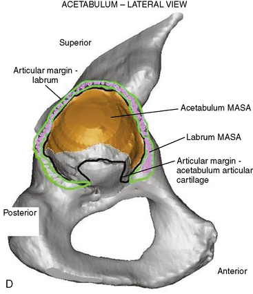
The acetabular labrum is made predominantly of fibrocartilage supported by a collagen scaffold. It runs circumferentially around the acetabular rim and the fovea. This scaffold is oriented in both a longitudinal and radial direction, which confers stability to the femoroacetabular articulation. The sensory innervation of this scaffold provides the hip with both proprioception and nociceptive function, and it participates in hip stability and pressure distribution in the joint. The labrum attaches to the transverse acetabular ligament anteriorly and posteriorly; it has a vascular peripheral capsular surface with a less-vascular central articular margin.

The labrum and the capsule help to contain the femoral head in extreme ranges of motion, and they act as load-bearing structures during flexion. Therefore, patients who have undergone extensive labrectomy may complain of significant rotational instability or hypermobility. The capsulolabral disruption may result in transient micro motion, abnormal load distribution, and redundant capsular tissue. This allows for redundancy of the anterior capsule, which can then lead to microinstability. Recent studies have shown that the labrum contains neural structures of both proprioceptive and nociceptive function. In addition, it has been shown that the majority of these mechanoreceptors and sensory nerve endings are most highly concentrated anteriorly, which is where the majority of symptomatic hip pathology is seen. Excessive debridement of the labral tissue not only involves the mechanical implications discussed previously, but it may also result in altered joint proprioception, which may further lead to joint derangement.

It has been suggested that the labrum enhances stability by maintaining negative intra-articular pressure in the hip joint and that it acts as a tension band for preventing joint expansion as part of a normal gait. In addition, the negative pressure creates a suction-seal effect at the innermost aspect of the joint by forming a seal between the femoral head and the acetabular rim and thus enhancing hip stability. Fluid mechanics models have provided evidence that, in a hip with a discontinuous labrum, the joint contact pressure distribution is greatly diminished and can lead to decreased cartilage surface consolidation.

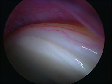
The obturator artery, with contributions from the inferior and superior gluteal vessels, provides the blood supply to the labrum (Figure 2-11). Histologic studies have shown that the blood supply to the labrum is analogous to that of the knee menisci. The majority of the microvascular supply proliferates the capsulolabral junction with a relative paucity of vascularization to the innermost aspect of the labrum. Therefore, like the meniscus, the labrum may have the greatest healing potential at the peripheral capsulolabral junction.

Magnetic resonance imaging is becoming more effective for detecting labral lesions. Most are encountered in the relatively avascular zone and thus warrant debridement. If the tear is located adjacent to the more vascular capsulolabral junction in a longitudinal peripheral orientation, then primary labral repair may be possible. However, many “labral repairs” that are currently performed are in fact labral refixation procedures after acetabular rim trimming in patients with pincer-type femoroacetabular impingement. Normal variants (e.g., a labral cleft) should not be interpreted as traumatic detachment.

The Cotyloid Fossa

The cotyloid fossa is roughly hemispheric. It has a wave-like profile of three peaks and three troughs, including the acetabular notch. The most clinically relevant anatomic structure within the cotyloid fossa is the ligamentum teres. The ligamentum teres is a highly variable structure, but it is generally described as a thin, flat ligament with a triangular cross section that originates from the fovea capitus on the femoral head. It is shrouded by synovium throughout its entirety, and it is often obscured by the prolific fat pad of the cotyloid fossa. The ligament is between 30 mm and 35 mm in length, and it consists structurally of two main bundles (i.e., anterior and posterior) that insert medially into the base of the cotyloid fossa adjacent to the transverse acetabular ligament. The function of the ligamentum teres in the adult is controversial. In the immature hip, it is clear that the ligamentum teres serves as a conduit for the arterial supply to the femoral head via the artery of the ligamentum teres, which is a branch of the obturator artery. However, as the human hip matures, the main blood supply to the femoral head is via the medial circumflex artery and the intracapsular perforating arteries, and the joint does not rely on direct vascularization from the ligamentum teres.

It has been suggested that the ligamentum teres contributes to the stability of the joint, especially in the presence of dysplasia. In addition, the bundles tighten in hip flexion, external rotation, and abduction, which is the common hip position during a dashboard injury; it may therefore play a role as a secondary restraint to a posterior hip dislocation. Others have postulated that the ligamentum teres has a proprioceptive function. However, recent studies have shown a paucity of mechanoreceptors in the bundles. In addition, the surgical debridement of the ligament after rupture shows excellent results, even in high-level athletes, thus further undermining its possible role in proprioception.

Peripheral Compartment

The peripheral compartment is an area of the hip that is considered to be extra-articular yet intra-capsular; it lies along the anterior femoral neck. Loose bodies commonly collect here, and cam-type bony lesions are largely localized to this compartment. Labral surgery can also be performed here in an “outside-in” fashion. Therefore, a systematic examination of the compartment is routine. After the inspection of the central compartment is complete, the peripheral compartment is most easily viewed from the anterior portal and accessed by flexing and externally rotating the hip without traction, thus atraumatically avoiding the femoral head. Other techniques have described a “peripheral-first” method.

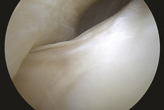
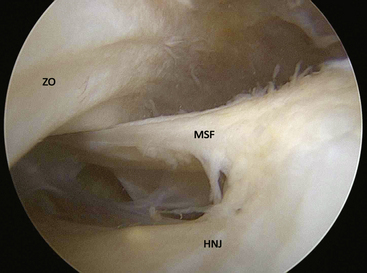
After entry into the peripheral compartment, the first anatomic landmark that can be readily identified is the medial synovial fold (Figure 2-12). The synovial folds are sheet-like collections of synovial tissue that run longitudinally in various parts of the peripheral compartment. The medial synovial fold is located at the anteromedial aspect of the femoral neck. The lateral synovial fold is located at the junction between the lateral and posterior femoral neck. It is important to closely inspect both of these structures, because their redundant tissue can easily obscure loose bodies. In addition, when significant synovitis is present, these synovial folds become inflamed and need to be thoroughly debrided as part of a synovectomy procedure.

The synovial folds can serve as important landmarks in the peripheral compartment. It is unusual for a cam-type lesion of the femoral head–neck junction to extend more medial than the medial synovial fold, so this can be used as a rough guide to the 6-o’clock position. The lateral synovial fold contains the lateral retinaculum and the intracapsular penetrating arteries from the lateral epiphyseal vessels. It is essential to employ great caution when working near and posterior to the lateral synovial fold to avoid iatrogenic injury to these vessels. The lateral synovial fold can also be used a rough landmark for the 12-o’clock position.

Another important landmark in the peripheral compartment is the zona orbicularis, which refers to the thickening of the hip capsule as it forms a ring around the circumference of the femoral neck. Although the exact function of the zona orbicularis is not entirely known, it has particular use as an arthroscopic anatomic landmark of the peripheral compartment. Just cranial to the zona orbicularis, by the anterior capsular recess, and in line with the medial synovial fold lies the psoas tendon. Anteromedially, the tendon may be covered by a thin, transparent capsule that leads directly to the iliopsoas bursa. In fact, in approximately 20% of the population, the iliopsoas bursa may be in direct communication with the peripheral compartment. This is of particular clinical relevance: coxa saltans interna (i.e., internal snapping hip) can be related to iliopsoas bursitis or iliopsoas tendonitis. This condition can be surgically treated by partially releasing or lengthening the iliopsoas tendon from the lesser trochanter. Arthroscopically, the tendon is visualized and released in the peripheral compartment just proximal to the zona orbicularis and deep to the thin anteromedial capsule in this region.

Open approaches to the hip joint

There are a number of open approaches to the hip that have been advocated for ideal anatomic exposure with minimal dissection and low patient morbidity. Surgeons who perform THA most commonly employ the anterolateral approach, the anterior approach, or the posterolateral approach, with all of these approaches now having new modifications. For acetabular access and reconstruction, the ilioinguinal and iliofemoral approaches are often recommended, whereas a surgical hip dislocation is now commonly performed for femoroacetabular impingement, arthroplasty, or fracture treatment.

Annotated references

Bardakos N.V., Villar R.N. The ligamentum teres of the adult hip. J Bone Joint Surg Br.. 2009;91(1):8-15.

Although once thought to be a developmental vestige, the ligamentum teres is now considered an important source of pain and mechanical symptoms. The authors, orthopaedic surgeons from The Richard Villar Practice, London, provide an update on the development, structure and function of the ligamentum teres, and its critical relevance to both arthroscopic and open hip procedures..

Barrack R.L., Butler R.A. Avoidance and management of neurovascular injuries in total hip arthroplasty. Instr Course Lect.. 2003;52:267-274.

This instructional review outlines operative techniques to avoid common neurovascular pitfalls during THA. In addition it discusses the mechanisms by which both the central and peripheral nervous system and vasculature may be damaged.

Byrd J.W. Avoiding the labrum in hip arthroscopy. Arthroscopy.. 2000;16(7):770-773.

The labrum is easily perforated during hip arthroscopy. The author of this short technical piece, an accomplished orthopaedic surgeon and founder of the Nashville Sports Medicine Centre, acknowledges that occasional damage is unavoidable. He highlights the importance of careful technique when establishing initial access into the capsule and provides surgeons with procedural guidance to avoid iatrogenic trauma of the labrum..

Byrd J.W. Labral lesions: an elusive source of hip pain case reports and literature review. Arthroscopy.. 1996;12(5):603-612.

This review, from the Nashville Orthopaedic Centre uses case reports and a literature review to discuss the relative contributions of clinical history, non-invasive imaging, and arthography to the diagnosis of labral pathology..

Byrd J.W., Pappas J.N., Pedley M.J. Hip arthroscopy: an anatomic study of portal placement and relationship to the extra-articular structures. Arthroscopy.. 1995;11(4):418-423.

This cadaveric anatomic study describes the proximity of the anterior, anterolateral and posterolateral arthroscopic hip portals to neurovascular structures. Although all measurements were made without distraction of the hip joint, the authors bring attention to the importance of negotiating anatomical structures, particularly the LFCN and sciatic nerve, when accessing the joint..

Clarke M.T., Arora A., Villar R.N. Hip arthroscopy: complications in 1054 cases. Clin Orthop Relat Res. (406); 2003:84-88.

This prospective study of a large sample of consecutive hip arthroscopies performed at one centre reports an overall complication rate of 1.4%. The authors are orthopaedic arthroscopy specialists, and the results are therefore perhaps not reflective of general practice. However, it is clear that with careful technique and via low-risk portals, arthroscopy of the hip has a low safety profile that is comparable to other orthopaedic procedures..

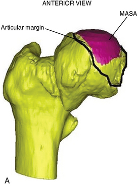
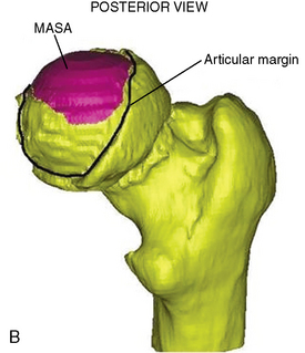
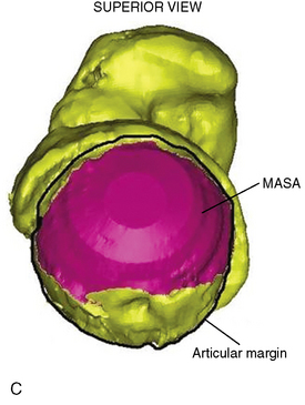
2008 Dandachli W., Kannan V., Richards R., Shah Z., Hall-Craggs M., Witt J. Analysis of cover of the femoral head in normal and dysplastic hips: new CT-based technique. J Bone Joint Surg Br.. 2008;90(11):1428-1434.

The femoroacetabular joint is a complex three-dimensional construct. This non-interventional case-control study uses novel 3D-CT methodology to describe femoral head coverage. The authors, from London, UK, are able to distinguish dysplastic and normal femoral head coverage – mean cover was 73% and 51% respectively. This is useful for anatomical understanding and has application in post-operative assessment of corrected hips..

2003 Ferguson S.J., Bryant J.T., Ganz R., Ito K. An in vitro investigation of the acetabular labral seal in hip joint mechanics. J Biomech.. 2003;36(2):171-178.

The authors harvest four human cadaveric pelvises and investigate the load- and stress-limiting role of the acetabular labrum in preventing cartilage layer consolidation and early joint degeneration. They demonstrate that the sealing mechanism of an intact labrum creates a hydrostatic fluid pressure in the intra-articular space, which may enhance joint lubrication. The labrum may be partially debrided prior to acetabular rim resection, or torn during acetabular trauma. This study emphasizes the importance of labral restoration in preventing functional deterioration of the hip joint ..

Ganz R., Gill T.J., Gautier E., Ganz K., Krugel N., Berlemann U. Surgical dislocation of the adult hip a technique with full access to the femoral head and acetabulum without the risk of avascular necrosis. J Bone Joint Surg Br.. 2001;83:1119-1124.

The authors describe their experience of surgical dislocation of the hip in 213 hips over seven years. Their technique is an anterior dislocation through a posterior approach with a ‘trochanteric flip’ osteotomy. The external rotator muscles are not divided and the medial femoral circumflex artery is protected by the intact obturator externus. Their results suggest adequate preservation of the vascularity of the femoral head can be achieved with little morbidity..

2000 Gautier E., Ganz K., Krugel N., Gill T., Ganz R. Anatomy of the medial femoral circumflex artery and its surgical implications. J Bone Joint Surg Br.. 2000;82(5):679-683.

The classic description of the approach and technique commenting on the advantages of a posterior exposure of the hip joint..

2001 Glick J.M. Hip arthroscopy. The lateral approach. Clin Sports Med.. 2001;20(4):733-747.

It is of the author’s opinion that the lateral approach is the safest, simplest and most versatile of arthroscopic hip approaches. This review presents technical operative direction for the arthroscopist, particularly highlighting techniques to prevent sciatic neuropraxia during traction..

Kagan A.2nd Rotator cuff tears of the hip. Clin Orthop Relat Res. (368); 1999:135-140.

Partial tears of the gluteus medius, near its attachment to the greater trochanter, are often misdiagnosed as greater trochanteric pain syndrome (GTPS). This seminal paper describes the diagnostic process and operative treatment of 7 patients with ‘rotator cuff tears of the hip’, and provides informative guidance for distinguishing this hip pathology. It is particularly illuminating given the recent advances in the peritrochanteric arthoscopic compartment..

Kelly B.T., Shapiro G.S., Digiovanni C.W., Buly R.L., Potter H.G., Hannafin J.A. Vascularity of the hip labrum: a cadaveric investigation. Arthroscopy.. 2005;21(1):3-11.

The function of the damaged hip labrum is notoriously difficult to restore. This study uses MRI-based ink-injection analysis to measure its vascularity. The authors found that although the labrum is a largely avascular structure, its capsular contribution is significantly more vascular. The authors propose that tears of the vascular portion may be more amenable to surgical repair and subsequent physiological healing, while tears in the articular, avascular portion are better debrided..

Mason J.B., McCarthy J.C., O’Donnell J., et al Hip arthroscopy: surgical approach, positioning, and distraction. Clin Orthop Relat Res. (406); 2003:29-37.

This is a comprehensive technical overview of operative techniques in hip arthroscopy. In addition to discussing the lateral approach, portals, patient positioning and portal placement, the paper provides a balanced review of the use of distraction in arthroscopy..

Murtha P.E., Hafez M.A., Jaramaz B., DiGioia A.M. 3rd. Variations in acetabular anatomy with reference to total hip replacement. J Bone Joint Surg Br.. 2008;90(3):308-313.

This descriptive study of 42 pelvises using three-dimensional CT imaging analyses the orientation of the human acetabulum. It rejects the gold standard of the Lewinnek’s ‘safe zone’ for the ideal position of the acetabular component in hip arthroplasty. It also shows that female acetabulae are more anteverted than their male counterparts. The paper raises questions about following the orientation of the native acetabulum for component placement, and whether gender-specific components better mimic normal anatomy..

Philippon M.J. The role of arthroscopic thermal capsulorrhaphy in the hip. Clin Sports Med.. 2001;20(4):817-829.

Hip instability is associated with capsular laxity. The author discusses the role of arthroscopic thermal shrinkage of type 1 collagen in the hip tissue to reduce capsular redundancy, restore stability and decrease the development of arthrosis. Although this practice is well established in the treatment of posterior shoulder instability, the author reviews its early results in the hip..

Rachbauer F., Kain M.S., Leuing M. The history of the anterior approach to the hip. Orthop Clin North Am. 2009;40(3):311-320.

The anterior approach to the hip has evolved since its description in 1881. This historical review details the changes and adaptations since its inception, and describes both the open and arthroscopic potential of this internervous and muscle sparing approach..

Robertson W.J., Kelly B.T. The safe zone for hip arthroscopy: a cadaveric assessment of central, peripheral, and lateral compartment portal placement. Arthroscopy.. 2008;24(9):1019-1026.

In addition to the three traditional arthroscopic portals to the hip, this cadaveric study measures the proximity of accessory portals to the central, peripheral and trochanteric compartments. A total of 11 portals were determined to provide viable access to the joint. The article provides a valuable anatomical insight into accurate and safe access for the arthroscopic hip surgeon..

Schmalzried T.P., Amstutz H.C., Dorey FJ. Nerve palsy associated with total hip replacement. Risk factors and prognosis. J Bone Joint Surg Am.. 1991;73(7):1074-1080.

In this retrospective analysis of 3126 consecutive operations performed over 17 years, the authors discuss the prevalence and aetiology of nerve palsy as a complication of total hip arthroplasty via an open lateral transtrochanteric approach. The study documents that while nerve palsy was the most debilitating of post-operative complications, there is a low prevalence rate of 1.7% and the sciatic nerve is involved in the majority of cases. The positioning of the limb and placement of the retractors play an important role in these injuries..

Strauss E.J., Campbell K., Bosco J.A. Analysis of the cross-sectional area of the adductor longus tendon: a descriptive anatomic study. Am J Sports Med.. 2007;35(6):996-999.

This descriptive anatomical cadaveric study demonstrates that the origin of the adductor magnus muscle has minimal tendinous content. The authors propose that this feature both contributes to the adductor longus’ propensity for damage during eccentric contraction and the relative difficulty experienced by surgeons when repairing it..

Vail T.P., Mariani E.M., Bourne M.H., Berger R.A., Meneghini R.M. Approaches in primary total hip arthroplasty. J Bone Joint Surg Am.. 2009;91(Suppl 5):10-12.

Following on from the review of the anterior approach to the hip, above, this three part multicentre paper compares the anterolateral and posterolateral approaches to THA. It presents novel data supporting the anterolateral approach, suggests a novel minimally invasive rethink of the classic posterolateral approach, and discusses the ongoing disagreement regarding the merits of different/minimally invasive approaches. The choice of approach in THA remains contentious, and the article’s moderator ultimately states that there is insufficient long-term data to support any particular preference..

Voos J.E., Ranawat A.S., Kelly B.T. The peritrochanteric space of the hip. Instr Course Lect.. 2009;58:193-201.

This instructional article discusses a recent addition to the hip arthroscopist’s armamentarium – the peritrochanteric space. It details the disorders, including tears of the gluteus maximus and recalcitrant trochanteric bursitis, which are amenable to endoscopic access and treatment. The authors discuss patient orientation and portal placement necessary for successful intervention, as well as outlining the relevant local anatomy..