[level-membership-for-emergency-medicine-category]
140 Arthropod Bites and Stings
 Key Points
• Anaphylaxis, typically caused by Hymenoptera stings, is the most serious complication of all arthropod encounters and should be treated with steroids, epinephrine, and antihistamines when needed.
• Massive envenomations (>10 stings per kilogram or >100 stings per person) by Hymenoptera merit close monitoring for systemic effects of the venom.
• Latrodectism should be treated with adequate analgesia and benzodiazepines, but antivenom should be considered for severe cases.
• Dapsone, hyperbaric oxygen, colchicine, and electric shock therapy are no more effective than supportive care for the treatment of true dermonecrotic arachnidism.
Perspective
Arthropods are the most diverse, widespread, and numerous of all animal phyla inhabiting the planet. Not surprisingly, their contact with humans is a common occurrence. In 2009, 40,657 calls related to arthropods were made to poison centers in the United States, and although most do not require hospital attention, many patients will still come to the emergency department (ED) complaining of a bite or sting from an unknown or unidentified insect.1 Fortunately, the vast majority of these patients can be treated with supportive care and medications for pruritus and pain; the challenge for the emergency physician (EP) is identifying more serious and rare complications of these encounters. The most clinically significant arthropods are summarized in Table 140.1.
Table 140.1 Common Clinical Manifestations of Arthropod Envenomation
| Bees and wasps | Urticarial eruptions, anaphylaxis, rhabdomyolysis, ARF, ARDS (after massive envenomations) |
| Widow spiders | Pain, muscle spasm, local diaphoresis, tachycardia, hypertension |
| Recluse spiders | Dermonecrosis; hemolysis, DIC, ARDS (rarely) |
| Scabies | Migratory pruritus, secondary infections |
| Ants | Urticarial and papular dermatitis, anaphylaxis risk |
| Scorpions | Pain, tingling, cranial neuropathy, ataxia, pancreatitis, DIC, ARDS (exotic species) |
| Caterpillars | Painful dermatitis, ocular and mucosal irritation |
| Mites | Papular urticarial dermatitis |
| Ticks | Local tissue reaction, tick paralysis, infectious complications |
| Reduviid bug | Bullous lesions, infectious complications |
| Lice and fleas | Papular urticarial dermatitis |
| Mosquitoes | Urticaria, pruritus, infectious complications |
| Tarantulas | Local pain (bite), urticarial dermatitis, ocular irritation (hairs) |
| Centipedes | Local pain |
| Millipedes | Skin discoloration from oily extractions |
ARDS, Acute respiratory distress syndrome; ARF, acute renal failure; DIC, disseminated intravascular coagulation.
Hymenoptera
Pathophysiology
Apidae (bees), Vespidae (wasps, yellow jackets, hornets), and Formicidae (ants) are the most clinically significant groups of arthropods for two reasons. First, the incidence of Hymenoptera venom allergy has been estimated to be 0.8% to 5% in the general population and is increasing, particularly in young people.2 Second, because of their complex social organization, multiple stings are more likely to occur during Hymenoptera encounters than with arthropods that do not build nests or hives.
Recent research indicates that the major allergens in Hymenoptera venom are phospholipases and hyaluronidases, as well as mellitin, a peptide that causes degranulation of mast cells.3 Hymenoptera venom is delivered via an ovipositor stinger and gland, although some anatomic variation does exist. Male bees have no stingers and are incapable of stinging when threatened. Females have barbed stingers that become lodged in human skin and eviscerate the bee after venom delivery. The retained stinger and venom sac can be removed with tweezers. Africanized “killer” bees deserve special mention in that (1) they are far more aggressive and territorial than the more docile domesticated varieties, (2) are known to pursue perceived threats for up to 1 km, and (3) do so in much larger swarms. Africanized bees are difficult to distinguish morphologically from domesticated bees, but fortunately, this distinction is of little clinical significance because of venom homology between Hymenoptera Apidae. In contrast to bees, vespids (wasps, yellow jackets, and hornets) have the ability to withdraw their stinger from the victim and deliver multiple stings. Most severe allergic reactions to Hymenoptera are due to encounters with vespids, particularly wasps and yellow jackets.3,4
Presenting Signs and Symptoms
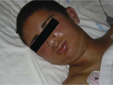
Massive envenomations are considered those in which the victim sustains more than 100 stings or more than 10 stings per kilogram (Fig. 140.1). Such cases merit special respect and victims should be considered for admission because of an increased risk for systemic symptoms, including nausea, vomiting, diarrhea, edema, dyspnea, hypotension, and rhabdomyolysis. Rarely, glomerulonephritis, acute renal failure, and acute respiratory distress syndrome can occur.5–7
Diagnostic Testing
No laboratory testing is necessary in cases limited to cutaneous symptoms from submassive envenomations. Massive envenomations or systemic reactions require investigation to evaluate for rhabdomyolysis, renal failure, or cardiac ischemia.5,6,8 Appropriate testing should include a basic chemistry panel and creatine phosphokinase (CPK) level.
Anaphylaxis and Allergic Reactions
It has been estimated that 40 deaths occur per year in this country as a result of anaphylaxis from Hymenoptera stings.4 Anaphylaxis is an IgE-mediated type I hypersensitivity reaction that leads to mast cell and basophil degranulation of vasoactive mediators, cytokines, prostaglandins, and platelet-activating factor. Some initial symptoms can be mild and include itchy eyes, urticaria, or cough. However, the symptoms can progress rapidly to shortness of breath, stridor, angioedema, and shock. Treatment should be initiated immediately and includes epinephrine, steroids, antihistamines, and bronchodilators (if bronchospasm is present). All available data suggest that failure or delay in the administration of epinephrine increases the chance for death from anaphylaxis. The risk for anaphylaxis with any event is dependent on the severity of the patient’s previous reaction, and it seems to be proportional to the rate of symptom onset. Once the symptoms have been controlled, patients should be observed for at least 2 hours to ensure resolution of the symptoms. Patients with persistent cardiopulmonary symptoms should be admitted to the hospital. An outline of anaphylaxis treatment is found in Box 140.1 and Table 140.2.2–5
Box 140.1 Treatment of Anaphylaxis Caused by Arthropod Venom or antivenom Therapy
Symptoms of allergy and anaphylaxis may be variable and include perioral or pharyngeal tingling, shortness of breath, tachypnea, bronchospasm and wheezing, stridor, chest pain, sudden tachycardia, hypotension, angioedema, and urticaria.
Bees and wasps are the most common sources of insect allergic reactions. Because animal-derived antibody products can also result in allergic reactions or anaphylaxis, each patient receiving antivenom must be monitored carefully. The antivenom infusion must be stopped immediately if allergic symptoms such as those listed develop.
Skin testing is a very imperfect (not sensitive, not specific) predictor of subsequent allergic reactions to antivenom.
Pretreatment includes antihistamines (e.g., diphenhydramine, 25 to 50 mg intravenously [IV], plus ranitidine, 50 mg IV) and antipyretics (acetaminophen, 500 mg or 15 mg/kg orally [PO]).
Treatment of any significant allergic reaction is prompt administration of epinephrine.
Steroids are recommended to prevent delayed allergic effects.
Pretreatment with steroids can be done in high-risk patients requiring antivenom.
Nebulized bronchodilators and supplemental oxygen can be used for bronchospasm.
Warn patients about the risk and signs of serum sickness, which occurs within 7 to 10 days of the envenomation or administration of antivenom. Serum sickness is characterized by a diffuse macular or urticarial rash, arthralgias, back pain, and sometimes hematuria. Therapy is a 10- to 14-day course of prednisone, 1 to 2 mg/kg/day PO, with tapering.
| ARTHROPOD | SIGNS AND SYMPTOMS | TREATMENT |
|---|---|---|
| Fleas and mites | Pruritic, erythematous, red papules | Oral or topical antihistamines, topical steroid cream Antimicrobials for secondary infections |
| Scabies | Significant nocturnal pruritus, intertriginous skin thickening, papules. The diagnosis can be made with microscopy of skin scrapings. Finger web spaces, wrists, elbows, and unscratched skin are the most productive sites for sampling | Topical and oral antihistamines |
| Topical scabicides: 5% permethrin cream applied once for 8-14 hr, then washed off. May be repeated in 1 wk; treatment failure typically results from incorrect application | ||
| Lindane cream no longer recommended | ||
| Ivermectin, 200 mcg/kg orally once, second dose recommended 14 days later. Only for topical treatment failure | ||
| Norwegian scabies | Severe scabies infection (thousands of organisms); typically indicates immunocompromised status | As above |
| Check for underlying immunodeficiency | ||
| Caterpillars | Stinging, pruritic lesions caused by contact with setae (spines) distributed in a pattern similar to those of the hairlike projections on the caterpillar. Mucosal and ophthalmic irritation, bronchospasm | Spine removal from the skin with tweezers, adhesive tape, or topical white school glue |
| Topical or oral steroid therapy for severe dermatitis | ||
| Ocular irrigation with 0.9% normal saline | ||
| Inhaled bronchodilators for bronchospasm | ||
| Antivenom for Brazilian Lonomia species | ||
| Centipedes | Carnivorous arthropod. Painful bite. Rare systemic reactions: nausea, vomiting, diaphoresis | Supportive care, analgesics, antihistamines |
| Millipedes | Herbivorous arthropod. Exoskeleton secretions can cause cutaneous irritation and discoloration | Wash the site copiously with soap and water, oral and/or topical antihistamines |
Follow-Up, Next Steps in Care, and Patient Education
Patients treated for anaphylaxis should be educated on avoidance of the allergen and use of an epinephrine autoinjector. Wearing a medical bracelet is also advisable. Finally, patients should be referred to an allergist for sensitivity testing and possible venom immunotherapy.3
Arachnids—Spiders, Scorpions, and Ticks
Spiders (Araneae)
Widow Spiders (Latrodectus Species)
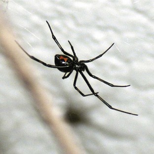
With its distinct shiny black color, bulbous abdominal segment, and ventral orange-red markings, Latrodectus species are some of the most identifiable of all spiders. Latrodectus mactans (black widow) is perhaps the best known of the genus and possesses an hourglass-shaped ventral marking (Fig. 140.2). As is the case with many arthropods, females are the larger of the two sexes, and males do not have fangs capable of piercing human skin. Latrodectus venom contains a potent neurotoxin, α-latrotoxin, a well-characterized protein that induces massive release of neurotransmitters from presynaptic neurons. The resultant effect of these neurotransmitters is activation of the autonomic and somatic nervous systems.9,10
Presenting Signs and Symptoms
The bite itself is typically painful, and this discomfort can spread throughout the affected extremity. Local erythema and diaphoresis occur early at the site with a resultant macule developing into a target lesion. The development of painful cramping in large muscle groups and autonomic instability is known as latrodectism. This systemic syndrome has been described as being so severe as to mimic an acute abdomen, even when the site of envenomation is remote from the center mass; the exact pathophysiology of this phenomenon is not well understood. Hypertension, tachycardia, headache, nausea, priapism, and dyspnea have also been described in association with Latrodectus bites.9
Treatment
IgG antivenom for Latrodectus envenomation is available in the United States, but this treatment is often reserved for the rare patients whose pain and muscle spasms are refractory to large doses of analgesics and relaxants. This antivenom is a whole-antibody horse-derived IgG preparation, and anaphylactoid reactions and serum sickness are potential consequences of its administration. Therefore, it should be administered slowly and with caution.10
Spiders Causing Dermonecrotic Arachnidism
Loxosceles, or recluse, spiders cause the syndrome of dermonecrotic arachnidism. These spiders, as their name suggests, prefer dark and isolated spaces such as attics, basements, and closets. They bite only when threatened or disturbed. In contrast to Latrodectus, Loxosceles reclusa is more difficult to identify. Many possess a violin-shaped dorsal marking (hence in some regions they are called “fiddleback” spiders), but this is not a diagnostic feature of all L. reclusa. The only unifying feature of all Latrodectus spiders is that they possess only three pairs of eyes, as opposed to the four pairs found in most spiders. Hobo spiders in the Pacific Northwest and several other species are also theorized to cause necrotic complications following bites, but these associations are more controversial. The venom of recluse spiders is a complex mixture of hyaluronidase, ribonucleases, lipases, and sphingomyelinase D, the latter being thought to be responsible for the necrosis.11,12
Presenting Signs and Symptoms
The most common clinical effect of true recluse spider bites is a progressively necrotic eschar. The lesion evolves from a nonspecific erythematous wheal into an eschar with surrounding rings of blanching and erythema (“red, white, and blue” sign) over a period of days to weeks. The slow destruction of skin structure occurs as a result of the effects of venom on the dermal microcirculation, and it can extend into adjacent tissues in a gravity-dependent manner. These lesions can be cosmetically disfiguring and stressful for patients because they expand and worsen over a period of many days to weeks. It is important to stress to patients that lesions from recluse spiders sometimes progress despite medical treatment.11,12 It is also important to appreciate the rarity of these injuries in comparison with the frequency with which they are reportedly perpetrated; many cutaneous infections are mistaken for recluse bites. In one study, spider bites were confirmed in only 3.8% of all individuals with the chief complaint of a spider bite.13 Rare cases of severe hematologic toxicity with hemolytic anemia, thrombocytopenia, and coagulopathy have been described in association with Loxosceles bites, particularly in Brazil.14,15
Treatment
A Loxosceles antivenom has been developed for use in Brazil, but based on animal models, it must be given within 24 hours to be effective. Because most patients are seen 2 to 3 days after being bitten, this type of antivenom is not typically a useful adjunct in the treatment of Loxosceles spider bites.15 Symptomatic and intensive supportive care is warranted in the rare case of disseminated intravascular coagulation, hemolytic anemia, renal failure, or acute respiratory distress syndrome following the bite of a recluse spider.
Tarantulas
Tarantulas are distributed worldwide and recognizable by their large size and prominent hairlike projections. Large or aggressive species can bite; the effects can range from relatively painless to deep throbbing pain with a febrile reaction that requires analgesics and antipyretics. By rubbing their hind legs against their abdominal wall, tarantulas can “flick” their hairs in the direction of perceived threats, and both dermal and ocular injuries have occurred from the highly irritating “urticating” hairs of tarantulas. The presence of urticaria, hives, intense pruritus, and mild erythema characterizes dermal lesions; ocular exposure to the urticating hairs has resulted in corneal abrasions, iritis, uveitis, and chronic granulomatous reactions (ophthalmia nodosum).16
Scorpions
Scorpions are easily recognized by the taillike abdominal segment that forms into a venom-filled bulb with a stinger (telson). In the United States, scorpions are commonly encountered hazards in the southwest, where Centruroides exilicauda (formerly Centruroides sculpturatus), or the bark scorpion, is endemic. They commonly hide in dark spaces such as closets and shoes; the exoskeleton’s ability to fluoresce under ultraviolet light is sometimes helpful in locating these creatures. Worldwide, species that represent significant hazards to human health include Tityus species in Trinidad and Brazil and Buthus and Parabuthus species in India, Africa, and the Middle East. Most scorpion stings occur when the creature feels threatened or alarmed.17,18
Presenting Signs and Symptoms
Local effects of erythema and tingling may be present, but these findings may be quite subtle initially. Tapping the site of discomfort gently accentuates the reported symptoms, even in the absence of visible skin lesions. Systemic findings are often more dramatic than the local effects and peak around 5 hours after the sting; signs and symptoms commonly include hypertension, tachycardia, convulsions, cranial neuropathies, roving ophthalmoplegia (also known as oculogyric crisis), ataxia, abdominal cramps, and respiratory failure from neuromuscular dysfunction.17,18
Treatment
The majority of patients respond to supportive care and aggressive pain management with analgesics and muscle relaxants. Continuous infusion of benzodiazepines may be considered in well-monitored patients to decrease agitation and abnormal motor activity.19 Short-acting antihypertensives such as esmolol or nitroprusside are also appropriate in the setting of severe hypertension and tachycardia.
Scorpion antivenom directed against different species has been produced for research or clinical use in more than 10 countries, and recommendations for their use are variable. In the United States, a recent study demonstrated improved outcome in critically ill children after treatment with C. exilicauda antivenom.18
Ticks (Ixodes, Dermacentor, Others) and Tick Paralysis
Follow-up, Next Steps in Care, and Patient Education
Removal of the offending tick rapidly leads to resolution of the symptoms and decreases the likelihood of tick-borne disease transmission. It can be achieved by grasping the tick near the skin with fine-point tweezers and pulling straight outward with steady, gentle traction. All patients with ascending paralysis should be admitted to the hospital, regardless of the cause. Once the tick has been removed and the neurologic symptoms are clearly improving, the patient can be safely discharged.20,21
Caterpillars
Caterpillars are the wormlike immature forms of butterflies and moths.22 Of the 165,000 total species, only 12 families worldwide account for human injuries. In 2009, 1422 exposures were reported to poison centers in the United States. Most of these exposures occur in individuals younger than 18 years. The numerous hairlike projections of these organisms are called setae and, in some species, are actually hollow connections to venom glands capable of piercing the skin and result in envenomation on contact. These solid setae are highly irritating to the skin on contact and are light enough to be dispersed by the wind. In fact, dry weather and strong winds facilitated the dispersion of setae and resulted in an epidemic of dermatitis among Shanghai residents in 1972.23
Several illness syndromes caused by caterpillars or butterflies (order Lepidoptera) are recognized. The most common injuries are dermal lesions, sometimes referred to as erucism or cutaneous lepidopterism. In the United States, the most common form of lepidopterism is dermatitis caused by the puss caterpillar, also known as the woolly slug. This flat, fuzzy caterpillar is found in the southern United States from Maryland to Texas. The related flannel moth caterpillar is endemic to New England and the eastern U.S. seaboard. Other species that cause dermatitis include the Automeris io, Megalopyge opercularis, and saddleback caterpillars. All these species induce a stinging, itchy, or painful lesion on contact with the setae. Characteristic lesions are often teardrop shaped in a gridlike pattern and mimic the shape of the offending caterpillar.24 The woolly slug induces a dull aching pain at the site of parallel papular eruptions. Caterpillar setae can occasionally irritate the eyes or respiratory passages on direct exposure to these surfaces. Distinguishing features and treatment of these lesions are summarized in Table 140.2.
Centipedes and Millipedes
Centipedes and millipedes both possess multiple body segments. The front pair of legs in the centipede is modified into a hollow fanglike appendage called a maxillepede, which expresses venom from a muscular sac. This digestive aid is also capable of piercing human skin. These nocturnal creatures range from 3 to 250 mm in length and prefer moist, warm climates. Centipedes are carnivores and can cause painful bites. Rare systemic reactions include nausea, vomiting, and diaphoresis.25 Solitary case reports have described more severe complications such as rhabdomyolysis and renal failure.1
In contrast to centipedes, millipedes are vegetarians but can induce dermal irritation injuries because they express a toxic substance onto the exoskeleton when threatened. This oily residue can cause ocular irritation and discoloration of the skin that can last for months.26 Distinguishing features and treatment of these bites are summarized in Table 140.2.
Other Arthropods—Scabies, Fleas, Lice, and Bedbugs
Mites, fleas, lice, and bedbugs are small arthropods that reside in a wide variety of environments. Various mites thrive naturally in or on grains, pets, rodent pests, feathers, furniture, house floors, and straw. Fleas and lice are ectoparasites that feed on the skin surface, whereas the scabies mite is an arachnid endoparasite that burrows under the skin. Fleas and lice are probably more important from an infectious standpoint because of the zoonotic diseases that they can transmit, such as plague and typhus, respectively (Box 140.2). Bedbugs, in particular, have gained much publicity in recent years because of their increasing prevalence. Adults are oval shaped and resemble small (less than 5 mm) cockroaches. Bites from all four can produce self-limited pruritic papules at the feeding site, but scabies is more apt to cause a persistent dermatitis secondary to shedding and leaving fecal droppings embedded in the burrowed skin.27
The worldwide prevalence of scabies has been estimated to be approximately 300 million cases annually.28 Most mites are transmitted via intimate interpersonal contact, but adult forms of the mite can survive remote from human tissue for 24 to 36 hours in bedding, clothing, and furniture. Dogs and cats can host other variants of the scabies mite that cannot complete their life cycle in humans but are able to survive up to 96 hours in human skin. Contact with infected pets can cause self-limited illness, papules, and urticaria in humans.
Severe pruritus and erythematous papules are the most characteristic symptoms of all these bites. Distinguishing flea, lice, and mite bites from one another is very difficult without the offending arthropod present for microscopic examination. All clothing and linen must be laundered in hot water, and potential contacts (prolonged skin-to-skin contact) must be treated simultaneously to avoid reinfection. Scabies-affected pets should also be treated with a scabicide. Features and treatment of these bites are summarized in Table 140.2.
Clark RF, Wethern-Kestner S, Vance MV, et al. Clinical presentation and treatment of black widow spider envenomations: a review of 163 cases. Ann Emerg Med. 1992;21:782–787.
Edlow JA, McGillicuddy DC. Tick paralysis. Infect Dis Clin North Am. 2008;22:397–413.
Gibly R, Williams M, Walter FG, et al. Continuous intravenous midazolam infusion for Centruroides exilicauda scorpion envenomation. Ann Emerg Med. 1999;34:620–625.
Greeman TM. Hypersensitivity to hymenoptera stings. N Engl J Med. 2004;351:1978–1984.
Vetter RS. Spider research; brown recluse ID. 2009. Available at http://spiders.ucr.edu/recluseid.html
1 Bronstein AC, Spyker DA, Cantilena LR, Jr., et al. 2009 Annual Report of the American Association of Poison Control Centers’ National Poison Data System (NPDS): 27th annual Report. Clin Toxicol. 2010;48:979–1178.
2 Simons FE. Anaphylaxis. J Allergy Clin Immunol. 2010;2 Suppl. 2:S161–S181.
3 Przybilla B, Ruëff F. Hymenoptera allergy. J Dtsch Dermatol Ges. 2010;8:114–127. quiz 128–30
4 Greeman TM. Hypersensitivity to hymenoptera stings. N Engl J Med. 2004;351:1978–1984.
5 Ellis AK, Day JH. Clinical reactivity to insect stings. Curr Opin Allergy Clin Immunol. 2005;5:349–354.
6 Betten DP, Richardson WH, Tong TC, et al. Massive honey bee envenomation–induced rhabdomyolysis in an adolescent. Pediatrics. 2006;117:231–235.
7 Reisman RE. Unusual reactions to insect stings. Curr Opin Allergy Clin Immunol. 2005;5:355–358.
8 Vetter RS. Mass envenomation by honey bees and wasps. West J Med. 1999;170:223–227.
9 Clark RF, Wethern-Kestner S, Vance MV, et al. Clinical presentation and treatment of black widow spider envenomations: a review of 163 cases. Ann Emerg Med. 1992;21:782–787.
10 Ushkaryov YA. Alpha-latrotoxin and its receptors. Handb Exp Pharmacol. 2008;184:171–206.
11 da Silva PH, da Silveira RB, Appel MH, et al. Brown spiders and loxoscelism. Toxicon. 2004;44:693–709.
12 Murray LM, Seger DL. Hemolytic anemia following a presumptive brown recluse spider bite. J Toxicol Clin Toxicol. 1994;32:451–457.
13 Suchard JR, Benoit R. Demographic survey of emergency department patients with “spider bite” lesions. Clin Toxicol. 2007;50:S12.
14 Rogers KM, Klotz CR, Jack M, et al. Systemic loxoscelism in the age of community acquired methicillin-resistant Staphylococcus aureus. Ann Emerg Med. 2011;57:138–140.
15 Pauli I, Puka J, Gulbert IC, et al. The efficacy of antivenom in loxoscelism treatment. Toxicon. 2006;48:123–137.
16 Belyea DA, Tuman DC, Ward TP, et al. The red eye revisited: ophthalmia nodosa due to tarantula hairs. South Med J. 1998;91:565–567.
17 Gateau T, Bloom M, Clark R. Response to specific Centruroides sculpturatus antivenom in 151 cases of scorpion stings. Clin Toxicol. 1994;32:165–171.
18 Boyer LV, Theodorou AA, Berg RA, et al. Antivenom for critically ill children with neurotoxicity from scorpion stings. N Engl J Med. 2009;360:2090–2098.
19 Gibly R, Williams M, Walter FG, et al. Continuous intravenous midazolam infusion for Centruroides exilicauda scorpion envenomation. Ann Emerg Med. 1999;34:620–625.
20 Diaz JH. A 60-year meta-analysis of tick paralysis in the US: a predictable, preventable, and often misdiagnosed poisoning. J Med Toxicol. 2010;6:15–21.
21 Edlow JA, McGillicuddy DC. Tick paralysis. Infect Dis Clin North Am. 2008;22:397–413.
22 Diaz JH. The evolving global epidemiology, syndromic classification, management, and prevention of caterpillar envenoming. Am J Trop Med Hyg. 2005;72:347–357.
23 Hossler EW. Caterpillars and moths. Dermatol Ther. 2009;22:353–366.
24 Eagleman DM. Envenomation by the asp caterpillar (Megalopyge opercularis). Clin Toxicol. 2008;46:201–205.
25 Balit CR, Harvey MS, Waldock JM, et al. Prospective study of centipede bites in Australia. J Toxicol Clin Toxicol. 2004;42:41–48.
26 Hendrickson RG. Images in clinical toxicology: millipede exposure. Clin Toxicol. 2005;43:211–212.
27 Goddard J, deShazo R. Bed bugs (Cimex lectularius) and clinical consequences of their bites. JAMA. 2009;310:1358–1366.
[/level-membership-for-emergency-medicine-category][not-level-membership-for-emergency-medicine-category]
140 Arthropod Bites and Stings
• Anaphylaxis, typically caused by Hymenoptera stings, is the most serious complication of all arthropod encounters and should be treated with steroids, epinephrine, and antihistamines when needed.
• Massive envenomations (>10 stings per kilogram or >100 stings per person) by Hymenoptera merit close monitoring for systemic effects of the venom.
• Latrodectism should be treated with adequate analgesia and benzodiazepines, but antivenom should be considered for severe cases.
• Dapsone, hyperbaric oxygen, colchicine, and electric shock therapy are no more effective than supportive care for the treatment of true dermonecrotic arachnidism.
Perspective
Arthropods are the most diverse, widespread, and numerous of all animal phyla inhabiting the planet. Not surprisingly, their contact with humans is a common occurrence. In 2009, 40,657 calls related to arthropods were made to poison centers in the United States, and although most do not require hospital attention, many patients will still come to the emergency department (ED) complaining of a bite or sting from an unknown or unidentified insect.1 Fortunately, the vast majority of these patients can be treated with supportive care and medications for pruritus and pain; the challenge for the emergency physician (EP) is identifying more serious and rare complications of these encounters. The most clinically significant arthropods are summarized in Table 140.1.
Table 140.1 Common Clinical Manifestations of Arthropod Envenomation
| Bees and wasps | Urticarial eruptions, anaphylaxis, rhabdomyolysis, ARF, ARDS (after massive envenomations) |
| Widow spiders | Pain, muscle spasm, local diaphoresis, tachycardia, hypertension |
| Recluse spiders | Dermonecrosis; hemolysis, DIC, ARDS (rarely) |
| Scabies | Migratory pruritus, secondary infections |
| Ants | Urticarial and papular dermatitis, anaphylaxis risk |
| Scorpions | Pain, tingling, cranial neuropathy, ataxia, pancreatitis, DIC, ARDS (exotic species) |
| Caterpillars | Painful dermatitis, ocular and mucosal irritation |
| Mites | Papular urticarial dermatitis |
| Ticks | Local tissue reaction, tick paralysis, infectious complications |
| Reduviid bug | Bullous lesions, infectious complications |
| Lice and fleas | Papular urticarial dermatitis |
| Mosquitoes | Urticaria, pruritus, infectious complications |
| Tarantulas | Local pain (bite), urticarial dermatitis, ocular irritation (hairs) |
| Centipedes | Local pain |
| Millipedes | Skin discoloration from oily extractions |
ARDS, Acute respiratory distress syndrome; ARF, acute renal failure; DIC, disseminated intravascular coagulation.
Hymenoptera
Pathophysiology
Apidae (bees), Vespidae (wasps, yellow jackets, hornets), and Formicidae (ants) are the most clinically significant groups of arthropods for two reasons. First, the incidence of Hymenoptera venom allergy has been estimated to be 0.8% to 5% in the general population and is increasing, particularly in young people.2 Second, because of their complex social organization, multiple stings are more likely to occur during Hymenoptera encounters than with arthropods that do not build nests or hives.
Recent research indicates that the major allergens in Hymenoptera venom are phospholipases and hyaluronidases, as well as mellitin, a peptide that causes degranulation of mast cells.3 Hymenoptera venom is delivered via an ovipositor stinger and gland, although some anatomic variation does exist. Male bees have no stingers and are incapable of stinging when threatened. Females have barbed stingers that become lodged in human skin and eviscerate the bee after venom delivery. The retained stinger and venom sac can be removed with tweezers. Africanized “killer” bees deserve special mention in that (1) they are far more aggressive and territorial than the more docile domesticated varieties, (2) are known to pursue perceived threats for up to 1 km, and (3) do so in much larger swarms. Africanized bees are difficult to distinguish morphologically from domesticated bees, but fortunately, this distinction is of little clinical significance because of venom homology between Hymenoptera Apidae. In contrast to bees, vespids (wasps, yellow jackets, and hornets) have the ability to withdraw their stinger from the victim and deliver multiple stings. Most severe allergic reactions to Hymenoptera are due to encounters with vespids, particularly wasps and yellow jackets.3,4
Presenting Signs and Symptoms
Massive envenomations are considered those in which the victim sustains more than 100 stings or more than 10 stings per kilogram (Fig. 140.1). Such cases merit special respect and victims should be considered for admission because of an increased risk for systemic symptoms, including nausea, vomiting, diarrhea, edema, dyspnea, hypotension, and rhabdomyolysis. Rarely, glomerulonephritis, acute renal failure, and acute respiratory distress syndrome can occur.5–7
Diagnostic Testing
No laboratory testing is necessary in cases limited to cutaneous symptoms from submassive envenomations. Massive envenomations or systemic reactions require investigation to evaluate for rhabdomyolysis, renal failure, or cardiac ischemia.5,6,8 Appropriate testing should include a basic chemistry panel and creatine phosphokinase (CPK) level.
Anaphylaxis and Allergic Reactions
It has been estimated that 40 deaths occur per year in this country as a result of anaphylaxis from Hymenoptera stings.4 Anaphylaxis is an IgE-mediated type I hypersensitivity reaction that leads to mast cell and basophil degranulation of vasoactive mediators, cytokines, prostaglandins, and platelet-activating factor. Some initial symptoms can be mild and include itchy eyes, urticaria, or cough. However, the symptoms can progress rapidly to shortness of breath, stridor, angioedema, and shock. Treatment should be initiated immediately and includes epinephrine, steroids, antihistamines, and bronchodilators (if bronchospasm is present). All available data suggest that failure or delay in the administration of epinephrine increases the chance for death from anaphylaxis. The risk for anaphylaxis with any event is dependent on the severity of the patient’s previous reaction, and it seems to be proportional to the rate of symptom onset. Once the symptoms have been controlled, patients should be observed for at least 2 hours to ensure resolution of the symptoms. Patients with persistent cardiopulmonary symptoms should be admitted to the hospital. An outline of anaphylaxis treatment is found in Box 140.1 and Table 140.2.2–5
Box 140.1 Treatment of Anaphylaxis Caused by Arthropod Venom or antivenom Therapy
Symptoms of allergy and anaphylaxis may be variable and include perioral or pharyngeal tingling, shortness of breath, tachypnea, bronchospasm and wheezing, stridor, chest pain, sudden tachycardia, hypotension, angioedema, and urticaria.
Bees and wasps are the most common sources of insect allergic reactions. Because animal-derived antibody products can also result in allergic reactions or anaphylaxis, each patient receiving antivenom must be monitored carefully. The antivenom infusion must be stopped immediately if allergic symptoms such as those listed develop.
Skin testing is a very imperfect (not sensitive, not specific) predictor of subsequent allergic reactions to antivenom.
Pretreatment includes antihistamines (e.g., diphenhydramine, 25 to 50 mg intravenously [IV], plus ranitidine, 50 mg IV) and antipyretics (acetaminophen, 500 mg or 15 mg/kg orally [PO]).
Treatment of any significant allergic reaction is prompt administration of epinephrine.
Steroids are recommended to prevent delayed allergic effects.
Pretreatment with steroids can be done in high-risk patients requiring antivenom.
Nebulized bronchodilators and supplemental oxygen can be used for bronchospasm.
Warn patients about the risk and signs of serum sickness, which occurs within 7 to 10 days of the envenomation or administration of antivenom. Serum sickness is characterized by a diffuse macular or urticarial rash, arthralgias, back pain, and sometimes hematuria. Therapy is a 10- to 14-day course of prednisone, 1 to 2 mg/kg/day PO, with tapering.
| ARTHROPOD | SIGNS AND SYMPTOMS | TREATMENT |
|---|---|---|
| Fleas and mites | Pruritic, erythematous, red papules | Oral or topical antihistamines, topical steroid cream Antimicrobials for secondary infections |
| Scabies | Significant nocturnal pruritus, intertriginous skin thickening, papules. The diagnosis can be made with microscopy of skin scrapings. Finger web spaces, wrists, elbows, and unscratched skin are the most productive sites for sampling | Topical and oral antihistamines |
| Topical scabicides: 5% permethrin cream applied once for 8-14 hr, then washed off. May be repeated in 1 wk; treatment failure typically results from incorrect application | ||
| Lindane cream no longer recommended | ||
| Ivermectin, 200 mcg/kg orally once, second dose recommended 14 days later. Only for topical treatment failure | ||
| Norwegian scabies |
