Arrhythmias

Published on 02/03/2015 by admin

Filed under Basic Science

Last modified 22/04/2025

Print this page

rate 1 star rate 2 star rate 3 star rate 4 star rate 5 star
Your rating: none, Average: 0 (0 votes)

This article have been viewed 2592 times

22 Arrhythmias

Normal cardiac electrophysiology

The normal cardiac rhythm, sinus rhythm, is characterised by contraction of first the atria and then the ventricles (systole) followed by relaxation (diastole) during which the heart refills with blood before the next cardiac cycle begins. This orderly sequence of contraction and relaxation is regulated by the heart’s electrical activity. Heart muscle cells (myocytes) are electrically active and capable of generating action potentials, which initiate contraction of the myocyte through a process known as excitation–contraction coupling. Adjacent myocytes form electrical connections through protein channels called gap junctions. An action potential in one myocyte causes current flow between itself and adjacent myocytes which in turn generate their own action potentials and in this way an ‘activation wavefront’ spreads though the myocardium, resulting in a wave of contraction.

Cardiac action potential

An understanding of the ionic basis of the cardiac action potential is important because drugs used in the treatment of cardiac arrhythmias act by altering the function of trans-membrane ion channels. Inherited abnormalities of ion channel function (‘channelopathies’) are an important cause of sudden cardiac death due to arrhythmia and are increasingly implicated in the pathogenesis of other arrhythmias including atrial fibrillation (AF).

The phospholipid membrane of cardiac myocytes is spanned by numerous proteins known as ion channels, whose permeability to specific ions varies during the cardiac cycle resulting in a resting (diastolic) membrane potential, diastolic depolarisation in cells with pacemaker activity, and action potentials.

The resting membrane potential of -60 to -90 mV occurs because the intracellular potassium (K+) concentration is much higher than the extracellular K+ concentration as a result of a transmembrane pump known as Na+–K+–ATPase, which pumps K+ ions into the cell in exchange for sodium (Na+) ions. K+ ions diffuse out of the cell through selective K+ channels (the inward rectifier current or IK1) unaccompanied by anions, resulting in a net loss of charge and thus a negative resting, diastolic or phase 4, transmembrane potential (Fig. 22.1A).

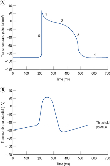
Certain specialised myocytes form the cardiac conduction system and these cells have pacemaker activity, that is, they are capable of generating their own action potentials due to gradual depolarisation of the transmembrane potential during diastole (phase 4), referred to as the pacemaker potential (Fig. 22.1B). The pacemaker potential occurs as a result of (i) a gradual reduction in an outward K+ current called the delayed rectifier (IK) current, (ii) increasing dominance of an inward current of Na+ and some Ca2+ ions known as If (f stands for ‘funny’) and (iii) an inward calcium current ICa through voltage-gated calcium channels. As a result of the pacemaker potential, the transmembrane potential gradually becomes less negative until a threshold potential is reached at which an action potential is triggered. The rate of depolarisation of the pacemaker potential, and hence the heart rate, is influenced by the autonomic nervous system. Sympathetic nervous system activation and circulating catecholamines increase the heart rate by binding to ß1-adrenoreceptors leading to an increase in intracellular cyclic AMP, which results in changes to the permeability of the various ion channels responsible for the pacemaker current. Parasympathetic nervous system activation, mediated by muscarinic cholinergic receptors, has the opposite effect.

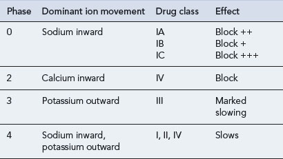
The rapid depolarisation of the cardiac action potential (Fig. 22.1A, phase 0) occurs because of a rapid increase in the permeability of the cell membrane to Na+ ions, which enter rapidly through ‘fast’ Na+ channels in a current known as INa. The INa current is brief as the ‘fast’ Na+ channels inactivate rapidly. The early phase of repolarisation (phase 1) is due to closure of the fast Na+ channels, an outward K+ current known as Ito (to – transient outward) and a further K+ current known as the ultra-rapid component of the delayed rectifier current or IKur. The plateau phase (phase 2) of the cardiac action potential occurs because the inward movement of Ca2+ ions (ICa) is balanced by the outward movement of K+ ions. Repolarisation (phase 3) occurs as ICa diminishes and two further components of the delayed rectifier (IK) current known as the rapid (IKr) and slow (IKs) components predominate, with an important contribution from IK1.

There is considerable variation in the expression of trans-membrane ion channels in different parts of the heart, with corresponding variation in the morphology of the action potential. The most marked example is that myocytes in the sinus and AV nodes contain few Na+ channels. The upstroke of the action potential in these cells is due, predominantly, to the influx of Ca2+ ions and, therefore, is considerably slower than the upstroke in other myocytes (Fig. 22.1B). The variation in ion channel expression throughout the heart is essential for normal cardiac function, helps to explain the pathophysiology of many inherited and acquired diseases complicated by cardiac arrhythmia and accounts for the relative selectivity of antiarrhythmic and other drugs for certain parts of the heart.

Normal cardiac conduction

During normal sinus rhythm (Fig. 22.2), an activation wavefront begins in the sinus node, a group of cells with pacemaker activity on the upper free wall of the right atrium. The rate of diastolic depolarisation and hence the rate of discharge of the sinus node is increased by sympathetic nerve stimulation, circulating catecholamines or sympathomimetic drugs mediated by ß1-adrenoreceptors on the cell membranes of the sinus node myocytes. Parasympathetic (vagus) nerve stimulation exerts the opposite effect, mediated by muscarinic cholinergic receptors.

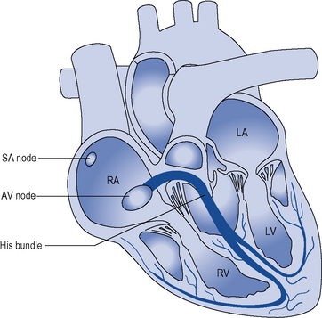
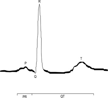
An activation wavefront spreads across the atrial myocardium, leading to atrial contraction and generating the P wave on the surface electrocardiogram (ECG; Fig. 22.3). The last part of the atria to be activated is the atrioventricular (AV) node, the electrical and structural properties of which result in a slow conduction velocity, allowing atrial emptying to be completed before ventricular contraction begins and represented by the PR interval on the ECG. Conduction velocity in the AV node is increased by sympathetic nerve stimulation, circulating catecholamines or sympathomimetic drugs, mediated by ß1-adrenoreceptors while parasympathetic (vagus) nerve stimulation exerts the opposite effect via muscarinic cholinergic receptors.

The atria and ventricles are electrically isolated from each other by the annulus fibrosus, the electrically non-conductive fibrous tissue forming the valve rings. In the normal heart, there is just one electrical connection between the atria and ventricles, the bundle of His, which conveys the activation wavefront from the AV node and penetrates the annulus fibrosus before dividing into the right and left bundle branches. The bundle branches ramify into a sub-endocardial network of Purkinje fibres, which convey the activation wavefront rapidly across the ventricles ensuring near-simultaneous contraction of the ventricular myocardium, and are represented by the narrow QRS complex of the ECG. Finally, the activation wavefront spreads from endocardium to epicardium. A wave of repolarisation then spreads across the ventricles resulting in the T wave. The QT interval on the ECG, therefore, represents the duration of ventricular depolarisation and repolarisation. There is an inverse relationship between the time to activation of different areas of the ventricular myocardium and APD such that the latest areas to be activated have the shortest APD. The purpose of this relationship is that repolarisation is rapid and uniform throughout the ventricular myocardium, which serves to maintain electrical stability.

Arrhythmia mechanisms

Cardiac arrhythmias occur because of abnormalities of impulse formation or propagation.

Abnormal impulse formation

Abnormal automaticity

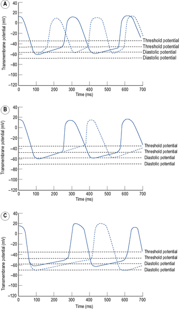
Automaticity is another term for pacemaker activity, a characteristic possessed by all cells of the specialised cardiac conduction system during health and, potentially, by other cardiac myocytes during certain disease states. The rate of firing of a pacemaker cell is largely determined by the duration of the phase 4 diastolic interval (Fig. 22.4). This in turn is determined by (i) the maximum diastolic potential following repolarisation of the preceding action potential, (ii) the slope of diastolic depolarisation due to pacemaker currents and (iii) the threshold potential for generation of a new action potential. In the healthy state, there is a hierarchy of firing rates within the specialised conduction system with the highest rate in the sinus node followed by the AV node and then the His–Purkinje system. The sinus node is, therefore, the dominant pacemaker and determines the heart rate, while the pacemaker activity in the distal conduction system is ‘overdriven’ by the sinus node. Abnormal automaticity describes either accelerated pacemaker activity in cells of the distal cardiac conduction system such that they escape from overdrive suppression by the sinus node, or the development of pacemaker activity in cells that do not form part of the cardiac conduction system.

Triggered activity

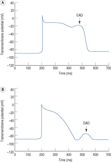
Triggered activity describes impulse formation dependent upon afterdepolarisations. Early afterdepolarisations (EADs) occur during phase 2 or 3 of the cardiac action potential whereas delayed afterdepolarisations (DADs) occur during phase 4 (Fig. 22.5). In both cases, afterdepolarisation may reach the threshold potential required for generation of a new action potential.

EADs are characteristic of the congenital and acquired long QT syndromes. The prolonged APD promotes reactivation of the inward calcium current ICa which may directly cause EADs during phase 2. Furthermore, action potential prolongation and ß-adrenoreceptor stimulation promote calcium overload in the sarcoplasmic reticulum. This in turn leads to the spontaneous release of calcium in bursts by the sarcoplasmic reticulum. The resultant increase in intracellular calcium concentration activates the transmembrane Na+/Ca2+ exchanger which moves one calcium ion out of the myocyte in exchange for three sodium ions and, therefore, results in an EAD during phase 3. In the long QT syndromes, an EAD may initiate a form of polymorphic ventricular tachycardia (VT) known as Torsade de Pointes. EADs are more prominent at slow heart rates.

DADs are seen during reperfusion following ischaemia, heart failure, digitalis toxicity and in catecholaminergic polymorphic VT. They occur because of spontaneous release of calcium in bursts by the sarcoplasmic reticulum, activating the Na+/Ca2+ exchanger as described for EADs and resulting in a DAD during phase 4. A DAD may result in a single extrastimulus (‘ectopic beat’) or in repetitive firing, that is, tachycardia. DADs are more prominent at rapid heart rates and during sympathetic nervous stimulation of ß-adrenoreceptors.

Abnormal impulse propagation

Re-entry

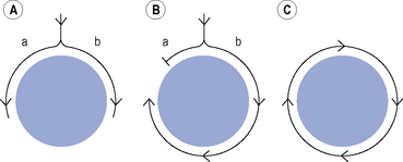
Many clinically important arrhythmias are due to re-entry, in which an activation wavefront rotates continuously around a circuit. Re-entry depends upon a trigger in the form of a premature beat, and a substrate, that is, the re-entry circuit itself. A precise set of electrophysiological conditions must be met in order for re-entry to occur (Fig. 22.6): (i) there must be a central non-conducting obstacle around which the re-entry circuit develops, (ii) a premature beat must encounter unidirectional conduction block in one limb (a) of the re-entry circuit, (iii) conduction must proceed slowly enough down the other limb (b) of the re-entry circuit that electrical excitability has returned in the original limb (a), allowing the activation wavefront to propagate in a retrograde direction along that limb, and (iv) the circulating activation wavefront must continue to encounter electrically excitable tissue. This is a function of the length of the re-entry circuit, the conduction velocity of the activation wavefront and the effective refractory period of the myocardium throughout the circuit. Class I antiarrhythmic drugs block sodium channels and, therefore, reduce the amplitude and rate of rise of the cardiac action potential and in so doing, reduce the conduction velocity of an activation wavefront. Class I antiarrhythmic drugs may exert their major antiarrhythmic effect by abolishing conduction altogether in areas of diseased myocardium forming part of a re-entry circuit in which conduction is already critically depressed. Class III antiarrhythmic drugs prolong cardiac APD and hence the refractory period. If previously activated cells in a re-entry circuit (the ‘tail’) remain refractory when the re-entrant wavefront (the ‘head’) returns to that area, conduction will fail and re-entry will be abolished. Drug-induced prolongation of the refractory period may, therefore, terminate and/or prevent re-entrant arrhythmias.

Clinical problems

Patients with a cardiac arrhythmia may present with a number of symptoms:

Arrhythmias may aggravate heart failure in two ways: (i) the haemodynamic effect of the arrhythmia may precipitate heart failure or aggravate existing heart failure and (ii) prolonged tachycardia of any type may lead to tachycardia-induced cardiomyopathy.

Diagnosis

A detailed history should be obtained, covering all of the symptoms listed above. A characteristic of cardiac arrhythmias is their random onset. Symptoms occurring under specific circumstances are less likely to be due to arrhythmia, but there are exceptions including certain uncommon types of VT, some cases of supraventricular tachycardia (SVT) due to an accessory pathway and vasovagal syncope (faints). Other key features of the history include:

Physical examination is essential but often normal between episodes of arrhythmia. Mandatory investigation includes a 12-lead ECG and an echocardiogram to detect structural heart disease. Other investigations for structural and ischaemic heart disease may be indicated at this stage with the aim of detecting any underlying structural heart disease. If the history does not include sinister features such as syncope or a family history of sudden unexpected death at a young age, and the resting 12-lead ECG and echocardiogram are normal, then the patient can be reassured that they are extremely unlikely to have a serious heart rhythm disturbance. The extent of further investigation will be dictated by how troublesome the symptoms are.

The most certain way of reaching a firm diagnosis is a 12-lead ECG recorded during the patient’s symptoms demonstrating arrhythmia. As many arrhythmias occur intermittently, some form of ECG monitoring is often necessary. This may include a continuous ambulatory ECG (Holter) recording for up to 7 days at a time if the symptoms occur frequently or, for less frequent symptoms an event recorder, which may store ECG strips automatically if it detects an arrhythmia or if activated by the patient during their symptoms. An insertable loop recorder may be implanted subcutaneously and is an ECG event recorder with a battery life of about 3 years, making it a useful tool for the diagnosis of infrequent arrhythmias.

Management

Pathological tachycardia is conventionally defined as a resting heart rate over 100 beats/min and can be classified according to whether it arises in or involves the atria (supraventricular tachycardias) or the ventricle (ventricular tachyarrhythmias).

Supraventricular tachycardias

These are tachycardias arising from or involving the atria.

Atrial flutter

Atrial flutter is a right atrial tachycardia with a re-entry circuit around the tricuspid valve annulus. The atrial rate is typically 300 min–1. The long refractory period of the AV node protects the ventricles from 1:1 conduction: In the presence of a healthy AV node and the absence of AV node-modifying drugs, there is usually 2:1 AV conduction resulting in a regular narrow-complex tachycardia with a ventricular rate of 150 min–1.

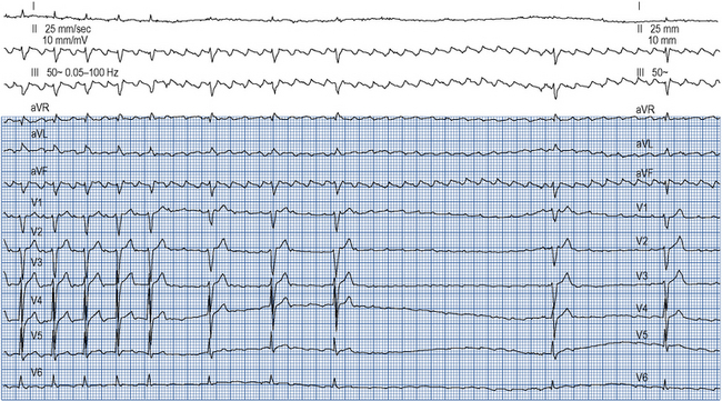
Atrial flutter confers a risk of thromboembolism similar to that of AF and this risk should be managed in the same way. Emergency management of atrial flutter is dictated by the clinical presentation but may include d.c. cardioversion or ventricular rate control with drugs which increase the refractory period of the AV node such as ß-blockers, verapamil, diltiazem or digoxin. ß-Blockers, verapamil and digoxin may be given intravenously. As the re-entry circuit is confined to the right atrium and does not involve the AV node, adenosine will not terminate atrial flutter but will produce transient AV block, allowing the characteristic flutter waves to be seen on the ECG (Fig. 22.7).

There is a limited role for antiarrhythmic drugs, whether used acutely to achieve chemical cardioversion or in the longer term to maintain sinus rhythm. Class Ic antiarrhythmic drugs such as flecainide should be used only in conjunction with AV node-modifying drugs such as ß-blockers, verapamil, diltiazem or digoxin because they may otherwise cause slowing of the atrial flutter circuit and 1:1 conduction though the AV node which may be life-threatening. Sotalol and amiodarone have been used to restore and maintain sinus rhythm and have the advantage of controlling the ventricular rate where rhythm control is incomplete. Catheter ablation of atrial flutter is highly effective and safe and is increasingly used in preference to long-term drug treatment.

Junctional re-entry tachycardia

The term ‘supraventricular tachycardia’ (SVT) is widely used to describe junctional re-entry tachycardias but is a misnomer because it implies any tachycardia arising from the atria. Junctional re-entry tachycardia is a more specific term and may be preferable.

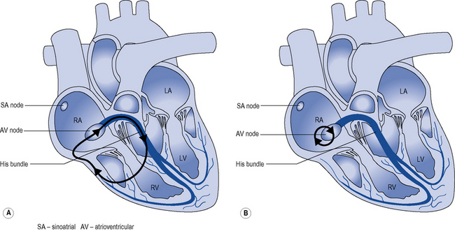
Two mechanisms account for most junctional re-entry tachycardias: both involve a macroreentry circuit (Fig. 22.8). AV nodal re-entry tachycardia (AVNRT) rotates around a circuit including the AV node itself and the so-called AV nodal fast and slow pathways, which feed into the AV node. Atrioventricular re-entry tachycardia (AVRT) comprises a re-entry circuit involving the atrial myocardium, the AV node, the ventricular myocardium and an accessory pathway, a congenital abnormality providing a second electrical connection between the atria and ventricles in addition to the His bundle, thus forming a potential re-entry circuit.

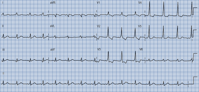
Many accessory pathways conduct only retrogradedly from the ventricles to the atrium. In these cases, the ECG during sinus rhythm appears normal and the accessory pathway is described as ‘concealed’. Other accessory pathways conduct anterogradely and retrogradely. In these cases, the ECG during sinus rhythm is abnormal and is described as having a Wolff–Parkinson–White pattern (Fig. 22.9). This abnormality is characterised by a short PR interval as the conduction velocity of an accessory pathway is usually faster than that of the AV node, and a delta wave, a slurred onset to the QRS complex which occurs because an accessory pathway inserts into ventricular myocardium which conducts more slowly than the His–Purkinje system.

Junctional re-entry tachycardias are characterised by a history of discrete episodes of rapid regular palpitation that start and stop suddenly and occur without warning and apparently at random. The peak age range at which symptoms begin is from the mid-teens to the mid-thirties and the condition is more common in women. There are no symptoms between episodes, and cardiac examination and investigation at these times are usually normal. The diagnosis is usually made on the basis of the history, ideally confirmed by an ECG recorded during an episode showing a regular narrow-complex tachycardia with no discernible P waves or P waves occurring in a 1:1 relationship with the QRS complexes.

Acute treatment of junctional re-entry tachycardia aims to terminate the tachycardia by causing transient conduction block in the AV node, an obligatory part of the re-entry circuit. Vagotonic manoeuvres such as carotid sinus massage, a Valsalva manoeuvre or eliciting the diving reflex by immersion of the face in ice-cold water may all result in a brief vagal discharge sufficient to block conduction in the AV node, terminating tachycardia. The same effect may be achieved with intravenous adenosine given as a rapid bolus injection in doses up to 12 mg. Intravenous verapamil 5 mg also as a rapid bolus injection is a good alternative where adenosine is contraindicated.

Junctional re-entry tachycardia is often recurrent. There is a limited role for prophylactic drug treatment as this is generally not a dangerous condition affecting young and otherwise healthy people. Among other factors, the efficacy, toxicity and acceptability of what may be long-term drug treatment require careful consideration. Options for prophylactic drug treatment include ß-blockers, verapamil, flecainide and sotalol. Particular importance should be given to discussion about the management of junctional re-entry tachycardia during pregnancy. Catheter ablation is curative in one sitting in a majority of cases.

Atrial fibrillation

AF is the most common sustained arrhythmia, affecting about 1% of the population. AF is rare before the age of 50 but its prevalence approximately doubles with each decade thereafter such that about 10% of those over 80 are affected. AF is characterised by extremely rapid and uncoordinated electrical activity in the atria and variable conduction through the AV node, resulting in irregular and usually rapid ventricular contraction.

The clinical importance of AF results from:

AF may be classified as:

Paroxysmal: self-limiting episodes of AF lasting no more than 7 days

Persistent: AF lasting more than 7 days or requiring cardioversion

Longstanding persistent: continuous AF for more than 1 year

Permanent: where a decision has been made not to attempt cure of persistent AF.
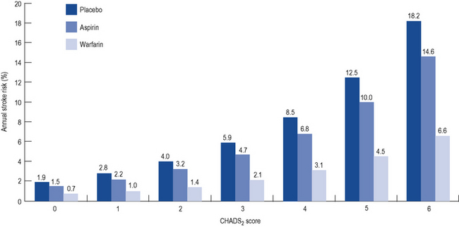
Stroke risk

All patients presenting with AF (or atrial flutter) should undergo assessment of their risk of stroke. Although various risk stratification schemes exist, they are exemplified by the CHADS2 score, which assigns one point each for Congestive cardiac failure, Hypertension, Age >75 years, and Diabetes mellitus and two points for if there is a history of previous Stroke. Surprisingly, the frequency of AF episodes does not seem to influence stroke risk. The risk of stroke is directly proportional to the CHADS2 score. Meta-analysis of numerous trials of stroke prevention in AF has suggested a relative risk reduction for stroke of 64% with warfarin and 22% with aspirin (Fig. 22.10).

Warfarin is more difficult to take than aspirin because of the need for monitoring of the international normalised ratio (INR) and because of the potential for dietary and drug interactions with warfarin. Warfarin also increases the risk of serious bleeding. The absolute benefit of warfarin over aspirin for the prevention of stroke in AF is proportional to the CHADS2 score and the point at which the benefit of warfarin is considered to outweigh the risk is an annual untreated stroke risk of 4%. For this reason, current guidelines recommend stroke prophylaxis with warfarin for those with a CHADS2 score of ≥2, warfarin or aspirin for a CHADS2 score of 1 and aspirin for those with a CHADS2 score of 0.

While CHADS2 is the most common guideline, in patients with AF and a CHADS2 score of 1, a lower incidence of stroke and/or death from all causes has been found among patients treated with vitamin K antagonists (VKAs) when compared with no antithrombotic therapy. In contrast, prescription of an antiplatelet agent was not associated with a lower risk of events compared with no antithrombotic therapy.

Identification of the ‘low risk’ category clearly needed to be improved, so patients can be truly identified as low risk. This can be achieved by using CHA2DS2–VASc [Cardiac failure, Hypertension, Age ≥75 (doubled), Diabetes, Stroke (doubled)–Vascular disease (prior myocardial infarction, peripheral artery disease, complex aortic plaque), Age 65–74 and Sex category (female =1, male = 0)]. This schema improves on CHADS2 as it classifies a low proportion of subjects into the ‘moderate risk’ category, and helps better determine the truly ‘low risk’ patients who have very low event rates and no need for anticoagulation.

There is no evidence that any form of treatment for AF, other than warfarin or aspirin, reduces the risk of stroke regardless of how apparently successful it is in maintaining sinus rhythm. Current guidelines, therefore, recommend indefinite stroke prophylaxis with warfarin or aspirin on the basis of the risk stratification.

It is often difficult to maintain INR levels within the therapeutic range of 2.0–3.0. There is evidence that the INR may be outside of this range up to half of the time. A subtherapeutic INR substantially increases risk of stroke or arterial thromboembolism. Conversely, a high INR increases the risk of bleeding. The approximate annual frequency of major and minor bleeding with warfarin is 3% and 10%, respectively. Patients’ and physicians’ concern about the use of warfarin has resulted in its under-utilisation, particularly among elderly people, who are at the greatest risk of stroke. These difficulties have led to a search for alternative agents. The most advanced of these achieve their anticoagulant effect by inhibiting a single activated clotting factor, either thrombin (factor IIA) or factor XA. These drugs have a more predictable pharmacological profile that negates the need for frequent monitoring and may represent a step forward in the care of patients with AF. These agents are expected to offer advantages of enhanced or similar efficacy compared with warfarin without an increased risk of major bleeding.

Rhythm control

Rhythm control can be considered in terms of either restoration or maintenance of sinus rhythm. The most rapid and effective means of restoring sinus rhythm is d.c. cardioversion. Where AF is of short duration, well tolerated and not associated with structural heart disease, class IC antiarrhythmic drugs such as flecainide and class III drugs such as amiodarone may be used intravenously in order to achieve chemical cardioversion. Stroke risk should be managed in the same way as described for the emergency management of AF.

Although sinus rhythm can be restored in most patients through d.c. cardioversion or antiarrhythmic drugs, alone or in combination, most patients will revert to AF without further treatment. The SAFE-T trial examined 665 patients with AF of at least 72 h duration. Sinus rhythm was restored with antiarrhythmic drugs (sotalol or amiodarone) or placebo, supplemented where necessary by d.c. cardioversion (Singh et al., 2005). Patients were then maintained on placebo, sotalol or amiodarone. By 2 years, that probability of remaining in sinus rhythm was 10% (placebo), 30% (sotalol) and 50% (amiodarone). Another similar study (Roy et al., 2000) demonstrated equivalent efficacy of sotalol and the class Ic antiarrhythmic propafenone in the maintenance of sinus rhythm (40% at 2 years) and confirmed the superiority of amiodarone (60%). Most heart rhythm specialists consider that the toxicity of amiodarone precludes its long-term use for the management of AF. Dronedarone, it was hoped, would provide efficacy similar to amiodarone but it has been shown to be less effective, although it has far fewer side effects.

Modern strategies for curative catheter ablation of AF followed the discovery in 1998 that paroxysmal AF is due, in most cases, to rapid firing by the musculature surrounding the pulmonary veins close to their junctions with the left atrium. The cornerstone of most current ablation strategies for paroxysmal AF is complete electrical isolation of all four pulmonary veins from the left atrium, using either radiofrequency ablation (cautery) or cryoablation to ablate in rings around the pairs of ipsilateral veins. Catheter ablation can cure a majority of paroxysmal AF but needs to be repeated in 30–40% of patients and carries risk including stroke (3/1000) and pericardial effusion (1–2/100). Catheter ablation has been shown in small randomised studies to be superior to antiarrhythmic drug therapy in maintaining sinus rhythm and improving symptoms and quality of life. Catheter ablation has also been shown in non-randomised studies to improve left ventricular ejection fraction and heart failure symptoms. No benefit has been demonstrated in terms of reduced stroke risk or mortality. The natural history of AF is for episodes of AF to increase in frequency and duration until persistent AF supervenes. This progression appears to occur as a result of atrial remodelling, a complex and incompletely understood process involving electrical and structural changes in the whole atrial myocardium predisposing to the development of AF independent of the pulmonary veins. Catheter ablation strategies for persistent AF are more complex than those for paroxysmal AF with a correspondingly higher rate of repeat procedures and a lower overall success rate. For all of these reasons, drug therapy remains the first line treatment of AF with catheter ablation reserved for patients with symptomatic AF that cannot be managed satisfactorily with drugs and whose symptoms trouble them enough to wish to undergo ablation.

Ventricular tachyarrhythmias

Ventricular tachycardia

VT is a rapid heart rhythm originating in the ventricles. VT may present with palpitation, chest pain, breathlessness, presyncope, syncope or sudden cardiac death (death occurring suddenly and unexpectedly within 1 h of the onset of symptoms from a presumed cardiac cause). It is clinically useful to subdivide VT in the following ways:

Ventricular fibrillation

Ventricular fibrillation (VF) comprises rapid and totally disorganised electrical activity in the ventricles such that effective contraction ceases and results in sudden death unless sinus rhythm is restored either spontaneously or by defibrillation. Acute myocardial ischaemia and infarction are probably responsible for most VF, although virtually any structural heart disease may also be complicated by VF. Other cases occur in the context of a group of conditions known as channelopathies:

Channelopathies

This is a group of inherited conditions characterised by abnormal function of the protein channels present in the cardiac myocyte cell membrane that regulate the flow of ions responsible for generating the resting transmembrane potential and the action potential. These include the long QT syndromes, short QT syndrome, early repolarisation syndrome, Brugada syndrome and catecholaminergic polymorphic VT. A detailed description of these conditions is beyond the scope of this chapter but there are certain key points:

Many drugs lengthen the QT interval (Box 22.1) and are contraindicated in patients with long QT syndromes. A list of drugs known to prolong the QT interval can be found at http://www.azcert.org.

Ongoing management of ventricular arrhythmias

Once stabilised, patients presenting with VT or VF should remain in hospital and their management should be discussed at an early stage with a specialist cardiac electrophysiology service. Investigations should be performed to establish the nature and extent of underlying heart disease, with emphasis on detecting structural heart disease, coronary artery disease, inducible myocardial ischaemia and consideration of channelopathies in those with structurally normal hearts.

Most patients with ischaemic heart disease should be treated with aspirin, statins, angiotensin converting enzyme (ACE) inhibitors or angiotensin II receptor antagonists and ß-blockers. ß-Blockers and ACE inhibitors reduce somewhat the risk of sudden cardiac death. Although these patients remain at high risk of sudden cardiac death due to recurrent ventricular tachyarrhythmias, there is no role for the routine use of antiarrhythmic drugs. In patients with complex ventricular ectopy and impaired left ventricular systolic function following acute myocardial infarction, class IC antiarrhythmic drugs including flecainide were shown in the CAST study (Echt et al., 1991) to increase mortality while in other high-risk groups amiodarone has been shown to have no effect on all-cause mortality (Cairns et al., 1997; Julian et al., 1997). All patients should be considered for an ICD. These devices have been shown to improve prognosis following:

Although ICDs treat further episodes of VT and VF, they do not prevent these arrhythmias from recurring and resulting in device therapies including shocks that are psychologically traumatic and lead to premature battery depletion. In the case of frequently recurring ventricular arrhythmias, patients should be on the maximum tolerated dose of a ß-blocker and there is a role for antiarrhythmic drugs including amiodarone and mexiletine. Catheter ablation of VT is an important adjunctive treatment in this situation.

Bradycardia

Bradycardia is conventionally defined as a resting heart rate below 60 min–1 when awake or 50 min–1 when asleep. Bradycardia can be classified as sinus bradycardia, where the sinus node discharges too slowly, or AV block (‘heart block’), where conduction between the atria and ventricles is impaired. AV block may be subdivided into three classes:

First degree. Every P wave conducts to the ventricles but takes longer than normal to do so. The PR interval on the ECG is prolonged to greater than 200 ms (one large square on a standard ECG).

Second degree. Some but not all P waves conduct to the ventricles. Progressive PR interval prolongation followed by a non-conducted P wave is referred to as Mobitz type I or Wenckebach heart block and implies block occurring in the AV node. Mobitz type II heart block is the term used when a non-conducted P wave is not preceded by progressive PR interval prolongation and implies block occurring in the conducting system below the level of the AV node.

Third degree (‘complete heart block’). No P waves conduct to the ventricles.

Bradycardia may be due to intrinsic cardiac disease or secondary to non-cardiac disease or drugs. In many cases, bradycardia due to intrinsic cardiac disease is idiopathic, that is, occurs without other identifiable heart disease. Bradycardia may also complicate acute myocardial infarction or virtually any form of structural heart disease and is also common following cardiac surgery. Non-cardiac causes of bradycardia include vasovagal syncope (faints), hypothyroidism, hyperkalaemia, hypothermia and raised intracranial pressure. Complete heart block may occur as a complication of Lyme disease (tick-borne borreliosis). Drugs commonly associated with bradycardia include ß-blockers, verapamil, diltiazem, digoxin and antiarrhythmic drugs of any class.

The management of bradycardia is as follows:

In an emergency situation, drugs may be used in an attempt to support the heart rate until trans-venous pacing can be established. The most useful drugs in this situation are atropine in 500 μcg boluses up to a total of 3 mg, adrenaline infused at a rate of 2–10 μcg/min or isoprenaline 1–10 μcg/min, titrated against heart rate. External pacing is another useful measure until transvenous pacing can be established. More detailed guidance on the emergency management of bradycardia may be viewed at http://www.resus.org.uk.

Drug therapy

Antiarrhythmic drug therapy is used to control the frequency and severity of arrythmias, with the aim of maintaining sinus rhythm where possible. Although antiarrhythmic drug treatment has been the mainstay of arrhythmia treatment, many of these drugs have limited efficacy and important toxicity. Many arrhythmias are now curable by catheter ablation. Implantable devices such as permanent pacemakers and ICDs have assumed an increasingly important role in the treatment of arrhythmias and, in many cases, antiarrhythmic drugs have an adjunctive role. Antiarrhythmic drugs can be grouped according to their electrophysiological effects at a cellular level, using the Vaughan–Williams classification. Alternatively, antiarrhythmic drugs may be classified according to their main sites of action within the heart.

Vaughan–Williams Classification

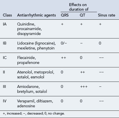
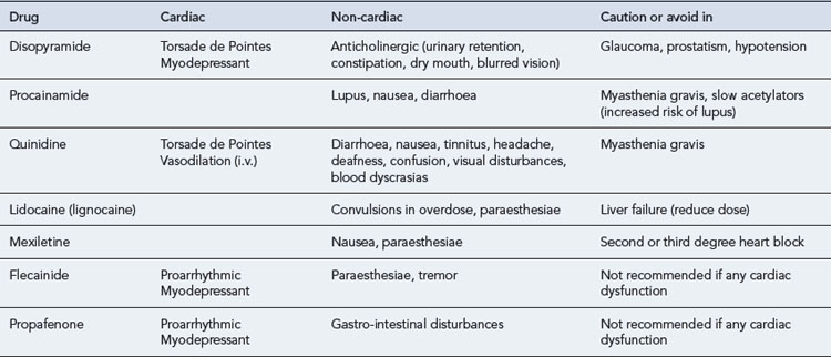
All antiarrhythmic drugs act by altering the movement of electrolytes across the myocardial cell membrane. The Vaughan–Williams classification groups drugs according to their ability to block the movement of one or more of these ions across the myocardial cell membrane. Most drugs have several modes of action, and their effectiveness as antiarrhythmic agents depends upon the summation of these effects. The effect of the different drug classes on the various phases of the action potential in His–Purkinje fibres are shown in Table 22.1. The choice of which drug to use is based upon the origin of the arrhythmia, regardless of its pattern. However, the preference of one class over another may vary, depending on a clinician’s experience with particular drugs, on the presentation of the arrhythmia and on patient characteristics. Such factors also govern the choice of drug within a class. The drug chosen should have the dosing schedule and adverse effect profile that best suit the patient or inconvenience them least (see Tables 22.222.4). Thus, for example, a patient with glaucoma or prostatism should not be given disopyramide which possesses marked anticholinergic properties, and a patient with obstructive airways disease should preferably not be prescribed a ß-blocker (class II), though if considered essential they could have a cardioselective agent.

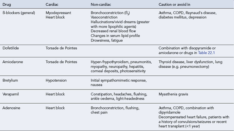
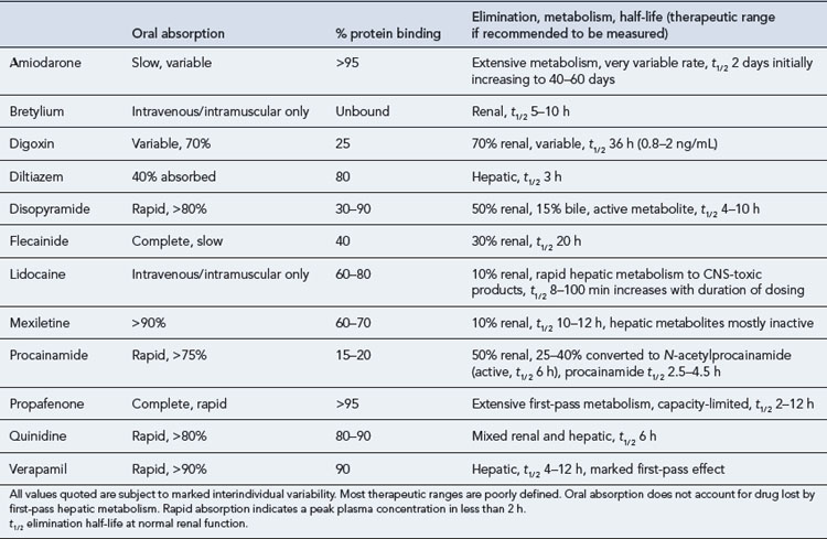
The pharmacokinetic profiles of selected antiarrhythmics are presented in Table 22.5.

Class I

Class I drugs act by blocking the fast sodium channels that are responsible for the rapid depolarisation phase of the cardiac action potential, thus reducing the rate of depolarisation (the slope of phase 0) and the amplitude of the action potential. The conduction velocity of an activation wavefront is determined partly by the slope and amplitude of the cardiac action potential and partly by the resistance to current flow through the myocardium. The effect of sodium channel blockade is a decrease in conduction velocity. Certain re-entrant arrhythmias such as VT complicating previous myocardial infarction depend upon slow conduction in part of the re-entrant circuit. Class I antiarrhythmic drugs may critically slow or even abolish conduction in these areas, thus terminating and/or preventing re-entry.

The action potential in the sinoatrial and AV nodes does not depend on fast sodium channels for depolarisation; instead, phase 0 depolarisation is carried by calcium channels. Class I antiarrhythmic drugs, therefore, have no direct effect on nodal tissue.

In addition to their effect on depolarisation, class I antiarrhythmic drugs may also alter the APD and hence the effective refractory period (ERP) via an effect on potassium channels responsible for action potential repolarisation. Class I antiarrhyhmic drugs are subdivided into three groups according to their effect on APD: class IA drugs increase the APD, class IB drugs shorten the APD, and class IC drugs have no effect on APD. These effects may be assessed by measurement of the QT interval on the ECG, which reflects average ventricular APD.

The properties of class I antiarrhythmic drugs may be summarised as follows:

Sodium channel blockade
IC > IA > IB
Effect on APD and ERP
IA prolong
IB shorten
IC no effect

Increasing the APD, and hence the effective refractory period, may terminate and prevent re-entry tachycardias by prolonging the duration that tissue is refractory and prevent re-entrant wavefronts from re-exciting the tissue. Although contributing to the antiarrhythmic effects of these drugs, APD prolongation is also responsible for one of their important adverse effects, Torsade de Pointes.

Class IA antiarrhythmic drugs have additional anticholinergic actions and oppose vagal activity. This can lead to both sinus tachycardia and a shortened refractory period of the AV node, as both the sinus and AV nodes are densely innervated and tonically inhibited by the vagus nerve. One conseqence of this effect on the AV node is a more rapid ventricular rate during AF, necessitating co-treatment with drugs such as digoxin, ß-blockers or calcium channel blockers.

Class IA agents

Class IA antiarrhythmic drugs have been used for the treatment of a variety of atrial and ventricular arrhythmias but are now rarely used because of their proarrhythmic (Torsade de Pointes) and non-cardiac side effects and potential for drug interactions.

Quinidine was one of the earliest antiarrhythmic drugs developed. Quinidine, an alkaloid derived from the cinchona tree bark, had a significant role in the treatment of many arrhythmias. After concerns about increased risk of ventricular arrhythmia and death with quinidine emerged, the use of quinidine fell dramatically in favour of newer antiarrhythmic medications.

Disopyramide has been used to treat a wide variety of supraventricular and ventricular arrhythmias. The drug is given orally and excreted by the kidneys, with a half-life of 6–8 h, necessitating frequent dosing. Disopyramide has strong affinity for muscarinic cholinergic receptors (40 times that of quinidine) and commonly causes anticholinergic side effects such as blurred vision, dry mouth, constipation and urinary retention. Disopyramide may precipitate acute glaucoma in predisposed individuals. Disopyramide also causes sympathetic inhibition resulting in vasodilation. Other adverse effects occur less frequently than with quinidine (e.g. gastro-intestinal, QRS prolongation, Torsade de Pointes and hypotension) and there is no interaction with digoxin.

Procainamide has been used in an initial attempt at the pharmacologic cardioversion of AF of recent onset. Procainamide may be given orally or intravenously. Procainamide is metabolised to n-acetyl procainamide. Both have antiarrhythmic activity and are excreted mainly by the kidneys. The half-life of procainamide is short (3–4 h), necessitating frequent dosing. The half-life of n-acetyl procainamide is considerably longer than that of procainamide. The risk of a lupus-like syndrome comprising a rash, fever and arthralgia (likeliest in slow acetylators) is about one in three of those patients treated for longer than 6 months. Hypotension due to an inhibitory effect on sympathetic ganglia, QRS and QT interval prolongation are common adverse effects with intravenous administration.

Class IB agents

As a group, class IB agents inhibit the fast sodium current (typical class I effect) while shortening the APD in non-diseased tissue. The former has the more powerful effect, while the latter might actually predispose to re-entrant arrhythmias, but ensures that QT prolongation does not occur. Class IB agents act selectively on diseased or ischaemic tissue, where they are thought to promote conduction block in slowly conducting tissue critical to the maintenance of re-entry, thereby interrupting re-entry circuits.

Lidocaine was previously the standard intravenous agent for the suppression of serious ventricular arrhythmias associated with acute myocardial infarction and following cardiac surgery but has now been almost completely superseded by ß-blockers. Lidocaine acts preferentially on ischaemic myocardium and is more effective in the presence of a high external potassium concentration. Therefore, hypokalaemia must be corrected for maximum efficacy. The kinetics of lidocaine is such that it is rapidly de-ethylated by the liver precluding oral administration. The two critical factors governing lidocaine metabolism, and hence its efficacy, are liver blood flow (decreased in old age and by heart failure and ß-blockade) and drugs that induce or inhibit the enzyme of the cytochrome P450 system. Lidocaine is rapidly distributed within minutes after an initial intravenous loading dose, requiring the need for a continuous infusion or repetitive dosing to maintain therapeutic blood levels. Lidocaine has no value in treating supraventricular tachyarrhythmias.

Mexiletine may be administered intravenously or orally to control VT. Frequent gastro-intestinal and central nervous system (CNS) side effects (dizziness, light-headedness, tremor, nervousness, difficulty with coordination) limit the dose and possible therapeutic benefit.

Class IC agents

The major electrophysioloical effects of these agents are that they are powerful inhibitors of the fast sodium channel causing a marked depression of the upstroke of the cardiac action potential. In addition, they may variably prolong the APD by delaying inactivation of the slow sodium channel and inhibition of the rapid component of the repolarising delayed rectifier current (Ikr) which may explain the prolongation of the QRS complex and QT interval. Class IC agents are potent antiarrhythmics used largely in the control of paroxysmal supraventricular and ventricular tachyarrhythmias resistant to other drugs, although they have acquired a particularly bad reputation as a result of the proarrhythmic effects seen in CAST (Cardiac Arrhythmia Supression Trial) and the CASH (Cardiac Arrest Study Hamburg) studies. Faster heart rates, increased sympathetic activity, and diseased or ischaemic myocardium all contribute to the proarrhythmic effects of these drugs. This has led to these drugs being contraindicated in patients with structural heart disease as poor systolic function exaggerates the proarrhythmic effects. However, flecainide is effective for the treatment of both supraventricular and ventricular arrhythmias in patients without structural heart disease and is moderately successful for maintenance of sinus rhythm after cardioversion of AF. Propafenone has mild ß-blocking properties, especially in higher doses so should be avoided in patients with reversible obstructive airways disease.

Class II agents: ß-Adrenoreceptor antagonists (ß-blockers)

ß1– and ß2-adrenoreceptors are present in the cell membranes of myocytes throughout the heart. Activation of ß-adrenoreceptors by norepinephrine released from postganglionic sympathetic neurones and circulating norepinephrine and epinephrine increases the rate of discharge of the sinus node (positive chronotropy), increases the conduction velocity and shortens the refractory period of the AV node (positive dromotropy) and increases the force of contraction (contractility) of myocytes (positive inotropy).

ß-Adrenoreceptors are coupled to G proteins, which activate adenylyl cyclase to form cAMP from ATP. Increased cAMP directly activates the pacemaker current If to increase the rate of diastolic depolarisation and hence to increase the sinus rate. cAMP also activates a cAMP-dependent protein kinase (PK-A) that phosphorylates L-type calcium channels, which causes increased calcium entry into the cell. Increased calcium entry during the plateau phase of the action potential leads to enhanced release of calcium by the sarcoplasmic reticulum and hence an increase in contractility. Intracellular calcium overload predisposes to the development of early or late afterdepolarisations which may result in arrhythmias due to triggered activity.

ß-Blockers prevent the normal ligand (norepinephrine or epinephrine) from binding to the ß-adrenoreceptor by competing for the binding site. The antiarrhythmic properties of ß-blockers are probably the result of several mechanisms: (i) reducing the likelihood of arrhythmias due to triggered activity, (ii) opposing the increased sympathetic activity in patients with sustained VT and in patients with acute myocardial infarction and (iii) indirectly preventing arrhythmia via their antihypertensive and anti-ischaemic effect.

ß-Blockers licensed for the treatment of arrhythmias include propranolol, acebutol, atenolol, esmolol, metoprolol and sotalol. The antiarrhythmic activity of the various ß-blockers is reasonably uniform, the critical property being ß1-adrenoreceptor blockade. Atenolol, metoprolol propranolol and esmolol are available for intravenous use. Esmolol, a selective ß1-adrenoreceptor antagonist, has a half-life of 9 min with full recovery from its ß-blockade properties within 30 min. Esmolol is quickly metabolised in red blood cells, independent of renal and hepatic function, and due to its short half-life, can be useful in situations where there are relative contraindications or concerns about the use of a ß-blocker. Sotalol has some class III activity as well as class II effects and bretylium is considered to have class II activity in addition to class III.

The use of ß-blockers is somewhat constrained by their adverse effects. ß2-Adrenoreceptors on bronchial smooth muscle are tonically activated by circulating catecholamines to cause bronchodilation. ß-Blockers can, therefore, cause bronchoconstriction and are contraindicated in patients with asthma and should be used with caution in chronic obstructive pulmonary disease. ß2-Adrenoreceptors are also found on vascular smooth muscle and are tonically activated by circulating catecholamines to cause vasodilatation. ß-Blockers may, therefore, cause vasoconstriction and exacerbate the symptoms of peripheral vascular disease.

Cardiac adverse effects of ß-blockers include sinus bradycardia, exacerbation of AV conduction block, reduced exercise capacity and exacerbation of acute heart failure. In patients with chronic, stable heart failure, however, due to mild to moderate LV systolic dysfunction and already treated by ACE inhibitors and diuretics, ß-blockers improve both symptoms and prognosis. Other adverse effects of ß-blockers include nightmares and impotence.

ß-Blockers vary in their lipid solubility. Agents such as propranolol and carvedilol are highly lipid-soluble whilst others such as atenolol and nadolol are more hydrophilic. Lipid solubility determines the degree of drug penetration into the CNS and the utility of haemodialysis or haemofiltration. High lipid solubility is associated with a larger volume of distribution and better CNS penetration. Lipophilic ß-blockers are primarily metabolised by the liver. Conversely, hydrophilic ß-blockers have a small volume of distribution and are eliminated essentially unchanged by the kidneys; this property allows hydrophilic ß-blockers to be removed by haemodialysis.

Class III agents

Class III antiarrhythmic drugs prolong cardiac APD by inhibiting repolarising outward potassium currents IKr and/or IKs. This action prolongs the effective refractory period, reducing the likelihood of arrhythmias due to re-entry. Prolongation of cardiac APD is reflected by QT interval prolongation on the ECG. Primary indications for class III agents are AF, atrial flutter and ventricular tachyarrhythmias.

Class III drugs include amiodarone, sotalol and bretylium. Amiodarone has additional class I, II and IV activity while sotalol has marked class II activity. An important limitation of class III agents is that action potential prolongation may be complicated by Torsade de Pointes. The development of Torsade de Pointes is attributed to a combination of triggered activity as a result of EADs and increased transmural dispersion of repolarisation within the ventricles as action potential prolongation is not uniform across the ventricular wall. Hypokalaemia, hypomagnesaemia or bradycardia increase the likelihood of Torsade de Pointes and, therefore, sotalol, with marked class II activity, may be uniquely arrhythmogenic.

Amiodarone

Amiodarone is a potent antiarrhythmic drug that is effective in treating a wide variety of atrial and ventricular arrhythmias but its use is constrained by complex pharmacokinetics and concern about toxicity. Many heart rhythm specialists would consider that the side effect profile of amiodarone precludes its use for the long-term treatment of atrial arrhythmias. Amiodarone may be extremely effective in the emergency treatment of VT and ventricular fibrillation, especially where recurrent. Amiodarone may also reduce the likelihood of recurrent ventricular arrhythmias when taken on a long-term basis but confers no prognostic benefit and should be considered as an adjunct to treatment with an ICD.

When rapid control of an arrhythmia is needed, the intravenous route is preferred, with 300 mg given over 30 min to an hour followed by 900 mg over 23–24 h, administered through a central vein. Higher loading doses may cause hypotension. A concurrent oral loading regimen of up to 2400 mg daily in two to four divided doses is usually given for 7–14 days and then reduced to a maintenance dose of 200 mg daily or less.

During the early stages of therapy with amiodarone (whether intravenous or oral), the kinetics of the drug are different from those after chronic administration. Amiodarone is highly lipid soluble and so has a very large volume of distribution. As the slowly equilibrating tissue stores are penetrated to a minimal extent during the early days of therapy, the effective elimination half-life (t1/2) is initially dependent upon a more rapidly exchanging compartment, with a t1/2 of 10–17 h, substantially shorter than the t1/2 seen during chronic administration. The short t1/2 becomes important during the acute phase and any intravenous to oral changeover period because the absorption of oral amiodarone is very slow, taking up to 15 h. The combination of a relatively fast elimination and a poor rate of absorption could lead to a significant fall in serum amiodarone levels if intravenous therapy is stopped abruptly when oral therapy is initiated, with the period of maximum risk being the first 24 h of oral therapy. It is, therefore, advisable to phase out intravenous therapy gradually and allow an intravenous/oral overlap period of at least 24 h. Once amiodarone has reached saturation, amiodarone is eliminated very slowly, with a half-life of about 25–110 days. Due to amiodarone’s long terminal half-life, there is a potential for drug interactions to occur several weeks (or even months) after treatment with it has been stopped. Common interactions include antibacterials, other antiarrhythmics, lipid-regulating drugs and digoxin.

Amiodarone has been associated with toxicity involving the lungs, thyroid gland, liver, eyes, skin, and peripheral nerves. The incidence of most adverse effects is related to total amiodarone exposure (i.e. dosage and duration of treatment). Therefore, practitioners must consider carefully the risk–benefit ratio of the use of amiodarone in each patient, use the lowest possible dose of amiodarone, monitor for adverse effects and, if possible, discontinue treatment if adverse effects occur.

Corneal microdeposits (reversible on withdrawal of treatment) develop in nearly all adult patients given prolonged amiodarone; these rarely interfere with vision, but drivers may be dazzled by headlights at night. However, if vision is impaired or if optic neuritis or optic neuropathy occur, amiodarone must be stopped to prevent blindness. Long-term administration of amiodarone is associated with a blue-grey discoloration of the skin. This is more commonly seen in individuals with lighter skin tones. The discoloration may revert upon cessation of the drug. However, the skin color may not return completely to normal.

Individuals taking amiodarone may become more sensitive to the harmful effects of UV-A light. Using sunblock that also blocks UV-A rays appears to prevent this side effect. Amiodarone contains iodine and can cause disorders of thyroid function. Both hypothyroidism and hyperthyroidism may occur. Clinical assessment alone is unreliable and laboratory tests should be performed before treatment and every 6 months including tri-iodothyronine (T3), T4 and thyroid stimulating hormone (TSH). A raised T3 and T4 with a very low or undetectable TSH concentration suggests the development of thyrotoxicosis. Amiodarone-associated thyrotoxicosis may be refractory to treatment and amiodarone should usually be withdrawn, at least temporarily, to help achieve control, although treatment with carbimazole is often required. Hypothyroidism can be treated safely with replacement therapy without the need to withdraw amiodarone if amiodarone is considered essential. Amiodarone is also associated with hepatotoxicity and treatment should be discontinued if severe liver function abnormalities or clinical signs of liver disease develop.

The most serious adverse effect of amiodarone therapy is pulmonary toxicity, typically acute pneumonitis or more insidious pulmonary fibrosis. In early studies, the frequency of pulmonary toxicity during amiodarone therapy was 2–17%. More recent studies have shown a lower incidence in patients receiving dosages of 300 mg/day or less. Although acute pneumonitis may respond to corticosteroids, pulmonary fibrosis is largely irreversible.

Class IV agents

The plateau phase of the cardiac action potential results from the inward movement of Ca2+ ions balanced by the outward movement of K+ ions. Class IV agents (calcium channel blockers) block the inward movement of calcium ions during phase 2 by binding to L-type calcium channels on cardiac myocytes. The effect of class IV antiarrhythmic drugs is most marked in the sinoatrial and AV nodes, in which depolarisation is dependent upon calcium channels. Class IV antiarrhythmic drugs, therefore, cause sinus bradycardia (negative chronotropic effect), and, by reducing the conduction velocity and prolonging the AV nodal effective refractory period, reduce the ventricular rate during atrial tachyarrhythmias such as AF and flutter (negative dromotropic effect). By reducing intracellular calcium concentration class IV antiarrhythmic drugs also exert a negative inotropic effect. Only the non-dihydropyridine calcium channel blockers (verapamil and diltiazem) have direct cardiac effects.

Verapamil possesses a chiral carbon and is marketed as a racemic mixture of R- and S-stereoisomers. In humans, both isomers share qualitatively similar negative chronotropic and dromotropic effects on the sinoatrial and AV nodes, respectively, but the S-stereoisomer is 10–20 times more potent than the R with respect to these effects. Hence, the S-stereoisomer determines the negative chronotropic and dromotropic effects of verapamil, while the R-stereoisomer is of minor importance.

Verapamil also undergoes extensive stereoselective first-pass hepatic metabolism. S-verapamil is more rapidly metabolised than R-verapamil after oral administration, resulting in a lower bioavailability of the S-stereoisomer and a proportionally higher concentration of the R-stereoisomer in the systemic circulation (20% and 50%, respectively). However, because Cmax is higher with the immediate-release formulation and S-verapamil is 10–20 times more potent than R-verapamil, it is unsurprising that this difference is also clinically significant. With the immediate-release formulation, a plot of PR-interval change versus time has the same shape as the concentration–time curve. The extended-release formulation does not have the same concentration–time effect relationship. This has been attributed to the difference in oral input rates, to the concentration-related saturable first-pass hepatic metabolism, or both.

Since the formulation of verapamil may play a role in the drug’s complex pharmacokinetics and efficacy, one formulation of verapamil cannot be safely substituted for another. Immediate release preparations are preferred to maximise bioavailability of the S-stereoisomer.

Class IV antiarrhythmic drugs should be avoided in sick sinus syndrome or second- or third-degree heart block unless the patient has a permanent pacemaker. Combined therapy with a calcium channel blocker and ß-blocker should be instituted with caution because of the risk of excessive AV block, and should be used where only where monotherapy is insufficient to control ventricular rate during atrial flutter or fibrillation. Verapamil causes greater arterial vasoodilation than diltiazem and may be especially useful in patients with hypertension or angina. Both agents have a negative inotropic effect and are thus contraindicated in heart failure. Adverse effects are mostly predictable and include ankle oedema, flushing, dizziness, light-headedness and headache. Constipation is common in patients receiving verapamil whilst a rash is common with diltiazem.

Adenosine

The potassium channel opener adenosine and its pro-drug adenosine triphosphate (ATP) act as indirect calcium antagonists and resemble verapamil in their antiarrhythmic activity. In cardiac tissue, adenosine binds to adenosine type 1 (A1) receptors, which are coupled to Gi proteins. Activation of this pathway opens transmembrane potassium channels, which hyperpolarises the cell. Activation of the Gi protein also decreases cAMP, which inhibits L-type calcium channels and, therefore, calcium entry into the cell. In cardiac pacemaker cells located in the sinoatrial node, adenosine acting through A1 receptors inhibits the pacemaker current (If), which decreases the slope of phase 4 of the pacemaker action potential thereby decreasing its spontaneous firing rate (negative chronotropy). Inhibition of L-type calcium channels also decreases conduction velocity of the AV node (negative dromotropy). Finally, adenosine, by acting on presynaptic purinergic receptors located on sympathetic nerve terminals, inhibits the release of norepinephrine.

The ultra-short duration of action (<10 s) of intravenous adenosine makes it very suitable as a diagnostic aid and for interrupting supraventricular arrhythmias in which the AV node is part of the re-entry pathway. Adenosine is, however, a bronchoconstrictor and causes dyspnoea, flushing, chest pain and further transient arrhythmias in a high proportion of patients, and its metabolism is inhibited by dipyridamole, a vasodilator drug that blocks adenosine uptake by cells, thereby reducing the metabolism of adenosine.

Digoxin

Digitalis compounds are potent inhibitors of transmembrane Na+/K+-ATPase. This ion transport system moves sodium ions out of the cell in exchange for potassium ions. The consequent rise in the intracellular sodium concentration increases the activity of a transmembrane Na+/Ca+ exchanger in cardiac myocytes as well as many other cells, which moves sodium out of the cell in exchange for calcium. The resulting increased intracellular calcium concentration stimulates calcium release from the sarcoplasmic reticulum which increases contractility (positive inotropic effect).

Digoxin also acts on the autonomic nervous system to increase vagal tone and reduce sympathetic nervous activity, reducing conduction velocity and increasing the effective refractory period of the AV node. Digoxin, therefore, reduces the ventricular rate during persistent atrial flutter and AF and may be particularly useful in patients with these arrhythmias in the context of congestive cardiac failure. Digoxin has limited efficacy in controlling the ventricular rate in situations where the sympathetic nervous system predominates such as during exercise. It is, therefore, only useful as monotherapy in sedentary patients. ß-Blockers and calcium channel blockers are more useful for controlling the ventricular rate on exertion as well as at rest.

Digoxin is no longer indicated for the treatment of paroxysmal AF as it has no direct antiarrhythmic effect and neither terminates an episode of AF nor reduces the likelihood of further episodes of AF occurring. Furthermore, digoxin has limited efficacy for ventricular rate control at the start of an episode of AF where sympathetic nervous system activity is often high.

The positive inotropic effect of digoxin has long been used in the treatment of patients with systolic heart failure and sinus rhythm. As evidence emerged that other positive inotropic agents such as milrinone increase mortality in heart failure the role of digoxin was reexamined. Perhaps the largest and best designed of these trials, the Digitalis Investigation Group (1997) trial, established that digoxin had no effect on all-cause mortality in patients with stable congestive cardiac failure, a left ventricular ejection fraction of under 45% and sinus rhythm but significantly reduced a combined endpoint of CHF mortality and hospitalisation due to heart failure. With a large body of evidence attesting to the morbidity and mortality benefits of ACE inhibitors, ß-blockers and spironolactone the role of digoxin has diminished. Digoxin may still be useful in patients who remain symptomatic despite comprehensive therapy with these drugs.

The long half-life of digoxin (about 36 h) warrants special consideration when treating arrhythmias as several days of constant dosing would be required to reach steady-state. Therefore, loading doses of up to 1.5 mg may be used rapidly to increase digoxin serum levels. Digoxin is given once daily thereafter, usually in 125 or 250 μcg doses and has a narrow therapeutic window with the ideal blood concentration regarded as 1–2 μcg/L. Since digoxin is excreted predominantly by the kidney (70% renal elimination in normal renal function), renal function is the most important determinant of the daily digoxin dosage. Importantly, in severe renal insufficiency, there is also a decrease in the volume of distribution of digoxin and, therefore, lower loading doses should be used.

Both the therapeutic and toxic effects of digoxin are potentiated by hypokalaemia and hypercalcaemia. There are also numerous drug interactions (Table 22.6), some of which are pharmacokinetic and some of which are pharmacological.

Table 22.6 Interactions involving digoxin

Effect Offending agent or condition
Serum level increased by Amiodarone, verapamil, diltiazem, quinidine, propafenone, clarithromycin, broad-spectrum antibiotics (erythromycin, tetracyclines), decreased renal blood flow (ß-blockers, NSAIDs), renal failure, heart failure
Serum level decreased by Colestyramine, sulfasalazine, neomycin, rifampicin, antacids, improved renal blood flow (vasodilators), levothyroxine (thyroxine)
Therapeutic effect increased by Hypokalaemia, hypercalcaemia, hypomagnesaemia, antiarrhythmic classes IA, II, IV, diuretics that cause hypokalaemia, corticosteroids, myxoedema, hypoxia (acute or chronic), acute myocardial ischaemia or myocarditis
Therapeutic effect decreased by Hyperkalaemia, hypocalcaemia, thyrotoxicosis

The occurrence of adverse drug reactions is common, owing to the narrow therapeutic index of digoxin. Adverse effects are concentration-dependent, and are rare when serum digoxin concentration is less than 0.8 μcg/L. Common adverse effects include loss of appetite, nausea, vomiting and diarrhoea as gastro-intestinal motility increases. Other common effects are blurred vision, visual disturbances (yellow-green halos and problems with colour perception), confusion and drowsiness. The often described adverse effect of digoxin, xanthopsia, the disturbance of colour vision (mostly yellow and green colour) is rarely seen.

Patient care

Patients with arrhythmias may experience considerable anxiety about the possibility that they will have a serious arrhythmia at any moment and may, therefore, require considerable reassurance with regular follow-up. The patient’s family and friends may need to be advised on what to do in the event of an acute arrhythmia. An individual’s anxiety may not be helped by the fact that most antiarrhythmic drugs work in only a proportion of patients and several treatment options may be tried before the most appropriate one is identified.

Patients should give informed consent for all interventions, and prescribers must be prepared for a patient to have a different view on the use of a medicine compared to their own. This was illustrated in a study of patients’ and prescribers’ attitudes to the use of aspirin and warfarin for stroke prevention in AF. Not only did prescribers differ markedly on the balance of risks between stroke prevention and bleeding caused by treatment but patients feared a stroke more than doctors. Prescribers should seek and respect patients’ views on such treatment choices, rather than assume all patients are the same or that they will always agree with their own views.

Examples of some common therapeutic problems that may occur during the management of arrhythmias are set out in Table 22.7.

Table 22.7 Common therapeutic problems in the management of arrhythmias

Problem Comment
All antiarrhythmics are proarrhythmic Prevention is better than cure. Minimise the requirement for drugs by careful attention to precipitating factors. Consider use of pacemakers or non-pharmacological therapies if appropriate
Nausea and vomiting with blurred vision and visual discolouration on digoxin Symptoms and signs of digoxin toxicity noting digoxin has a narrow therapeutic range. Poor renal function may have also contributed
ß-Blockers are generally contraindicated in bronchial and peripheral vascular disease Consider verapamil or diltiazem
Calcium channel blockers-induced constipation If it occurs, give regular osmotic laxatives
Torsade de Pointes may be precipitated by taking other medication with amiodarone or disopyramide Patients should remind members of health care team that they are taking antiarrhythmic drugs, as well as consider electrolyte disturbance such as hypokalaemia
Patients experiencing myopathy Healthcare professionals involved in screening prescriptions for antiarrhythmics should be aware of the clinically relevant interactions and how to manage these. Patients who are taking statins will need regular monitoring for signs of myopathy, particularly those on high-intensity statin therapy
Severe asthmatic patient admitted with SVT Adenosine is contraindicated due to the risk of bronchospasm. Verapamil is a suitable alternative
Amiodarone is commonly associated with an increased tendency to sunburn Warn all patients to stay covered up when outdoors, use sun block or stay indoors
Acutely treated patient with AF cardioverted initially with i.v. amiodarone, but on converting to oral therapy, converted back to AF While amiodarone has a long terminal half-life once saturated, amiodarone has a very large volume of distribution and since tissue stores are penetrated to a minimal extent during the early days of therapy, the effective elimination half-life (t1/ 2) is initially dependent upon a more rapidly exchanging compartment, with a t1/ 2 of 10–17 h, substantially shorter than the t1/ 2 seen during chronic administration. The shorter t1/ 2 becomes important during the acute phase and any intravenous to oral changeover period because the absorption of oral amiodarone is very slow, taking up to 15 h. The combination of a relatively fast elimination and a poor rate of absorption could lead to a significant fall in serum amiodarone levels if intravenous therapy is stopped abruptly when oral therapy is initiated, with the period of maximum risk being the first 24 h of oral therapy. It is, therefore, advisable to phase out intravenous therapy gradually and allow an intravenous/oral overlap period of at least 24 h

SVT, supraventricular tachycardia; AF, atrial fibrillation.

Case studies

References

AFFIRM investigators. Relationship between sinus rhythm, treatment, and survival in the atrial fibrillation follow-up investigation of rhythm management (AFFIRM) study. Circulation. 2004;109:1509-1513.

Cairns J.A., Connolly S.J., Roberts R., et al. Randomised trial of outcome after myocardial infarction in patients with frequent or repetitive ventricular premature depolarisations: CAMIAT Canadian Amiodarone Myocardial Infarction Arrhythmia Trial Investigators. Lancet. 1997;349:675-682.

Crouch M.A., Limon L., Cassano A.T. Drug-related QT interval prolongation: drugs associated with QT prolongation. Pharmacotherapy. 2003;23:802-805.

Digitalis Investigation Group. The effect of digoxin on mortality and morbidity in patients with heart failure. N. Engl. J. Med.. 1997;336:5325-5333.

Echt D.S., Liebson P.R., Mitchell L.B., et al. Mortality and morbidity in patients receiving encainide, flecainide, or placebo: the Cardiac Arrhythmia Suppression Trial. N. Engl. J. Med.. 1991;324:781-788.

Julian D.G., Camm A.J., Frangin G., et al. Randomised trial of effect of amiodarone on mortality in patient with left ventricular dysfunction after recent myocardial infarction: EMIAT. European Myocardial Infarct Amiodarone Trial Investigators. Lancet. 1997;349:667-674.

Roy D., Talajic M., Doran P. Amiodarone to prevent recurrence of atrial fibrillation. N. Engl. J. Med.. 2000;342:913-920.

Singh B.N., Singh S.N., Reda D.J. Amiodarone versus sotalol for atrial fibrillation. N. Engl. J. Med.. 2005;352:1861-1872.