between the transverse duodenum and aorta at the site of the graft-aortic suture line
  .
.
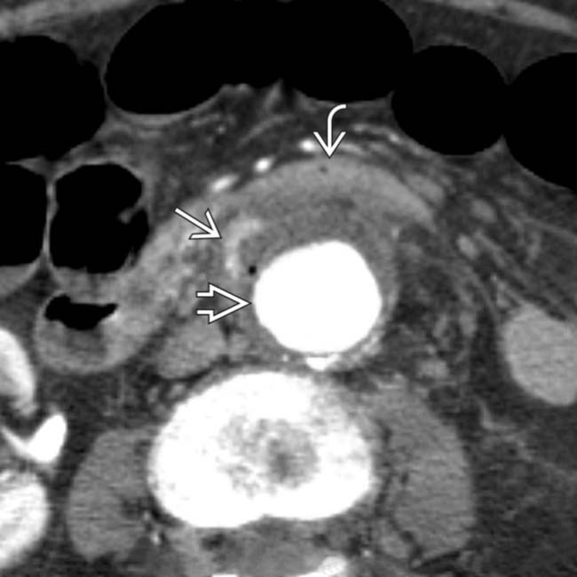
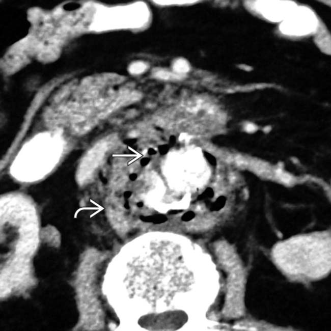
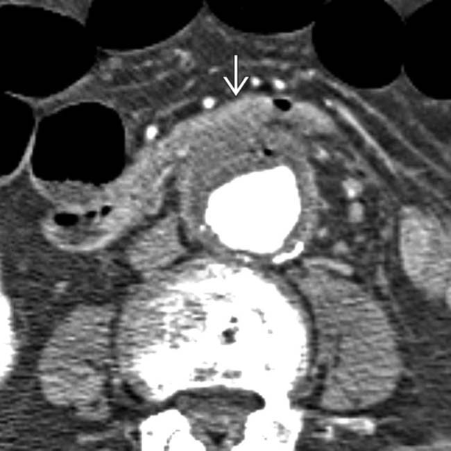
 wrapped around a synthetic graft. A gas collection is noted between the graft and aortic wall
  , indicating infection or fistula. Note the soft tissue density surrounding the aorta and 3rd portion of duodenum
  .
.
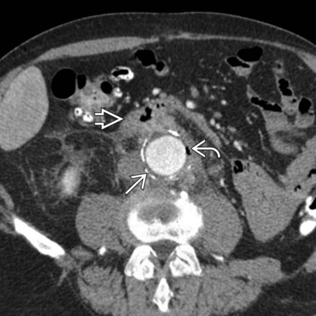
 . At the level of the 3rd portion of the duodenum, the duodenal wall
  appears to be adherent to the aorta. Note an enhanced focus
  that may represent active bleeding or inflammation.
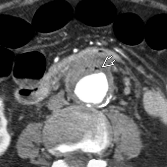
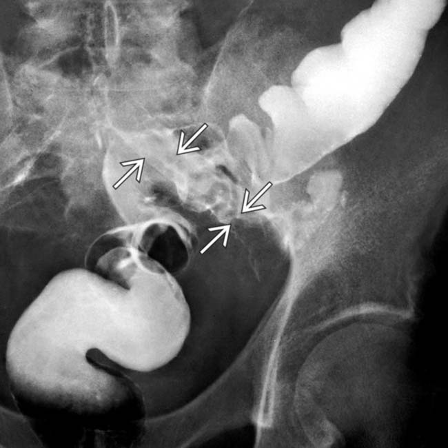
 ; aortoenteric fistula with underlying infection was surgically confirmed.


 and abnormal perigraft soft tissue indicating infection
  . (Courtesy F. Hughes, MD.)
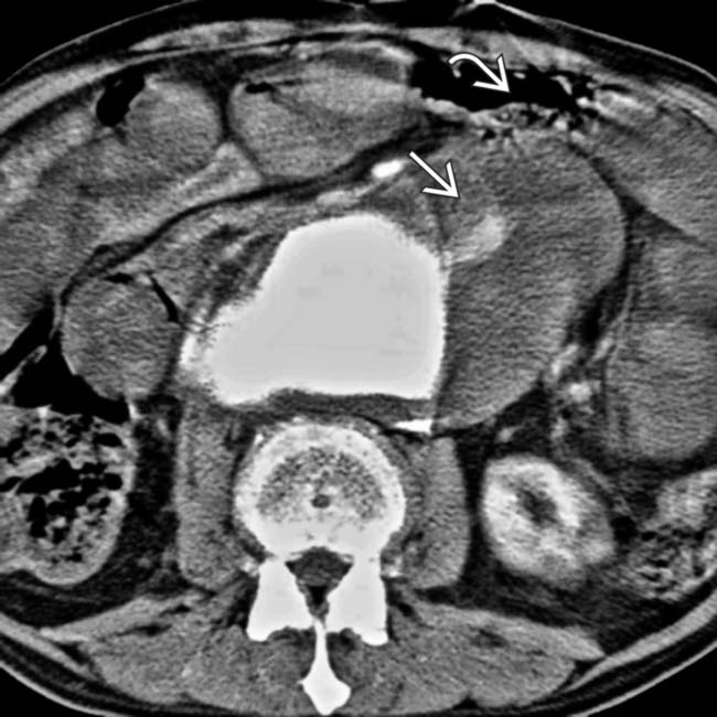
 . The transverse duodenum
  is draped over the hematoma.
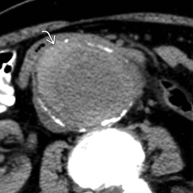
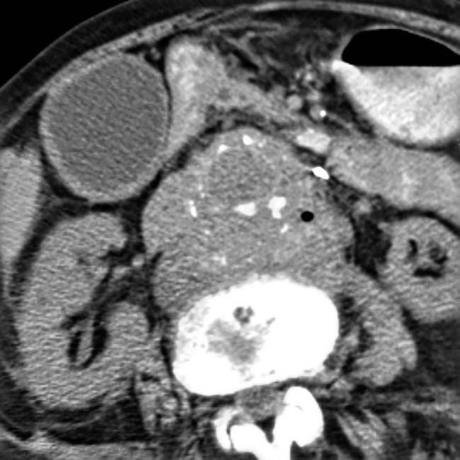
 of higher density between the aortic intimal calcification and patent lumen.
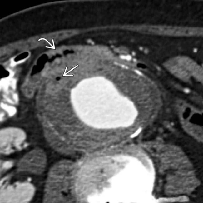
 within the thrombus, suggesting infection &/or communication to the bowel lumen. The 3rd portion of the duodenum stretches over and appears to adhere to the AAA
  .
.
 .
.
 .
.