Antiviral Therapy, Susceptibility Testing, and Prevention

Published on 08/02/2015 by admin

Filed under Basic Science

Last modified 22/04/2025

Print this page

rate 1 star rate 2 star rate 3 star rate 4 star rate 5 star
Your rating: none, Average: 4 (1 votes)

This article have been viewed 6554 times

Antiviral Therapy, Susceptibility Testing, and Prevention

Antiviral Therapy

Antiviral therapy has expanded over the past several years as a treatment for a number of viral infections. Although most of the population is susceptible to such treatments, overuse of these agents has led to the emergence of drug-resistant strains, especially in immunocompromised patients. Resistance is known to develop to all agents and may be detected in vitro by using antiviral susceptibility testing. Virology laboratories are increasingly being asked to perform in vitro testing of antiviral agents when a patient’s infection fails to respond clinically to antiviral therapy, but testing for antiviral resistance is not currently available in many clinical settings. This chapter provides an overview of the viral diseases in which antiviral resistance has emerged, the need for in vitro susceptibility testing, and the phenotypic and genotypic susceptibility testing methods currently available.

Antiviral Resistance

Antiviral resistance means that a virus has changed in such a way that the antiviral drug is less effective in preventing illness. Antiviral resistance is indicated if a patient is taking an antiviral drug that has been proven in vitro to be effective against a virus, but the patient shows no improvement and continues to deteriorate clinically. Drug resistance must be distinguished from clinical resistance. With clinical resistance, the viral infection fails to respond to the antiviral therapy because of factors other than a change in the virus; such factors may include the patient’s immunologic status, the pharmacokinetics of the antiviral drug in the individual patient, and, if a combination of drugs is administrated, potential antagonism and interference with the absorption of one or more drugs. Other patient factors that also affect the success of drug therapy include nonadherence to or intolerance of a specific drug and prescriptive errors, such as inappropriate doses or route of administration. In addition, infections in immunocompromised patients may fail to respond to therapy that has proven effective in immunocompetent individuals.

Very few standards have been established for antiviral susceptibility testing. The development of such protocols began in 2004 with the establishment of an approved standard for susceptibility testing for herpes simplex virus (HSV) by the Clinical and Laboratory Standards Institute (CLSI).

The final result of antiviral susceptibility testing is determined by many variables, and these variables also hinder the standardization of antiviral susceptibility testing. Some of these variables include the following:

Each of these categories in turn has variables that affect the final results. For example, if the inoculum quantity is too large, a susceptible isolate may appear resistant; if the inoculum quantity is too small, the isolate may appear susceptible. The complexity of all these variables makes it imperative that established control strains also be tested when antiviral susceptibility testing is performed. Controls should include both drug-resistant and drug-susceptible isolates that have been well characterized. Several research laboratories across the nation can provide reference and drug-resistant strains of a virus for susceptibility testing; they include the National Institute of Allergy and Infectious Diseases AIDS Research and Reference Reagent Program (niaid.nih.gov) and the American Type Culture Collection (atcc.org). Pharmaceutical companies also are a source.

Methods of Antiviral Susceptibility Testing

The purpose of antiviral susceptibility testing is to evaluate new antiviral chemoprophylaxis, to test for cross resistance or cross reactivity to alternate agents, and to determine how frequently drug-resistance viral mutations occur.

The two general types of antiviral susceptibility testing are phenotypic testing and genotypic testing. Phenotypic susceptibility assays measure viral replication in the presence of antiviral agents; they measure the inhibitory effect of an antiviral agent on the entire virus population in a clinical isolate. Genotypic susceptibility assays use polymerase chain reaction (PCR) to detect genes known to be responsible for resistance, coupled with molecular sequencing to determine whether genome alterations associated with resistance have occurred. These assays use the virus’s nucleic acid to determine whether the virus has mutations capable of causing viral drug resistance. Alternately, a combination of the two general types of antiviral susceptibility, known as a virtual phenotype resistance assay, may be performed. This assay is a characterization of the patient’s virus genotype compared to a data base that includes paired genotypic and phenotypic information. This information is then used to estimate the most likely phenotype of the patient’s virus. The success of this approach varies with the virus type and antiviral drugs examined.

Each of these types of susceptibility testing has unique properties that can be used to complement each other. Phenotypic assays are better used to assess the combined effect of multiple-resistance mutations on drug susceptibility, but they are labor intensive, expensive, and have lengthy end result times. Genotypic assays have a shorter turnaround time and are less expensive than phenotypic assays, but they can detect only defined viral mutations.

Phenotypic Assays

Phenotypic assays use a variety of end-point measurements to determine whether a virus is inhibited by an antiviral drug or demonstrates drug resistance. Some of these end-point measurements include a reduction in the number of plaques, inhibition of viral DNA synthesis, a reduction in the yield of a viral structural protein, or a reduction of the enzymatic activity of a functional protein. As mentioned, an advantage of phenotypic assays is that they are much better for assessing the combined effect of multiple-resistance mutations on drug susceptibility. This is useful for assaying viruses such as hepatitis B virus (HBV), human immunodeficiency virus type 1 (HIV-1), and human cytomegalovirus (HCMV), which acquire resistance mutations in multiple genes. The disadvantages of phenotypic assays are that they are labor intensive, expensive, and require weeks to perform.

Genotypic Susceptibility Assays

Genotypic susceptibility assays use PCR to detect genes known to be responsible for resistance, coupled with genetic sequencing to determine whether genome alterations associated with resistance have occurred. Genotypic assays use DNA sequencing by automated sequencers, PCR amplification and restriction enzyme digestion of the products, and hybridization to microarrays containing multiple oligonucleotide probes. These assays are rapid, because isolation of the virus in culture is not necessary for testing.

The response to an antiviral agent is also measured by quantitative monitoring of the viral load (by means of the nucleic acid concentration) in the patient’s blood. Such testing is common in patients infected with HBV, hepatitis C virus (HCV), and cytomegalovirus (CMV). The viral load should diminish significantly after addition of an antiviral agent to which the virus is susceptible. Using molecular testing (e.g., quantitative PCR) to measure the amount of virus in serum is a surrogate test for resistance to antiviral agents. The viral load rises quickly when resistance appears.

Pyrosequencing

DNA sequencing is among the most important testing methods for the study of biologic entities. Pyrosequencing, which is relatively new, is a sequence-based detection method that allows rapid, accurate quantification of sequence variation. It allows rapid acquisition of short reads (100 to 200 bp) of genomic sequence to identify known mutations. It is based on the technology of detection of released pyrophosphate (PPi) during DNA synthesis. In a sequence of enzymatic reactions, a enzyme (polymerase) catalyzes the addition of nucleotides into a nucleic acid chain. As a result of this addition, a PPi molecule is released and converted to adenosine triphosphate (ATP) by the ATP enzyme sulfurylase. Visible light is produced when a luciferin molecule is oxidized during the luciferase reaction. The visible light or signal strength generated is proportional to the number of nucleotides incorporated into the final product.

The two types of pyrosequencing methods currently available are solid-phase pyrosequencing and liquid-phase pyrosequencing. Solid-phase pyrosequencing involves a three-enzyme system that uses immobilized DNA, and a washing step is performed to remove excess substrate after each nucleotide addition. In liquid-phase pyrosequencing, a fourth nucleotide-degrading enzyme (made from potato) is added. The advantage of the liquid-phase system is that it eliminates the need for solid support and the intermediate washing step, allowing the reaction to be performed in a single tube.

Because of its rapid, accurate quantification of sequence variation, pyrosequencing is an adaptable tool that can be used for a wide range of applications. Automation with pyrosequencing is made possible by the liquid-phase methodology. Pyrosequencing signals are quantitative, which allows a large number of people to be screened through examination of the allelic frequency in a population. This technique also is used taxonomically to group different organisms into strains or subtypes, and it can be applied to bacteria, yeasts, and viruses. It is currently the fastest method for sequencing a PCR product and can be applied to the resequencing of PCR-amplified disease genes for mutation screening. It also is used to screen clinical isolates for the genes that confer resistance to antiviral therapy, such as for analysis of influenza specimens for the adamantine resistance mutation.

Human Immunodeficiency Virus

Patients infected with HIV frequently develop resistance to the antiretroviral drugs, which often results in treatment failure. Testing for antiretroviral resistance is crucial to the assessment of a regimen of drugs intended to suppress HIV replication and to testing for cross resistance to alternative antiretroviral drugs. The U.S. Department of Health and Human Services and a European panel of experts have developed guidelines and established protocols to monitor patients with acute and chronic HIV infection. Susceptibility testing should proceed as follows:

The recombinant virus assay (RVA) is a phenotypic type of susceptibility test for HIV. It is used to test the reaction of HIV-1 isolates to nucleoside analog reverse transcription (RT) inhibitors. RVA uses reverse transcription PCR (RT-PCR) amplification of the RT and pathogenesis-related (PR) gene coding sequences directly from the patient’s plasma. An advantage of this assay is that in a single test, a virus’s ability to replicate in the presence of various levels of an antiretroviral drug is measured by the detection of luciferase activity in the target cells. Two types of commercial kits are available for this method of testing.

Genotypic susceptibility testing has become a routine component of the management of patients infected with HIV. Genotypic assays for mutations that confer resistance are useful because of their rapid turnaround time. Several genotypic methods and commercial assays are available to test for these mutations in HIV; they include sequencing, selective PCR, oligonucleotide-specific hybridization, microarray hybridization, and reverse hybridization.

Influenza

Currently, two main approaches are used in health care to control the spread of influenza: vaccination and the use of antiviral drugs. The influenza virus has the unique capability of being able to change its antigenic makeup; this mechanism, known as antigenic drift, occurs with all three types of influenza virus (A, B, and C). Influenza A shows the greatest rate of antigenic change. Antigenic drift is caused by sequential point mutations in the hemagglutination (HA) or NA genes that arise during viral ribonucleoprotein (RNP) replication and immune selection, giving rise to new strains; this gives the virus the ability to reinfect “nonimmune” susceptible hosts each season. Another phenomenon, antigenic shift, is manifested only by the influenza A virus. It involves complete reassortment of the segmented viral genome during a co-infection with a nonhuman animal, which results in major antigenic change and periodic worldwide outbreaks (pandemics) of a never before circulated type of influenza A virus. Influenza B undergoes antigenic change very slowly.

Antigenic drift requires the reformulation of the influenza vaccine each year to ensure maximum efficacy against the currently circulating strains of influenza A and influenza B, because the vaccine is only as efficient as the influenza strains selected for it. This is accomplished by global surveillance of the yearly influenza epidemics to evaluate the strains that are circulating and provide early detection of viruses that may have pandemic potential. The World Health Organization (WHO) coordinates a influenza surveillance program in more than 80 countries. In the United States, the surveillance program established by the Centers for Disease Control and Prevention (CDC) includes monitoring of pneumonia and influenza deaths above a calculated “epidemic threshold.” It also includes tallying pediatric deaths, assessment of weekly virology data, and typing of influenza virus isolates submitted by reference laboratories. This extensive surveillance system provides the data for determining and predicting the influenza strains likely to be circulating in the upcoming winter, and vaccine components are chosen annually by WHO based on the analysis of these strains. The summer months are used to manufacture the vaccine so that it is ready for early autumn distribution to health care providers. In the United States, the vaccine is prepared from viruses grown in embryonated chicken eggs; this is a trivalent vaccine containing two influenza A strains with the newest HA and NA surface antigens and a current type B strain.

Currently two types of vaccine are used to prevent influenza infection: the trivalent inactivated influenza vaccine (TIV) and the live attenuated influenza virus vaccine (LAIV). The TIV is a noninfectious vaccine administered intramuscularly. It currently is approved in the United States for individuals 6 months or older, including those with chronic medical conditions. It is 70% to 100% effective in preventing infection among healthy adults and 30% to 60% effective in the elderly and pediatric populations. The LAIV contains live whole infectious virus. It is administered intranasally and currently is approved in the United States for healthy individuals 2 to 49 years of age. Because it contains live virus, it is not recommended for immunocompromised individuals, the elderly, or people with reactive airway disease. The LAIV causes shedding of the virus that is detectable in rapid antigen assays for about a week.

Two classes of antiviral drugs, the adamantanes and the neuraminidase inhibitors, currently are used to treat influenza infections. The adamantanes, which include the drugs amantidine and rimantadine, were the first antiinfluenza class of antiviral treatment developed. Their mechanism of viral defense is blockage of the virion M2 ion channel, which prevents the virus from uncoating. This class of drugs is effective only at treating influenza A infections; it has never had any effect on influenza B infections. The neuraminidase inhibitors include the drugs zanamivir (Relenza) and oseltamivir (Tamiflu). Both of these drugs inhibit the viral protein neuraminidase, which prevents release of the virus from infected cells. The neuraminidase inhibitors are used to treat both influenza A and influenza B infections, although oseltamivir has been reported to have lower efficacy against influenza B. Both classes of drugs have proven to be most effective when administrated within 48 hours of symptoms. The drugs shorten the duration of the infection and reduce complications.

The need for effective influenza antiviral susceptibility surveillance has increased around the world, and its importance is validated by the emergence of universal resistance to the adamantine antiviral therapy for influenza A (H3N2). Samples of viruses collected from around the United States and worldwide are studied to determine whether they are resistant to any of the four influenza antiviral drugs approved by the U.S. Food and Drug Administration (FDA). The CDC, in collaboration with state public health departments and WHO, conducts ongoing surveillance and performs testing of influenza viruses to monitor for antiviral resistance. The number of surveillance sites, both domestically and globally, are being increased, and the data from this surveillance are used to make public health policy recommendations on the use of these antiviral medications. The CDC is constantly improving its methods of rapidly detecting and monitoring antiviral resistance. Laboratory methods for testing also are being improved, and the number of laboratories capable of testing for antiviral resistance is rising.

Antiviral resistance to the adamantanes among circulating influenza A (H3N2) viruses rapidly increased worldwide beginning in the 2003-2004 influenza season. Data from the CDC’s World Health Organization (WHO) Collaborating Center for Surveillance, Epidemiology and Control of Influenza reports that the percentage of influenza A (H3N2) virus isolates submitted from around the world that were adamantine resistant increased from 0.4% in the 1994-1995 season to 12.3% in the 2003-2004 season. This resistance continued to increase; during the 2005-2006 influenza season, the CDC reported that of 209 isolates, 193 (92%) of the influenza A (H3N2) isolates carried a change at amino acid 31 in the M2 gene that confers resistance to the adamantanes. At the end of the 2008-2009 influenza season, 100% of influenza A H3N2, along with novel 2009 influenza A H1N1, were resistant to the adamantanes.

Resistance to oseltamivir appeared in the seasonal influenza A/H1N1 virus subtype during the 2007-2008 season. Oseltamivir resistance can result from a number of mutations in the neuraminidase gene, and for the 2007-2008 season, the CDC reported a nationwide resistance of 10.9% of the isolates submitted. This resistance also continued to increase; at the end of the 2008-2009 influenza season, the CDC reported that of 825 isolates of seasonal influenza A H1N1, 820 (99.4%) were resistant to oseltamivir. None of the other strains of influenza (i.e., influenza H3N2, novel 2009 influenza A H1N1, and influenza B) showed any resistance to the neuraminidase inhibitors (neither oseltamivir nor zanamivir).

For the 2010 influenza season, resistance to the adamantanes remained high; both circulating influenza A viruses (H3N2 and 2009 H1N1) showed high levels of resistance to the these drugs. These viruses are still susceptible to the neuraminidase inhibitors, and this class of antiviral medication is the current therapy of choice for antiviral treatment and for chemoprophylaxis of current circulating influenza A virus strains (Table 67-1).

TABLE 67-1

Antiviral Agents

Virus Mode of Action Target Examples of Common Drugs
CMV Nucleoside analog Viral DNA Ganciclovir
HIV* Nucleoside analog
Nucleotide analog
Nonnucleoside analog
Protease inhibitor
Fusion inhibitor
Viral DNA
Viral DNA
Reverse transcriptase
Viral protease
Virus, host cell membrane
Efavirenz
Tenofovir disoproxil fumarate
Emtricitabine
HSV/VZV Nucleoside analog
Pyrophosphate analog
Viral DNA
DNA polymerase
Acyclovir
Foscarnet
Hepatitis B Nucleoside analog
Nucleotide analog
Reverse transcriptase
DNA polymerase
Lamivudine
Adefovir dipivoxil
Influenza A Inhibit penetration and uncoating of virus Host cell membrane Amantadine, imantadine
Influenza A and B Prevent release of virus Neuraminidase inhibitors Zanamivir, oseltamivir
RSV Inhibit expression of viral mRNA and protein synthesis Viral mRNA Ribavirin
HCV Inhibit expression of viral mRNA; increase resistance to virus Viral mRNA or neighboring host cells Ribavirin plus interferon-alpha
Picornaviruses (enteroviruses and rhinoviruses) Inhibit attachment and uncoating of virus Binds to virus Pleconaril

image

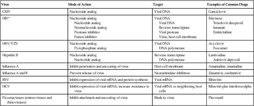
CMV, Cytomegalovirus; HIV, human immunodeficiency virus; HSV, herpes simplex virus; VZV, varicella-zoster virus; RSV, respiratory syncytial virus; HCV, hepatitis C virus.

*More than 20 antiretroviral drugs in six different mechanistic classes are available to design treatment regimens. See the most recent guidelines at http://aidsinfo.nih.gov/guidelines

At the end of the 2009-2010 season, almost all (98.9%) of the 2009 H1N1 isolates characterized at the CDC were susceptible to oseltamivir (Tamiflu), and all (100%) were susceptible to oseltamivir (Relenza). The rare 2009 H1N1 oseltamivir-resistant influenza A viruses shared a single genetic mutation, causing them to be resistant to this antiviral medication. Many of the influenza A H5N1 strains (avian influenza) are resistant to the adamantanes, so oseltamivir is the current antiviral of choice. Early treatment with oseltamivir improves the chance of survival in individuals infected with this type of influenza virus, but the mortality rate for the disease remains high.

Prevention of Other Viral Infections

Vaccination

Control of many viral diseases has been accomplished by vaccination. Since Jenner developed the first vaccine against smallpox 200 years ago, attenuated-live or inactivated-dead viral vaccines have been used successively to prevent yellow fever, poliomyelitis, measles, mumps, rubella, hepatitis B, and influenza (Table 67-2). Smallpox was eliminated in 1977 by an effective vaccination program. Additional vaccines continue to appear. New smallpox vaccines with fewer side effects are being developed to prevent outbreaks in the event of bioterrorism. A live-attenuated varicella (chickenpox) vaccine is now recommended for all children, and an inactivated hepatitis A vaccine is available for travelers and others entering areas of higher endemicity. Rotavirus vaccines are approved by the FDA and are now available. Recombinant vaccines are also available for the prevention of HPV infection.

TABLE 67-2

Examples of Vaccines for Preventing Viral Diseases

Disease Type of Vaccine
Yellow fever Attenuated-live
Poliomyelitis Attenuated-live and inactivated
Measles Attenuated-live
Mumps Attenuated-live
Rubella Attenuated-live
Hepatitis B Inactivated
Influenza Inactivated
Smallpox Attenuated-live
Chickenpox Attenuated-live
Hepatitis A Inactivated
Rabies Inactivated
Rotavirus Attenuated-live

Immune Prophylaxis and Therapy

Immune prophylaxis is used to prevent serious viral infection in patients who are immunocompromised or functionally compromised. Instead of actively immunizing an individual with an antiviral vaccine, limited protection can be conferred by intramuscular inoculation of human immunoglobulin. Pooled human immunoglobulin contains antibody against all common viruses. Specific high-titered immunoglobulin can be collected from patients recovering from a specific infection to ensure maximum antibody levels. Immune prophylaxis should be considered an emergency procedure. Table 67-3 lists immune prophylaxis available for viral infections.

TABLE 67-3

Immune Prophylaxis or Therapy for Viral Diseases

Disease Circumstances of Use
Prophylaxis  
Hepatitis A Traveler to developing country
Hepatitis B Newborns of infected mothers or unimmunized laboratory worker following needlestick
Rabies After bite from potentially rabid animal
Measles Unimmunized close contact with infected individual
Varicella Newborns of infected mothers at time of delivery
Respiratory Infants younger than 2 years of age with underlying lung syncytial disease virus
Therapy  
Lassa fever During disease to reduce severity

Passive immunoprophylaxis of respiratory syncytial virus (RSV) infection in infants younger than 2 years who have underlying lung disease resulting from premature birth or congenital heart disease is particularly effective at preventing life-threatening bronchiolitis and pneumonia in this patient group. The drug, palivizumab (Synagis), is a manufactured antibody to RSV. It is used in certain infants and young children to prevent RSV infections of the breathing tubes and lungs; it cannot be used to treat a child already sick with RSV.

Passive immunization occasionally is effective as therapy for viral infection (see Table 67-3). Therapy with immune serum for some hemorrhagic fevers, such as Lassa fever, has also been successful in reducing mortality associated with the disease.

Eradication

Global eradication of a viral disease has occurred only with smallpox. Factors that result in eradication of any viral disease include no animal reservoir, a lack of recurrent infectivity, one or few stable serotypes, and an effective vaccine. Viral diseases currently considered candidates for eradication include measles and poliomyelitis. Poliomyelitis has been known and feared by humans for thousands of years. Infection with the poliovirus causes an acute flaccid paralysis that can affect the ability to breathe. Years ago, it was often seen in children. In the mid-1950s, Jonas Salk developed the first polio vaccine from dead virus, and in 1960, Sabin developed an oral polio vaccine using a live-attenuated virus. These developments allowed the United States to launch a massive vaccination program against polio, and the last case of indigenous polio was reported in the United States in 1979 (other reports of polio cases were due to vaccination or occurred in individuals who had emigrated from other countries).

In 1988 WHO resolved to eradicate acute paralytic poliomyelitis from the rest of the world and staged a massive vaccination campaign to accomplish this. At the time, poliomyelitis was endemic in 125 countries on five continents and was responsible for an estimated 350,000 cases annually. The success of this program reduced the number of polio-endemic countries to six by 2003, and by 2006 the disease remained endemic in only four countries: Afghanistan, India, Nigeria, and Pakistan. The strategies used to eradicate the disease included surveillance of acute flaccid paralysis, routine vaccination with the oral polio vaccine, and supplementary immunization activities.