ANTIBACTERIAL THERAPY: THE OLD, THE NEW, AND THE FUTURE

Published on 10/03/2015 by admin

Filed under Critical Care Medicine

Last modified 22/04/2025

Print this page

rate 1 star rate 2 star rate 3 star rate 4 star rate 5 star
Your rating: none, Average: 0 (0 votes)

This article have been viewed 1052 times

CHAPTER 97 ANTIBACTERIAL THERAPY: THE OLD, THE NEW, AND THE FUTURE

Infections remain the leading cause of death in hospitalized patients, and antimicrobial therapy is a mainstay of treatment. However, widespread overuse and misuse of antibiotics have led to an alarming increase in multiple-drug-resistant (MDR) pathogens. New agents may allow shorter courses of therapy and prophylaxis, which are desirable for cost control and control of microbial flora. Moreover, antibiotics are second only to analgesic agents in the number of adverse drug reactions.

PRINCIPLES OF PHARMACOKINETICS

The goal of pharmacotherapy is an effective response with no toxicity. The prescriber must have knowledge of the principles of drug absorption, distribution, and elimination. The dose-response relationship is influenced by dose, dosing interval, and route of administration. The plasma drug concentration is influenced by absorption, distribution, and elimination—which in turn depend on drug metabolism and excretion. The plasma concentration may not reflect tissue concentrations, as penetration into individual tissues is variable. Finally, the relationship between local drug concentration and effect is defined by several pharmacodynamic (PD) principles (see following discussion).

A few basic concepts of pharmacokinetics (PK) are useful to the practitioner. Bioavailability is defined as the percentage of an administered dose of a drug that reaches the systemic circulation. By definition, bioavailability is 100% after intravenous administration. However, this varies among drugs after oral administration, being affected by absorption (a function of product formulation and gastric emptying time), intestinal transit time, and the degree of hepatic first-pass metabolism.

Half-life refers to the amount of time required for the drug concentration to reduce by half, and thus is a hybrid of consider ations of both clearance and volume of distribution. Half-life is useful to estimate when a steady-state drug concentration will be achieved. If a “loading dose” is not administered intravenously, thereby creating instantaneously a desired drug concentration to be maintained throughout therapy, four to five half-lives must elapse to achieve a steady state. Changes in dosage and changes in half-life owing to disease state (e.g., renal failure) must be accounted for. Interpretation of drug concentration data is difficult if the patient is not at a steady state, especially so in critical illness characterized by fluctuating organ function and volume of distribution.

Volume of distribution (VD) is a proportionality constant that relates to plasma concentration and the amount of drug in the body. VD is useful for estimating achievable plasma drug concentrations that result from a given dose. It is a derived parameter that is independent of a drug’s clearance or half-life. It does not have particular physiologic significance, but pathophysiologic conditions can alter VD substantially. A reduction of VD will result in a higher plasma drug concentration for a given dose. However, the “third space” extravascular volume redistribution, fluid overload, and hypoalbuminemia (with decreased drug binding) of surgical illness act to increase VD, all of which makes dosing a complex matter.

Clearance refers to the volume of liquid from which drug is eliminated completely per unit of time (whether by distribution to tissues, metabolism, or elimination) and is important for determining the amount of drug necessary to maintain a steady-state concentration. Drug elimination may be by metabolism, excretion, or dialysis. Most drugs are metabolized by the liver to polar compounds that can then be excreted by the kidney, but metabolism does not imply inactivation. For example, metronidazole is metabolized to a bactericidal metabolite with a prolonged half-life that has dosing implications. The kidneys are most important for excretion of metabolized drugs, although some drugs are metabolized or conjugated by the kidneys. Renal excretion may occur by filtration or by active or passive transport. The degree of filtration is determined by molecular size and charge and by the number of functional nephrons. In general, if greater than 40% of administered drug or its active metabolites is eliminated unchanged in the urine, decreased renal function will require a dosage adjustment. Active reabsorption and concentration of aminoglycosides by proximal tubular cells is a likely component of its well-recognized nephrotoxicity.

PRINCIPLES OF PHARMACODYNAMICS

The variable responses to drugs administered to a heterogeneous patient population can be described and perhaps reduced by an understanding of PD, the relationship of a drug to its intended effect. The PD of antibiotic therapy is especially complex because drugpatient, drug-microbe, and microbe-patient interactions must be accounted for. Knowledge of how patient characteristics influence absorption, distribution, and elimination of a drug—and how an antibiotic interacts with the targeted microbe—can increase the likelihood of a salutary clinical response. In turn, antimicrobial effects on bacteria are highly variable. Microbial physiology, inoculum size, microbial growth phase, intrinsic and extrinsic mechanisms of resistance, microenvironmental factors such as the pH at a local site of infection, and the patient’s immune response are important factors. In the case of antimicrobial therapy, the key drug interaction is not with the host but with the microbe.

Because of microbial ability to alter the nature of the interaction with antimicrobial agents (principally via the development of resistance), mere delivery of drug may not be microbicidal. Factors that may contribute to the development of resistance are the production of drug-inactivating enzymes, alteration of cell surface receptor target molecules, and altered bacterial permeability to antimicrobial penetration. Critical to the microbe-patient interaction is the patient’s immune system. Also inseparable are drug-patient factors that may influence PK, such as hepatic and renal function, serum albumin concentration, and extracellular volume status.

Antibiotic PD is determined by laboratory analysis, and thus the extrapolation of in vitro results to the patient may be challenging because the interaction with the host immune system is isolated from the analysis of the drug-microbe interaction. Analyses from in vitro study include the minimal inhibitory concentration (MIC). The MIC is the minimal serum drug concentration necessary for inhibition of bacterial growth, expressed as the proportion of the inoculum inhibited (MIC90 refers to 90% inhibition). However, some antibiotics may have important effects on bacteria at subinhibitory concentrations. Moreover, MIC testing may not detect the presence of resistant bacterial subpopulations (a particular problem with “heteroresistance” of Gram-positive bacteria, particularly Staphylococcus aureus).

Sophisticated analytic strategies draw upon the principles of both PK and PD; for example, by determination of the peak serum concentration:MIC ratio, the duration of time plasma concentration remains above the MIC, and the area of the plasma concentration-time curve above the MIC (the “area under the curve,” or AUC). With some agents, antibacterial effects may persist for prolonged periods after the plasma drug concentration has become “subtherapeutic.” The persistent inhibition of bacterial growth (but not killing) that persists after the serum drug concentration has fallen below the MIC for the organism is known as the postantibiotic effect (PAE). Appreciable PAE can be observed with amino-glycosides and fluoroquinolones for Gram-negative bacteria, and with some β-lactam drugs (notably carbapenems) against S. aureus. Through analyses of this type, certain drugs (e.g., aminoglycosides) have been characterized as having concentration-dependent killing whereby a higher peak concentration increases the efficacy of bacterial killing (up to a point). Other agents (most β-lactam agents) exhibit bactericidal properties that are independent of concentration. Rather, efficacy is determined by the duration of time the plasma concentration remains above the MIC. Other agents (e.g., fluoroquinolones) exhibit both properties such that bacterial killing may increase as drug concentration increases up to a point of saturation, after which the effect becomes independent of concentration.

EMPIRIC ANTIBIOTIC THERAPY

The decision to administer empiric antibiotic therapy must be considered carefully. An injudicious approach could result in nontreatment of established infection or therapy when the patient has only sterile inflammation or colonization with bacteria. Inappropriate therapy (e.g., delay, therapy misdirected against usual pathogens, failure to treat MDR pathogens) leads unequivocally to increased mortality. Several questions should be asked in each circumstance where empiric therapy is being considered.

Are antibiotics indicated at all? The answer is ultimately often no, but the decision to start treatment of the unstable patient must often be made before definitive information becomes available. The decision to start antibiotics empirically is based on the likelihood of infection, its likely source, and whether the patient’s condition is sufficiently precarious that a delay will be detrimental. Outcome from serious infections is improved if antibiotics are started promptly, but on the other hand only about 50% of fever episodes in hospitalized patients are caused by infection. Many causes of the systemic inflammatory response syndrome are not due to infection (e.g., aspiration pneumonitis, burns, trauma, pancreatitis), although they may be complicated later by infection. Multiple organ dysfunction syndrome may progress even after an infectious precipitant has been controlled, due to a dysregulated host response.

Must antibiotics be started immediately? If the presumed infection is not destabilizing, this decision also depends on the overall status of the patient and should take into consideration such host factors as age, debility, renal and hepatic function, and immunosuppression. Culture yields are highest before antibiotics are administered, which for certain types of specimens (e.g., blood, cerebrospinal fluid) can be crucial. However, for many infections (e.g., bacteremia, intraabdominal infection, pneumonia) early appropriate therapy improves outcome.

Which organisms are the likely pathogens, and are they likely to be MDR? The clinical setting must be considered (e.g., nosocomial versus community-acquired infection, recent antimicrobial therapy), as must the patient’s environment (e.g., recent hospitalization, proximity to another infected patient, the presence of MDR pathogens in the unit) and any recent microbial cultures obtained from the patient.

Will a single antibiotic suffice? The likely diagnosis and the nature of the probable pathogens are crucial determinants. If a nosocomial Gram-positive pathogen is suspected (e.g., wound or surgical site infection, catheter-related infection, prosthetic device infection, pneumonia) and methicillin-resistant S. aureus (MRSA) is endemic, empiric vancomycin (or linezolid) is appropriate. Some authorities recommend dual-agent therapy for serious Pseudomonas infections (i.e., an antipseudomonal β-lactam drug plus an aminoglycoside). It is important to use at least two antibiotics for empiric therapy of any infection that may be caused by a Gram-positive or Gram-negative infection (e.g., nosocomial pneumonia).

Duration of Therapy

Perhaps the most difficult issue is identifying the endpoint. If bona fide evidence of infection is evident, treatment is continued as indicated clinically. Often, however, the cultures will return negative and the decision must be arbitrary. The decision is complicated further when the patient has had a clinical response to antibiotic therapy in the absence of corroborating evidence, which may be coincident with or a result of false-negative cultures. Moreover, the bias to do something to treat the patient (i.e., continue antibiotic therapy) can be compelling in a patient who is deteriorating.

It must be recognized that careful culture techniques and specimen handling, combined with current sophisticated microbiology laboratory support, make it unlikely that substantive pathogens will be missed. Therefore, continuing empiric antibiotic therapy beyond 48 hours becomes difficult to justify. There are two possible exceptions. One occurs when fungal infection is suspected because the organisms can be difficult to culture, and the other occurs when deep cultures are needed from areas that are inaccessible without radiologic-guided aspiration and some time is necessary to make appropriate arrangements (but is not an excuse for procrastination).

How long should a course of therapy be continued? Effective broad-spectrum antibiotics are widely available, and many infections can be treated with therapy lasting 5 days or fewer. It is important that every decision to start antibiotics must be accompanied by a decision regarding the duration of therapy. A reason to continue therapy beyond the predetermined endpoint must be compelling. Bacterial killing is rapid in response to effective agents, but the host response may not subside immediately. Therefore, the clinical response of the patient should not be the sole determinant for continuation of therapy. If a patient still has sepsis syndrome at the end of a defined course of therapy, it is more useful to stop therapy and obtain a new set of cultures to look for new sites of infection, resistant pathogens, and noninfectious causes of inflammation.

There is a clear trend toward shorter courses of antibiotics for established infections. Broad-spectrum antibiotics that achieve excellent tissue penetration have been an important clinical development, but they also carry morbidity. The worldwide emergence of MDR Gram-positive and Gram-negative bacteria, superinfections in immunosuppressed patients, and the increased mortality associated with nosocomial infections in general make it important that adequate therapy be provided rapidly and for the shortest possible duration. Unfortunately, duration of therapy is not well established in the literature—and new studies are seldom designed with duration of therapy as a primary endpoint. Much depends on expertise and clinical judgment, which is accumulating in favor of shorter courses of therapy. Nowhere is this clearer than for peritonitis and intra-abdominal abscess, for which the previous standard 7- to 10-day courses of therapy have been reduced to 5 days.

Infections that require 24 hours of therapy or less (sometimes just a single dose) include uncomplicated acute appendicitis or cholecystitis, uncomplicated bacterial cystitis (with some agents), and intestinal infarction without perforation. There is seldom justification to continue antibacterial therapy for more than 10 days. Examples of bacterial infections that require more than 14 days of therapy include tuberculosis of any site, endocarditis, osteomyelitis, and selected cases of brain abscess, liver abscess, lung abscess, some cases of postoperative meningitis, and some cases of endophthalmitis. Among the many reasons to limit therapy to only that which is needed is that antibiotic therapy has adverse consequences, despite a widespread perception that therapy is safe if not entirely benign. Adverse consequences of antibiotics include allergic reactions; development of nosocomial superinfections, including fungal infections, enterococcal infections, and Clostridium difficile–related disease; organ toxicity; promotion of antibiotic resistance; reduced yield from subsequent cultures; and induced vitamin K deficiency with coagulopathy or accentuation of warfarin effect.

CHOICE OF ANTIBIOTIC

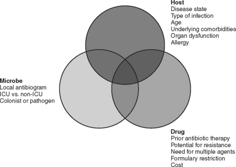
The choice of which antibiotic to prescribe is made based on several interrelated factors. Paramount is activity against identified pathogens, presuming that a distinction between infecting and colonizing organisms can be made and that narrow-spectrum coverage is always most desirable. Knowledge of antimicrobial resistance patterns, nationally and especially in one’s own institution and unit, is essential. Also important is an assumption regarding likely pathogens, which is paramount in cases where empiric therapy is necessary. Estimation of likely pathogens depends on the disease process believed responsible, whether the infection is community- or hospital-acquired, whether MDR organisms are present, and proximity to other infected patients. Also important are patient-specific factors, including age, debility, immunosuppression, intrinsic organ function, prior allergy or another adverse reaction, and recent antibiotic therapy. Institutional factors that may play a role include the existence of guidelines or practice parameters that may specify a particular therapy, or the availability of specific agents as defined by inclusion on the formulary or restriction by antibiotic control programs (Figure 1).

Development of Bacterial Resistance

In general, bacteria use four different mechanisms to develop resistance to antibiotics. Cell wall permeability to antibiotics is decreased by changes in porin channels (especially important for Gram-negative bacteria with complex cell walls, affecting aminoglycosides, β-lactam drugs, chloramphenicol, sulfonamides, tetracyclines, and possibly quinolones). Production of specific antibiotic-inactivating enzymes by plasmid-mediated or chromosomally mediated mechanisms affects aminoglycosides, β-lactam drugs, chloramphenicol, and macrolides. Alteration of the target for antibiotic binding in the cell wall affects β-lactam drugs and vancomycin, whereas alteration of target enzymes can inhibit β-lactam drugs, sulfonamides, quinolones, and rifampin. Drugs that bind to the bacterial ribosome (aminoglycosides, chloramphenicol, macrolides, lincosamides, streptogramins, and tetracyclines) are also susceptible to alteration of the receptor on the ribosome. Antibiotics may be extruded actively once entry to the cell is achieved in the case of macrolides, lincosamides, streptogramins, quinolones, oxazolidinones, and tetracyclines.

Cephalosporin resistance among Gram-negative bacilli can be the result of induction of chromosomal β-lactamases after exposure to the antibiotic. The extended-spectrum cephalosporins are rendered ineffective when bacteria such as enteric Gram-negative bacilli mutate to constitutively produce a β-lactamase that is normally an inducible enzyme. Although resistance to cephalosporins can occur by several mechanisms, the appearance of chromosomally mediated β-lactamases has been identified as a consequence of the use of third-generation cephalosporins. Resistance rates decline when use is restricted. The induction of an extended-spectrum β-lactamase (ESBL) in Klebsiella by ceftazidime was first reported approximately 20 years ago, but more than 200 mutations have now been described in several species of Gram-negative bacteria. The mutant bacteria develop resistance rapidly not only to all cephalosporins but to entire other classes of β-lactam antibiotics. It is therefore justifiable to restrict the use of ceftazidime, especially in institutions grappling with an ESBL-producing bacterium. The carbapenems generally retain useful microbicidal activity against ESBL-producing strains. Increasingly, Pseudomonas aeruginosa produces beta-lactamases of the ampC type.

Quinolone resistance, which is increasing rapidly, is for the most part chromosomally mediated, primarily by changes in the target sites for the antibiotic (DNA gyrase or topoisomerase IV). Changes in permeability or efflux may sometimes cause resistance to quinolones as well. Quinolone resistance is relatively easy to induce if a less-than-maximally effective drug is chosen for initial therapy. Resistance to one quinolone may also increase the MIC for the other quinolones against the organism, and thus if a quinolone is used, a highly active agent given in adequate dosage is essential.

ANTIBIOTIC SPECTRUM OF ACTIVITY

Susceptibility testing of specific organisms is necessary for management of serious infections (including all nosocomial infections). Recommended agents for specific organisms are guidelines only because in vitro susceptibilities may not correlate with clinical efficacy. The necessary concentration of antibiotics may not be achieved in tissue because of underdosing or poor tissue penetration. Moreover, bacterial killing correlates well with peak serum antibiotic concentrations for some drugs (e.g., aminoglycosides) and disorders (e.g., bacterial endocarditis) but correlates better with the duration of bactericidal drug concentrations for other antibiotics (e.g., β-lactam agents).

Penicillins

With the exception of carboxy- and ureidopenicillins, penicillins do not retain important activity against most strains of Gram-negative bacilli. Penicillin G (parenteral) and V (oral) are useful against most strains of aerobic and anaerobic streptococci (except for the increasingly important problem of penicillin-resistant pneumococci [PRSP, up to 40% of isolates] in bacteremia, recurrent otitis, and upper respiratory tract infections). Penicillins also have activity against Enterococcus faecalis (but not E. faecium), Corynebacterium diphtheriae, and Listeria monocytogenes. Gram-negative bacteria that are susceptible to penicillins include Neisseria meningitidis (highly resistant strains exist), some strains of Proteus mirabilis, and Pasturella multocida. In addition to anaerobic streptococci, penicillins are effective against other anaerobes, such as Bacteroides melaninogenicus (but not B. fragilis) and all clostridial species other than C. difficile.

The penicillinase-resistant semisynthetic penicillins include methicillin, nafcillin, oxacillin, cloxacillin, and dicloxacillin. Although these agents have useful activity against streptococci, C. diphtheriae, and anaerobic streptococci, the primary use of these agents is as therapy for sensitive strains of staphylococci. Hospitalized patients who need empiric therapy should not be treated with these agents because 60% of strains of S. aureus (MRSA), 90% of strains of S. epidermidis (MRSE), and virtually all enterococcal strains are resistant. However, these drugs are the treatment of choice for infections caused by susceptible isolates of S. aureus.

Activity against Gram-negative organisms was achieved initially by the addition of an amino group to the penicillin nucleus, thereby creating such drugs as ampicillin and amoxicillin. These drugs retain their antistreptococcal activity and a similar spectrum against most other Gram-positive pathogens, including anaerobic streptococci, but do not have appreciable activity against staphylococci. Ampicillin is highly effective against E. faecalis, including some vancomycinresistant strains (VRE), but only rarely effective against E. faecium. Useful activity remains against N. meningitidis, Moraxella catarrhalis, community-acquired strains of E. coli and Klebsiella spp., Salmonella and Shigella spp., and Proteus spp. Ampicillin remains reasonably effective against community-acquired strains of Hemophilus influenzae, but H. influenzae is increasingly important as a nosocomial pathogen and resistant strains are recognized.

The carboxypenicillins (ticarcillin and carbenicillin) and ureidopenicillins (azlocillin, mezlocillin, and piperacillin; sometimes referred to as acylampicillins) have enhanced activity against Gram-negative bacteria and some activity against P. aeruginosa. Ureidopenicillins have greater intrinsic activity against Pseudomonas, but with the advent of β-lactamase inhibitor combination drugs none of these agents is used widely anymore. Beta-lactamase inhibitors (sulbactam, tazobactam, and clavulanic acid) result in enzymatic inactivation and enhanced effectiveness of the antibacterial agent. The effectiveness of these drugs as antibacterial agents is primarily a function of the inherent antibacterial properties of the parent compound (ampicillin < ticarcillin < piperacillin), and to a lesser extent of the effectiveness of the inhibitor (sulbactam ∼ clavulanic acid < tazobactam). The spectrum of activity varies as a result, and the treating clinician needs to be familiar with each of the drugs in this class.

All of these drugs are effective against streptococci, methicillin-sensitive strains of S. aureus, Listeria monocytogenes, Salmonella, Proteus, and Providentia spp., P. multocida, and widely effective against anaerobes—including anaerobic cocci, B. fragilis, Bacteroides and Prevotella spp., and Clostridium spp. (except for C. difficile). Piperacillin/tazobactam has the widest spectrum of activity against Gram-negative bacteria, and the most potency against P. aeruginosa. Although ampicillin/sulbactam has excellent activity against community-acquired Gram-negative bacilli, it has major shortcomings against hospital-acquired strains of E. coli and Klebsiella (as many as 50% of strains may be resistant). However, sulbactam has useful activity against Acinetobacter spp., making ampicillin/sulbactam an option for therapy of infections caused by susceptible strains.

Cephalosporins

More than 20 antibiotics comprise this class of agents. The characteristics of the drugs thus vary widely when considered individually. It is useful to consider these drugs within four broad “generations” whose general characteristics are similar. For example, the firstgeneration agents retain useful activity against Gram-positive organisms—whereas the second-generation agents generally lose that activity in favor of antianaerobic activity. In contrast, the thirdgeneration agents generally have enhanced activity against Gram-negative bacilli—and some have specific antipseudomonal activity. However, most lack efficacy against Gram-positive organisms and none is effective against anaerobic bacteria.

Cefepime, the fourth-generation cephalosporin available in the United States, has enhanced antipseudomonal activity and has regained activity against most Gram-positive cocci but not MRSA. None of the cephalosporins, regardless of class, has clinically useful activity against any of the enterococci. Regardless, there is sufficient heterogeneity of spectrum (especially among the third-generation agents) such that the clinician should be familiar with all of these drugs. Collectively, they account for a majority of prescriptions for parenteral antibiotics. Ceftriaxone, a third-generation agent unique in its class for excellent activity against Gram-positive organisms and once-daily dosing, was at one time the most-prescribed injectable antibiotic worldwide.

Third-Generation Cephalosporins

Rightly or wrongly, third-generation cephalosporins dominate prescribing practices for parenteral antibiotics. These agents include cefoperazone, cefotaxime, cefpodoxime, cefprozil, ceftazidime, ceftibuten, ceftizoxime, ceftriaxone, and lorcarbicef. They are relatively resistant to β-lactamases, and therefore have an extended spectrum of activity against Gram-negative bacilli. Despite this, these agents lack efficacy against Gram-positive bacteria (except for ceftriaxone) and anaerobic bacteria. Activity is reliable against non-ESBL–producing species of Enterbacteriaceae, including Enterobacter, Citrobacter, Providencia, and Morganella. Activity is variable against Acinetobacter and the pseudomonads, with broad activity against Aeromonas, reasonable albeit variable activity against P. aeruginosa (cefoperazone and ceftazidime), but no activity against S. maltophilia. Ceftriaxone and ceftazidime have activity against Borrelia burgdorferi, the agent of Lyme disease.

Paradoxically, third-generation cephalosporins (particularly ceftazidime) have been associated with the induction of ESBLs among many of the Enterobacteriaceae. Production of ESBLs was first reported in strains of Klebsiella pneumoniae, but now is so well recognized that susceptible pathogens are now referred to commonly as “inducible enteric” bacteria. The resistance induced by ESBL production is not just against other third-generation cephalosporins but affects entire other classes of β-lactam antibiotics. Third-generation cephalosporins, especially ceftazidime, have also been implicated (in concert with the widespread overuse of vancomycin; see material following) in the emergence of VRE. Because resistance can be transferred between enterococci and staphylococci, staphylococci of intermediate susceptibility to glycopeptides (GISA) or resistant to vancomycin (VRSA) have now been reported. Because of the potential to induce resistance of hospital flora, many centers no longer use third-generation cephalosporins as empiric therapy but rather reserve them for directed narrow-spectrum monotherapy of known susceptible organisms.

Carbapenems

Carbapenems have a five-carbon ring attached to the β-lactam nucleus. The alkyl groups are oriented in a trans-configuration rather than the cis-configuration characteristic of other β-lactam agents, making these drugs resistant to β-lactamases. Four drugs (imipenem/cilastatin, meropenem, doripenem, and ertapenem) are available for clinical use in the United States, and other agents are in clinical trials. Imipenem/cilastatin does induce β-lactamase production, but because it is resistant itself to ESBLs the activity of the drug is undiminished and little cross-resistance develops. Cilastatin is irrelevant to the antibacterial activity of imipenem/cilastatin, but it inhibits renal dihydropeptidase I, thereby abrogating the profound nephrotoxicity of the parent compound.

Imipenem-cilastatin, meropenem, and doripenem have the widest antibacterial spectrum of any antibiotics, with excellent activity against aerobic and anaerobic streptococci, methicillin-sensitive staphylococci, and virtually all Gram-negative bacilli except Legionella, P. cepacia, and S. maltophilia. Activity against the Enterobacteriaceae exceeds that of all antibiotics, with the possible exceptions of piperacillin/tazobactam and cefepime—and activity of meropenem against P. aeruginosa is approached only by that of amikacin. All of the carbapenems are superlative antianaerobic agents, and thus there is no reason to combine a carbapenem with metronidazole except for example to treat concurrent C. difficile colitis in a patient with a life-threatening infection that mandates continuance of the carbapenem. Other differences in spectra between imipenem-cilastatin and meropenem are trivial except that imipenem is an effective drug against E. faecalis (but not E. faecium). Meropenem is ineffective against enterococci.

Meropenem and doripenem appear not to have the same potential for neurotoxicity that is recognized with imipenem-cilastatin, which is contraindicated in patients with active central nervous system disease or injury (excepting the spinal cord) because of the rare (∼0.5%) appearance of myoclonus or generalized seizures in patients who have received doses of more than 3 g/day (with normal renal function) or who have not had dosage reductions in the setting of renal insufficiency. With both drugs, the widespread disruption of the host microbial flora inherent in such broad-spectrum therapy may lead to superinfections (e.g., fungi, C. difficile, Stenotrophomonas, or resistant enterococci).

Ertapenem is not active against Pseudomonas spp., Acinetobacter spp., Enterobacter spp., or MRSA, but is a useful drug nonetheless by virtue of its long half-life and substantial PAE—permitting once-daily dosing. In addition, ertapenem is highly active against ESBL-producing Enterobacteriaceae and has less potential for neurotoxicity than imipenem-cilastatin.

Cell-Wall–Active Agents

Lipoglycopeptides

Vancomycin is a soluble lipoglycopeptide with a complex bactericidal mechanism of action. The drug inhibits synthesis and assembly of the second phase of cell wall peptidoglycan synthesis, and it may also injure protoplasts by altering the permeability of their cytoplasmic membrane. There is some evidence that RNA synthesis may be impaired as well. These multiple mechanisms, along with a lack of cross-resistance with other antibiotics, may explain the historic low resistance rate for Gram-positive bacteria. Vancomycin is rapidly bactericidal, but only on dividing organisms. A PAE persists for about 2 hours. Unfortunately, tissue penetration of vancomycin is poor for almost all tissues—which can limit its effectiveness.

Both S. aureus and S. epidermidis are susceptible to vancomycin, although MICs for S. aureus are increasing and may require higher doses for therapeutic effect. Streptococcus pyogenes, group B streptococci, S. pneumoniae (including penicillin-resistant strains), and C. difficile are also susceptible. Listeria monocytogenes, anaerobic cocci, other clostridial species, and Actinomyces are usually susceptible. Most strains of E. faecalis are inhibited (but not killed) by concentrations attainable in serum, but E. faecalis is increasingly resistant to vancomycin. Resistant enterococci have emerged because of prolonged or indiscriminate use of vancomycin (Table 1), occasioned by the ubiquity of MRSA/MRSE. Both GISA and strains of S. aureus fully resistant to vancomycin are recognized, but so far only in association with prolonged (i.e., weeks to months) exposure to vancomycin.

Table 1 Situations in Which Use of Vancomycin Is Discouraged

Vancomycin usage is often inappropriate, and it is important for the public health that inappropriate usage should be curtailed. Bona fide indications include serious infections caused by MRSA/MRSE, Gram-positive infections in patients with serious penicillin allergy, and oral therapy (or by enema in patients with ileus) for C. difficile-related colitis in patients who have failed or are intolerant to metronidazole. Parenteral vancomycin is now usually administered in a dose of 15 mg/kg actual body weight q12h. The infusion must be performed over the course of at least 1 hour. The dose must be reduced in renal failure, and monitoring of serum concentrations may be helpful in that circumstance. New high-flux hemodialysis membranes dialyze vancomycin partially, and a 500-mg dose should be given after each dialysis.

Dalbavancin is a second-generation lipoglycopeptide agent that has a mechanism of action similar to vancomycin, resulting in disruption of the bacterial cell wall. Advantages of dalbavancin over vancomycin include a long elimination half-life in human beings, which makes once-weekly dosing feasible. For example, a phase III randomized trial demonstrated that two doses of dalbavancin (1 g initially, followed by 500 mg 7 days later) in complicated skin infections can take the place of other antibiotics requiring up to 28 doses. An additional possible advantage is that dalbavancin is bactericidal, whereas vancomycin is bacteriostatic, against most Gram-positive cocci.

Cyclic Lipopeptides

Daptomycin is a cyclic lipopeptide antibiotic with potent bactericidal activity against most gram-positive organisms, including MDR strains. The unique structure of daptomycin consists of a 13-member amino acid cyclic lipopeptide with a decanoyl side chain. This distinctive structure confers a novel mechanism of action, believed to involve insertion of the lipophilic daptomycin tail into the bacterial cell membrane—causing rapid membrane depolarization and a potassium ion efflux. This is followed by arrest of DNA, RNA, and protein synthesis, resulting in bacterial cell death. The bactericidal effect of daptomycin is rapid, with greater than 99.9% of both MRSA and MSSA bacteria dead in less than 1 hour without appreciable bacterial cell lysis.

Daptomycin is effective in a concentration-dependent manner, has a long half-life (8 hours), and demonstrates a prolonged PAE (up to 6.8 hours). Once-daily dosing of daptomycin results in linear PK with minimal drug accumulation. A dosing regimen of 4 mg/kg once daily is recommended for complicated skin/skin structure infections (cSSSI). Daptomycin is excreted renally. Therefore, the dosing interval should be increased to every 48 hours in patients with a creatinine clearance of less than 30 ml/min. Because of daptomycin’s unique mechanism of action and because it is not metabolized by cytochrome p450 or other hepatic enzymes, no antagonistic drug interactions have been observed.

In vitro potency of daptomycin has been demonstrated against many aerobic and anaerobic Gram-positive bacteria, including MDR strains. Daptomycin’s spectrum of activity encompasses difficult-to-treat antibiotic-resistant Gram-positive cocci, including MRSA and VRE. Daptomycin demonstrates activity against vancomycin-resistant S. aureus, as well as against linezolid- and quinupristin/dalfopristin-resistant S. aureus and E. faecium. Furthermore, daptomycin is also effective against a variety of streptococci—including S. pyogenes (group A) and S. agalactiae (group B) as well as other Streptococcus spp.—and against a variety of anaerobic species, including Peptostreptococcus spp., C. perfringens, and C. difficile. Daptomycin’s efficacy is enhanced by the near absence thus far of antibiotic resistance, as verified by both in vitro and clinical studies. No transferable elements conferring daptomycin resistance have been isolated to date.

Daptomycin has been approved in the United States for the treatment of cSSSI associated with S. aureus (both MSSA and MRSA), S. pyogenes, S. agalactiae, S. dysgalactiae subsp. equisimilis, and E. faecalis (vancomycin-susceptible only) and for bacteremia caused by susceptible pathogens. Importantly, daptomycin must not be used for the treatment of pneumonia or empiric therapy when pneumonia is in the differential diagnosis (even when caused by a susceptible organism) because daptomycin penetrates lung tissue poorly and is inactivated by pulmonary surfactant.

PROTEIN SYNTHESIS INHIBITORS

Several classes of antibiotics, although dissimilar structurally and having widely divergent spectra of activity, exert their antibacterial effects via the similar mechanism of binding to bacterial ribosomes to inhibit protein synthesis. This classification is valuable mechanistically and serves to link several classes of antibiotics conceptually that have few clinically useful members.

Aminoglycosides

With a reputation as toxic agents that have been superceded by newer antibiotics, it is ironic that the resurgence of aminoglycoside use has occurred as resistance to these newer antibiotics (especially thirdgeneration cephalosporins and quinolones) has developed. Aminoglycosides exert their microbicidal activity by binding to the bacterial 30S ribosomal subunit, thereby inhibiting protein synthesis. With the exception of slightly better activity against Gram-positive cocci possessed by gentamicin, the spectrum of activity for the various agents is nearly identical. Differences among the agents are based on differences in toxicity, and efficacy is based on local resistance patterns. Gentamicin, tobramycin, and amikacin are still used frequently. Netilmycin is comparable in toxicity, but seldom used. Neomycin and kanamycin are quite toxic, and are now used only topically. Streptomycin is also quite toxic, but is still used in regimens for antimycobacterial therapy.

Nevertheless, the potential toxicity is real and aminoglycosides are seldom first-line therapy anymore except in a synergistic combination to treat a serious Pseudomonas infection, enterococcal endocarditis, or an infection caused by a MDR Gram-negative bacillus. As second-line therapy, these drugs are efficacious against the Enterobacteriaceae, M. catarrhalis, H. influenzae, Salmonella spp., and Shigella spp. Notably, there is somewhat less activity against Acinetobacter, and limited activity against P. cepacia, Aeromonas spp., S. maltophilia, and anaerobic organisms.

Aminoglycosides kill bacteria most effectively when the peak concentration of antibiotic is high. Therefore, a loading dose is necessary and serum drug concentration monitoring is often performed. Synergistic therapy with a β-lactam agent is theoretically effective because damage to the bacterial cell wall caused by the β-lactam drug enhances intracellular penetration of the aminoglycoside. However, evidence of improved clinical outcomes is scant. Serious infections require 5 mg/kg/day of gentamicin or tobramycin after a 2-mg/kg loading dose, or 15 mg/kg day of amikacin after a loading dose of 7.5 mg/kg. Clearance and volume of distribution are variable and unpredictable in critically ill patients, and doses that are higher still are sometimes necessary (e.g., burn patients). High doses (e.g., gentamicin 7 mg/kg/day) administered as part of a single-daily-dose protocol can obviate these problems in selected patients. Marked dosage reductions are necessary in renal failure, but the drugs are dialyzed and a maintenance dose should be given after each hemodialysis treatment.

Tetracyclines

Tetracyclines bind irreversibly to the 30S ribosomal subunit, but unlike aminoglycosides are bacteriostatic agents. Widespread resistance limits their utility in the hospital setting (with two exceptions), but they are still prescribed as oral agents. Short-acting oral tetracyclines include oxytetracycline and tetracycline HCl. Intermediate-acting oral agents of this class include demeclocycline, whereas those with a long half-life include the semisynthetic lipophilic congeners doxycycline and minocycline. Most pneumococci and H. influenzae are inhibited by achievable concentrations in serum. Thus, the tetracyclines may be used for management of sinusitis and acute exacerbations of chronic bronchitis. Gonococci and meningococci are quite susceptible. Unfortunately, penicillin-resistant gonococci tend also to be resistant to tetracycline. Outpatient urinary isolates of E. coli can be treated with tetracyclines, as can most infections caused by Vibrio spp. Most recently, doxycycline has been used with some success against VRE.

Tetracyclines are active against anaerobic pathogens. Actinomyces can be treated successfully. Doxycycline has activity against B. fragilis, but is it seldom used for the purpose. Many spirochetes are susceptible, including the Lyme disease pathogen Borrelia burgdorferi. The drugs can be used against ricckettsiae, Chlamydophila spp., mycoplasmas, and to some extent protozoa (Entamoeba histolytica).

Tigecycline is a novel glycycline antibiotic derived from minocycline. The drug shares with other tetracyclines its bacteriostatic mechanism of action and toxicities, including the contraindicated administration to children under the age of 8 years owing to dental toxicity. With the major exception of Pseudomonas spp., the spectrum of activity is broad—including many MDR Gram-positive and Gram-negative bacteria. Tigecycline is able to overcome typical bacterial resistance to tetracyclines because of modification at position 9 of its core structure. This enables it to bind to the bacterial 30S ribosomal unit with greater affinity than earlier-generation tetracyclines. The modification at position 9 provides additional steric hindrance, giving tigecycline a broader spectrum of activity than traditional tetracyclines. In vitro Gram-positive activity is directed against streptococci (including anaerobic species), staphylococci (including methicillin- and fully vancomycin-resistant strains), and enterococci (including VRE, E. avium, E. casseliflavus, and E. gallinarum). Activity against Gram-negative bacilli is directed against Enterobacteriaceae (including ESBL-producing strains), P. multocida, A. hydrophila, S. maltophila, E. aerogenes, and Acinetobacter spp. Activity against anaerobic bacteria is excellent.

Tigecycline has been approved in the United States for treatment of cSSSI and complicated intraabdominal infection caused by susceptible organisms. As clinical experience accrues, the utility of tigecycline for therapy of MDR organisms will become clear.

Oxazolidinones

Oxazolidinones bind to the 50S subunit of the prokaryotic ribosome, preventing it from complexing with the 30S subunit, mRNA initiation factors, and formylmethionyl-tRNA. The net result is to block assembly of a functional initiation complex for protein synthesis, thereby preventing translation of mRNA. This mode of action differs from that of existing protein synthesis inhibitors such as chloramphenicol, macrolides, lincosamides, and tetracyclines—which allow mRNA translation to begin but then inhibit peptide elongation. This difference may seem trivial, but is important in two respects. First, linezolid (the first oxazolidinone to be marketed) appears to be particularly effective in preventing the synthesis of staphylococcal and streptococcal virulence factors (e.g., coagulase, hemolysins, and protein A). Second, linezolid has a target that does not overlap with those of existing protein synthesis inhibitors. Consequently, its activity is unaffected by the rRNA methylases that modify the 23S rRNA so as to block the binding of macrolides, clindamycin, and group B streptogramins. Preventing the initiation of protein synthesis is no more inherently lethal than prevention of peptide elongation. Consequently, linezolid (similar to chloramphenicol, clindamycin, macrolides, and tetracyclines) is essentially bacteriostatic. The only protein synthesis inhibitors to achieve strong bactericidal activity are the aminoglycosides, which cause misreading of mRNA—leading to the manufacture of defective proteins that, among other effects, destabilize the membrane structure and cause leakage of cell content. The ribosomes of E. coli are as susceptible to linezolid as those of Gram-positive cocci. However, with minor exceptions Gram-negative bacteria are oxazolidinone-resistant—apparently because oxazolidinones are excreted by endogenous efflux pumps.

Linezolid is equally active against methicillin-susceptible and -resistant staphylococci; against vancomycin-susceptible enterococci and those with VanA, VanB, or VanC resistance determinants (VRE); and against pneumococci with susceptibility or resistance to penicillins or macrolides. Most Gram-negative organisms are resistant to linezolid, but susceptibility is observed for many Bacteroides spp., M. catarrhalis, and Pasteurella spp.

Linezolid exhibits excellent tissue penetration, and does not require a dosage reduction in renal insufficiency. Some class II and class III evidence suggests that linezolid may produce better outcomes compared with vancomycin for hospital-acquired pneumonia and cSSSI. Confirmation of these observations is required for linezolid to supplant vancomycin definitively as first-line therapy for serious infections caused by Gram-positive cocci.

The Macrolide-Lincosamide-Streptogramin Family

Macrolides and Ketolides

Azithromycin, clarithromycin, dirithromycin, and erythromycin (the available macrolide antibiotics) and telithromycin (the first ketolide) are characterized by a macrocyclic lactone ring. Clarithromycin was developed against atypical mycobacteria in immunosuppressed patients, for which it is indeed effective. However, the macrolides are now used broadly in the outpatient setting—largely for upper respiratory tract infections and sometimes for uncomplicated skin infections. Clarithromycin and telithromycin are only available orally. Erythromycin has been available for more than 40 years, but its toxicities (e.g., nausea, vomiting, diarrhea for the oral form and gastrointestinal upset, cholestasis, and phlebitis for the parenteral form) and an unfavorable drug interaction profile make low cost the only advantage erythromycin possesses over the other agents in the class. Azithromycin is also available orally and parenterally.

All of these agents have excellent activity against aerobic streptococci, but azithromycin and clarithromycin are better against methicillin-sensitive Staphylococcus aureus. There is no appreciable activity against coagulase-negative staphylococci or methicillinresistant strains of either organism. For Gram-positive organisms, susceptibility to erythromycin reflects activity of the newer drugs. Azithromycin is approved in the United States for treatment of sexually transmitted diseases caused by C. pneumoniae. The usefulness of these drugs for community-acquired upper respiratory tract infections is reflected by activity against M. catarrhalis and L. pneumophilia, but only azithromycin, clarithromycin, and telithromycin (especially) have useful activity against H. influenzae. Clarithromycin is extremely active against Helicobacter pylori. The penicillin-resistant pneumococci are almost always resistant to macrolides.

Macrolides inhibit the function of the cytochromes P450. Patients on theophylline should be monitored carefully when clarithromycin and erythromycin are used concurrently, but neither azithromycin nor dirithromycin alters the PK profile. Interactions between erythromycin or clarithromycin and other drugs that prolong the Qtc interval, such as quinolones, may precipitate ventricular dysrhythmias such as torsades de pointes. Serum concentration of the anticonvulsant carbamazepine must be monitored carefully during clarithromycin therapy.

Streptogramins

The streptogramin group is a separate family of antimicrobials within the macrolide-lincosamide-streptogramin (MLS) framework. Thus, they rarely exhibit cross-resistance with other anti-infective agents. Several compounds are known, but antimicrobial activity depends on a tertiary complex of two agents with the ribosome. Pristinamycin, one such combination, has been available in Europe for many years as an oral antistaphylococcal agent. Quinupristin/dalfopristin has been approved for clinical use in the United States. Quinupristin (a derivative of pristinamycin IA) and dalfopristin (a derivative of pristinamycin IIA) are admixed in a fixed 30:70 ratio for administration. Each component binds to a different site on the 50S ribosomal subunit to form the stable tertiary complex. The drug exhibits rapid bactericidal activity against Gram-positive cocci, and a prolonged PAE.

The in vitro activity of quinupristin/dalfopristin includes most Gram-positive pathogens, including methicillin-resistant S. aureus and S. epidermidis, penicillin- and macrolide-resistant pneumococci, and most strains of VRE (including the vanA and vanB phenotypes of E. faecium). Some Gram-negative respiratory tract pathogens are covered, including M. catarrhalis, N. meningitides, and H. influenzae and the intracellular respiratory pathogens Legionella spp., Mycoplasma pneumoniae, and Chlamydophila spp.

Both components are converted rapidly in the liver to active metabolites. Although the elimination half-lives for quinupristin and dalfopristin are ∼0.9 and 0.75 hours, respectively, the prolonged PAE is ∼10 hours for S. aureus and ∼9 hours for pneumococci. The clearance for both drugs is similar (0.7 l/kg), as is the volume of distribution (1 l/kg). Less than 20% is excreted by the kidneys. The usual adult dose is 7.5 mg/kg q8 hours. Dosage reductions for renal dysfunction are not needed, but are necessary in hepatic insufficiency. Musculo-skeletal toxicity or phlebitis may require cessation of therapy.

DRUGS THAT DISRUPT NUCLEIC ACIDS

Quinolones

The quinolones inhibit bacterial DNA synthesis rapidly by inhibiting DNA gyrase, which serves to fold DNA into a superhelix in preparation for the initiation of replication. These are potent antimicrobial agents with an unfortunate propensity to develop resistance rapidly. The fluoroquinolones enjoy a broad spectrum of activity, demonstrate excellent oral absorption and bioavailability, and are generally well tolerated.

Numerous quinolones are available, and more are in development. Oral agents include ciprofloxacin, gatifloxacin, gemifloxacin, levofloxacin, and moxifloxacin, whereas parenteral formulations are available for ciprofloxacin, levofloxacin, and moxifloxacin.

Currently available quinolones are most active against enteric Gram-negative bacteria, particularly the Enterobacteriaceae and Hemophilus spp. There is activity against P. aeruginosa, S. maltophilia, and Gram-negative cocci. Activity against Gram-positive cocci is variable, being least for ciprofloxacin and best for the so-called “respiratory quinolones” (gatifloxacin, gemifloxacin, and moxifloxacin). Among commonly prescribed fluoroquinolones, ciprofloxacin is most active against Gram-negative isolates, particularly P. aeruginosa. The in vitro susceptibility to moxifloxacin is comparable to metronidazole for B. fragilis, and acceptable for bacteria of the B. fragilis group. However, rampant overuse (particularly in the outpatient setting) is leading to rapidly increasing resistance that may limit severely the future usefulness of these agents.

Rifampin

The rifamycins, of which rifampin is widely used clinically, inhibit DNA-dependent RNA polymerase at the β-subunit—which prevents chain initiation. Rifampin, a zwitterion that is soluble in acidic aqueous solution, is highly diffusable through lipid membranes. It penetrates well almost all body tissues. Rifampin has a unique ability to penetrate living neutrophils and to kill phagocytosed intracellular bacteria. Rifampin is available both orally and parenterally, and is active against a wide range of pathogens. Oral bioavailability approaches 100% with the usual dose of 600 mg once daily. Unfortunately, the rapid development of resistance relegates this agent to combination therapy in virtually all circumstances.

Rifampin is active against staphylococci (including some activity against MRSA) and against other Gram-positive and Gram-negative cocci, including the gonococcus and the meningococcus. Among the Gram-negative bacilli, it is most active against Hemophilus influenzae, with little activity against the Enterobacteriaceae. It is the most active known agent against Legionella spp., more so than the macrolides (which are the drugs of choice). It is as active as vancomycin in vitro against C. difficile, and is useful against M. tuberculosis and C. pneumoniae.

In addition to antituberculosis chemotherapy, rifampin is used for meningococcal meningitis prophylaxis of close contacts, synergistic therapy of MSSA endocarditis (this is controversial because of questions about antagonism and a propensity to develop resistance), the staphylococcal carrier state (including MRSA), chronic staphylococcal arthritis or osteomyelitis, synergistic therapy of Legionnaire’s disease, brucellosis, and staphylococcal prosthetic device infections. Synergistic therapy with rifampin and vancomycin is controversial for MRSA endocarditis, and there are no data to support synergistic therapy for other MRSA infections.

Rifampin is a potent inducer of the hepatic microsomal enzyme system. Reduced oral bioavailability and decreased serum half-life occurs for a number of drugs, including barbiturates, benzodiazepines, calcium channel blockers, chloramphenicol, cyclosporine, digitalis, estrogens, fluconazole, haloperidol, histamine H2-antagonists, metoprolol, phenytoin, prednisone, propranolol, quinidine, theophylline, and warfarin (Table 3).

Table 3 Antimicrobial Interactions with Oral Anticoagulants

Potentiated Effect of Oral Anticoagulants

Attenuated Effect of Oral Anticoagulants

CYTOTOXIC ANTIBIOTICS

Metronidazole

Metronidazole is active against nearly all anaerobic infections, and against many protozoa that are human parasites. Against anaerobes, metronidazole has the best bactericidal activity of all—including activity against B. fragilis, Prevotella spp., Clostridium spp. (including C. difficile), and anaerobic cocci. The most notable exception to the antianaerobic efficacy of metronidazole is a lack of activity in actinomycosis. Potent bactericidal activity is characterized by killing often at the same concentration required for inhibition. Resistance has been reported, but it remains rare and of negligible clinical significance. Also sensitive are Campylobacter fetus, Gardnerella vaginalis, H. pylori, Giardia lamblia, Trichomonas vaginalis, and E. histolytica.

Metronidazole causes DNA damage after intracellular reduction of the nitro group of the drug. Acting as a preferential electron acceptor, it is reduced by low-redox potential electron transport proteins—decreasing the intracellular concentration of the unchanged drug and maintaining a transmembrane gradient that favors uptake of additional drug. Toxicity is mediated directly by short-lived intermediate compounds or free radicals.

The drug diffuses well into nearly all tissues, including the central nervous system—thus making it an effective agent for deep-seated infections, even against bacteria that are not multiplying rapidly. Absorption after oral or rectal administration is rapid and nearly complete. Historically, a loading dose of 15 mg/kg followed by 7.5 mg/kg every 6 hours by intravenous administration was recommended. However, the loading dose was seldom administered in practice. This short dosing interval is also difficult to reconcile considering that the half-life of the drug is 8 hours owing to the production of an active hydroxy metabolite. Increasingly, intravenous metronidazole is administered every 8–12 hours in recognition of the active metabolite.

No dosage reduction is required for patients with renal insufficiency, but the drug is dialyzed effectively and administration should be timed to follow dialysis if twice-daily dosing is used. PK studies of patients with hepatic impairment performed at higher doses indicated that dosage reduction of 50% was necessary, but this is probably not the case when twice-daily dosing is used.

Trimethoprim-Sulfamethoxazole

Sulfonamides exert bacteriostatic activity by interfering with bacterial folic acid synthesis, a necessary preliminary step in purine synthesis and ultimately in DNA synthesis. Resistance is widespread, and the agents are seldom used for infections other than of the urinary tract. The addition of sulfamethoxazole to trimethoprim, which prevents the conversion of dihydrofolic acid to tetrahydrofolic acid by the action of dihydrofolate reductase (downstream from the action of sulfonamides), accentuates the inherent bactericidal effects of trimethoprim.

Trimethoprim-sulfamethoxazole (TMP-SMX) is active in vitro against S. aureus, S. pyogenes, S. pneumoniae, E. coli, P. mirabilis, Salmonella, Shigella spp., Pseudomonas spp. (but not P. aeruginosa), Yersinia enterocolitica, S. maltophilia, L. monocytogenes, and Pneumocystis carinii. The combination is useful in urinary tract infections, acute exacerbations of chronic bronchitis, and Pneumocystis infections in immunocompromised patients, and is the treatment of choice for infections caused by S. maltophilia. The drug may be used as a second-line therapy for many other infections caused by susceptible organisms because tissue penetration is generally excellent.

A fixed-dose combination of TMP-SMX of 1:5 is available for parenteral administration. The standard oral formulation is 80:400 mg, but lesser- and greater-strength tablets are available. Oral absorption is rapid and bioavailability is nearly 100%. Ten ml of the parenteral formulation contains 160:800 mg drug. Full doses (15–30 mg/kg TMP in three to four divided doses) may be given as long as the creatinine clearance is greater than 30 ml/minute, but the drug is not recommended when the creatinine clearance is less than 15 ml/min.

ANTIBIOTIC TOXICITIES

Beta-Lactam Allergy

Allergic reaction, although less common than generally believed, is the most common toxicity of β-lactam antibiotics. The incidence is approximately 7–40/1000 treatment courses of penicillin. Reactions of four distinct types are recognized, but certain reactions are not easily classified. Immediate hypersensitivity reactions occur because of an interaction with preformed β-lactam-specific IgE antibodies bound to mast cells or circulating basophils via high-affinity receptors. Cytotoxic antibody reactions occur when β-lactam-specific IgG (usually) or IgM antibodies bind to red blood cells or renal interstitial cells that have bound to antigen, resulting in complement-dependent cell lysis.

Complement-independent toxicity may result from binding to neutrophil or macrophage cell membranes. Examples include leukopenia, thrombocytopenia, hemolytic anemia, and interstitial nephritis. Immune complex (Arthus) reactions occur when circulating antigenantibody (IgG, IgM) complexes fix complement and lodge in various tissue sites, causing serum-sickness–like reactions and possibly drug fever. The onset of these reactions is usually 7–14 days after therapy has begun, even if drug has already been stopped. In cell-mediated hypersensitivity, β-lactam antigen-specific T-cell receptors bind the antigen—causing cytokine release and lymphocyte proliferation. Contact dermatitis is the usual manifestation. Certain reactions do not fall under these classifications, including pruritis, maculopapular reactions, erythema multiforme, erythema nodosum, photosensitivity, and exfoliative dermatitis.

The immunochemistry of penicillin reactions has been well defined. Penicillin binds with tissue proteins to produce multivalent hapten-protein complexes, which are required for induction of immunity. The most common hapten form of penicillin in vivo is the penicilloyl derivative, which is called the major determinant. Accelerated (1–72 hours) and late reactions are usually in response to the major determinant. Small quantities of other minor determinants may be formed by metabolic activity, and these induce a variable response. Anaphylactic reactions are usually in response to a minor determinant.

Parenteral therapy causes more clinical allergic reactions, but this is a function of the dose administered. Most serious reactions occur in patients with no history of an allergic reaction, simply because a history of penicillin allergy is often sought specifically. Patients with a prior reaction have a four- to sixfold increased risk of another reaction compared to the general population. However, this risk decreases with time—from 80%–90% skin test reactivity at 2 months to 20% reactivity at 10 years. An estimated 5%–20% of patients give a history of penicillin allergy. The risk of cross-reactivity between penicillins and cephalosporins is 5%–10%, being higher for first-generation agents. There is a low incidence of cross-reactivity between carbapenems and penicillins, but negligible cross-reactivity to monobactams.

Nephrotoxicity

Aminoglycosides

The inherent potential of aminoglycosides for nephrotoxicity is related to the degree of positive electrical charge at physiologic pH. There is little if any clinical difference among commonly used agents in terms of potential nephrotoxicity. Aminoglycosides do not provoke inflammation, and thus there are no allergic components to this or any other manifestation of aminoglycoside toxicity.

The mechanisms of clinical toxicity relate to ischemia and to toxicity to of renal proximal tubular cell (PTC). Aminoglycosides cause afferent arteriolar vasoconstriction. Thus, ischemia is a prominent component of the response. Aminoglycosides bind to the brush border membrane of PTC after glomerular filtration, leading to enzymuria, excretion of calcium and magnesium, and internalization by pinocytosis. The consequence is perturbation of the phosphatidyl inositol “middle messenger” system, with membrane damage and increased excretion of membrane phospholipids. Subsequently, there is rapid perinuclear localization of drug—with disturbed protein synthesis and mitochondrial respiration. Ultimately, the injury is manifested by necrosis of the PTC, reduction of the glomerular filtration rate (GFR), and decreased creatinine clearance. Postulated mechanisms of reduced GFR include release of vasoconstrictive hormones, transepithelial back-leak of toxins, obstruction by necrotic cellular debris, or a change in glomerular fenestrae and the ultrafiltration coefficient.

The PTC is actually relatively resistant to injury, which is usually reversible. It generally takes several days of therapy to induce a clinically important injury.

Most patients develop a non-oliguric decrease in creatinine clearance. Progression to dialysis dependence is rare. Aminoglycoside nephrotoxicity is accentuated by frequent dosing, older age, sodium and volume depletion, acidemia, hypokalemia, hypomagnesemia, coexistent liver disease, and other nephrotoxic drugs. The risk of injury is ameliorated by single-daily-dose therapy. If renal function deteriorates, it is advisable to discontinue therapy. If necessary (i.e., life-threatening Pseudomonas infection), therapy may be continued.

Ototoxicity

Aminoglycosides

Aminoglycosides can cause cochlear and vestibular toxicity. Ototoxicity is usually irreversible, and may develop after the cessation of therapy. Repeated exposures create cumulative risk. Most patients develop cochlear toxicity or a vestibular lesion. Rarely are both organs injured. Cochlear toxicity can be a subtle diagnosis to make because baseline audiograms are virtually never available and formal screening programs are undertaken seldom. Few patients complain of hearing loss, yet when sought the incidence of cochlear toxicity may be more than 60%. Clinical hearing loss may occur in 5%–15% of patients.

The outer hair cells of the basal turn of the cochlea, where high-frequency detection is located, are most susceptible to aminoglycosides. Amikacin and netilmicin are less ototoxic than gentamicin and streptomycin, and tobramycin is intermediate in toxicity. Neomycin is extremely ototoxic, and caution must be used when the drug is administered topically or orally to patients with renal insufficiency. Risk factors include treatment duration, high serum drug concentrations, a large cumulative dose, concomitant ototoxic drug therapy (especially vancomycin or furosemide), hypovolemia, and renal or liver disease. Cochlear injury may be unilateral or bilateral, and may occur days to weeks after termination of therapy. There is no apparent correlation with the development of nephrotoxicity.

The target of vestibular toxicity is the type I hair cell of the summit of the ampullar cristae. The true incidence of vestibular toxicity has been impossible to determine, but the best estimate is about 5%. Whether different agents have different potential for injury is unknown. Patients can suffer considerable injury before the onset of symptoms, owing to the compensatory contribution of visual and proprioceptive cues (symptoms may therefore be worse at night). Complaints of nausea, vomiting, and vertigo are most common—and patients may exhibit nystagmus.

AVOIDING TOXICITY

Adjustment of Antibiotic Therapy in Hepatic Insufficiency

The liver is crucial for metabolism and elimination of drugs that are too lipophilic for renal excretion. This metabolism is carried out by several different sets of enzymes. For example, the cytochromes P450 (a gene superfamily consisting of more than 300 different enzymes) carry out oxidative reactions that convert lipophilic compounds to water-soluble products. Other enzymes convert drugs or metabolites by conjugating them with sugars, amino acids, sulfates, or acetate to facilitate biliary or renal excretion—whereas enzymes such as esterases and hydrolases act by other distinct mechanisms. Many of these functions are disrupted when liver function is impaired.

The clinical problem of drug dosing is complicated by several factors. The wide variability of severity of injury, the insensitivity for clinical assessments of liver function to quantify the degree of impairment, the fact that few if any hepatic clearance functions are performed at 100% capacity, and changing metabolism as the degree of impairment fluctuates (e.g., resolving cholestasis) must all be considered. Changes in renal function that develop as the liver becomes progressively impaired must also be taken into account. Renal blood flow is decreased in cirrhosis, and glomerular filtration is decreased in cirrhosis with ascites. Clinical studies indicate that adverse drug reactions are more frequent in patients with cirrhosis than in patients with other forms of liver disease or with renal disease.

Liver disease has the greatest effect on those drugs that undergo extensive oxidative metabolism. With such a multiplicity of factors involved, it is difficult to predict the effect of disease on drug disposition in individual patients. There is no useful clinically available test of liver function that can be used as a guide to dosage, such as glomerular filtration rate in the case of renal failure. A general rule is that dosage reduction should be up to 25% of the usual dose if hepatic metabolism is 40% or less and renal function is normal, the drug is given acutely, and has a large therapeutic index (see Table 2). Greater dosage reductions (up to 50%) are advisable if the drug is administered chronically, there is a narrow therapeutic index, protein binding is significantly reduced, or the drug is excreted renally and renal function is severely impaired. In circumstances where renally excreted therapeutic substitutes exist for patients with liver disease, such drugs should be used.

Adjustment of Antibiotic Therapy in Renal Insufficiency

Drug elimination by the kidneys depends on the GFR, tubular secretion, and reabsorption. Renal dysfunction may alter any or all of these parameters, which in turn may be influenced by nonrenal organ dysfunction. Different types of renal disease, or acute versus chronic renal failure, may result in different drug clearance rates among patients with the same GFR. The management of antibiotics in renal failure must be individualized because most antibiotics are excreted via the kidneys. Relatively precise estimates of renal function are especially important in patients with impaired renal function who have not yet come to dialysis because the clearance of many drugs by dialysis actually makes management easier.

Volume of distribution can change in renal failure due to fluid overload or hypoproteinemia. Antimicrobials known to have an increased volume of distribution in renal failure are aminoglycosides, azlocillin, cefazolin, cefoxitin, cefuroxime, cloxacillin and dicloxacillin, erythromycin, trimethoprim, and vancomycin. Few antimicrobials have a decreased volume of distribution in renal failure, but chloramphenicol and methicillin are notable examples.

Renal failure may affect hepatic as well as renal drug metabolic pathways. Drugs whose hepatic metabolism is likely to be disrupted in renal failure include aztreonam, cefmetazole, cefonicid, cefotaxime, ceftizoxime, erythromycin, and imipenem/cilastatin. Some potential for disruption exists for cefamandole and cefoperazone.

Factors influencing drug clearance by hemofiltration include molecular size, aqueous solubility, plasma protein binding, equilibration kinetics between plasma and tissue, and the apparent volume of distribution. Generally, drugs that have a molecular weight greater than 500 daltons are less efficiently dialyzed by standard dialysis membranes. However, the new high-flux polysulfone membranes can clear efficiently molecules up to 5 kD (the molecular weight of vancomycin is 1.486 kD) (Table 4).

Table 4 Dosing of Selected Parenteral Antibiotics Applied After Dialysis

Antibiotic Dose
Amikacin 2.5–3.75 mg/kg
Ampicillin 1 g
Azlocillin 3 g
Aztreonam 0.125 g
Cefamandole 0.5–1 g
Cefepime 0.5 g
Cefoxitin 1 g
Ceftazidime 1 g
Ceftizoxime 1–3 g
Cefuroxime 0.75 g
Chloramphenicol 1 g
Gentamicin 1.0–1.7 mg/kg
Imipenem/cilastatin 0.25–0.5 g
Meropenem 0.5 g
Mezlocillin 2–3 g
Netilmicin 2 mg/kg
Piperacillin 2 g
Piperacillin/tazobactam 2.25 g
Ticarcillin 3 g
Ticarcillin/clavulanic acid 3.1 g
Tobramycin 1.0–1.7 mg/kg
Trimethoprim/sulfamethoxazole 5 mg/kg trimethoprim
Vancomycin 0.5 g if using polysulfone dialysis membrane; otherwise no supplement

Cefaclor, cefoperazone, ceftriaxone, chloramphenicol, clindamycin, cloxacillin and dicloxacillin, doxycycline, erythromycin, linezolid, methicillin/nafcillin/oxacillin, metronidazole, rifampin, and tigecycline do not require dosage reductions in renal failure. Many penicillins and cephalosporins require a dosage reduction only when severe renal insufficiency (variously defined as a creatinine clearance <30–50 ml/min) exists (Table 5). Tetracyclines other than doxycycline and tigecycline are contraindicated in renal failure.

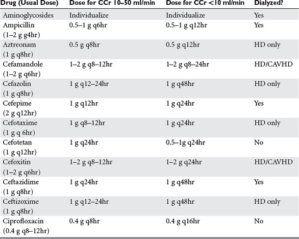
When adjusting therapy in renal failure, the dose can be reduced or the interval between doses can be prolonged. The initial dose should be the same regardless, in order to obtain adequate peak serum concentrations. It is preferred to maintain the dose and prolong the interval with aminoglycosides because of the importance of maintaining a high peak concentration. However, it makes sense to reduce dose but maintain the interval when administering β-lactam drugs (especially those with no PAE) in order to maintain a constant drug concentration. The need to dose patients during or after a renal replacement therapy treatment must be borne in mind. During continuous renal replacement therapy, the estimated creatinine clearance is 15 ml/minute in addition to the patient’s intrinsic clearance.

SUGGESTED READINGS

American Thoracic Society. Guidelines for the management of adults with hospital-acquired, ventilator-associated, and healthcare-associated pneumonia. Am J Respir Crit Care Med. 2005;171:388-416.

GM Anstead & AD Owens Recent advances in the treatment of infections due to resistant Staphylococcus aureus Curr Opin Infect Dis 17 549–555

Bartlett JG, Perl TM. The new Clostridium difficile—what does it mean? N Engl J Med. 2005;343:2503-2505.

Benko AS, Cappelletty DM, Kruse JA, et al. Continuous infusion versus intermittent administration of ceftazidime in critically ill patients with suspected Gram-negative infections. Antimicrob Agents Chemother. 1996;40:691-695.

Bosso JA. The antimicrobial armamentarium: evaluating current and future treatment options. Pharmacotherapy. 2005;25:55S-62S.

Carlet J, Ben Ali A, Chalfine A. Epidemiology and control of antibiotic resistance in the intensive care unit. Curr Opin Infect Dis. 2004;17:309-316.

Chastre J, Wolff M, Fagon JY, et al. Comparison of 15 vs. 8 days of antibiotic therapy for ventilator-associated pneumonia in adults: a randomized trial. JAMA. 2003;290:2588-2598.

Clark NM, Hershberger E, Zervosc MJ, et al. Antimicrobial resistance among Gram-positive organisms in the intensive care unit. Curr Opin Crit Care. 2003;9:403-412.

Dellinger EP. Duration of antibiotic treatment in surgical infections of the abdomen. Undesired effects of antibiotics and future studies. Eur J Surg. 1996;576:29-31. (Suppl)

DiPiro JT, Edmiston CE, Bohnen JMA. Pharmacodynamics of antimicrobial therapy in surgery. Am J Surg. 1996;171:615-622.

Evans RS, Pestotnik SL, Classen DC, et al. A computer-assisted management program for antibiotics and other antiinfective agents. N Engl J Med. 1998;338:232-238.

Fry DE. The importance of antibiotic pharmacokinetics in critical illness. Am J Surg. 1996;172:20S-25S. (Suppl)

Garnacho-Montero J, Garcia-Garmendia JL, Barrero-Almodovar A, et al. Impact of adequate empirical antibiotic therapy on the outcome of patients admitted to the intensive care unit with sepsis. Crit Care Med. 2003;31:2742-2751.

Gold HS, Moellering RC. Antimicrobial drug resistance. N Engl J Med. 1996;335:1445-1453.

Harbarth S, Ferriere K, Hugonnet S, et al. Epidemiology and prognostic determinants of bloodstream infections in surgical intensive care. Arch Surg. 2002;137:1353-1359.

Jones RN. Microbiological features of vancomycin in the 21st century: minimum inhibitory concentration creep, bactericidal/static activity, and applied breakpoints to predict clinical outcomes or detect resistant strains. Clin Infect Dis. 2005;42:S13-S24.

Kollef MH, Micek ST. Strategies to prevent antimicrobial resistance in the intensive care unit. Crit Care Med. 2005;33:1845-1853.

LeDell K, Muto CA, Jarvis WR, et al. SHEA guideline for preventing nosocomial transmission of multidrug-resistant strains of. Staphylococcus aureus and Enterococcus. Infect Control Hosp Epidemiol. 2003;24:639-641.

Livermore DM. Bacterial resistance: origins, epidemiology, and impact. Clin Infect Dis. 2003;36:S11-S23.

V Loo, L Poirier & MA Miller et al A predominantly clonal multi-institutional outbreak of Clostridium difficile-associated diarrhea with high morbidity and mortality. N Engl J Med 353 2442–2449

McDonald LC, Kilgore GE, Thompson A, et al. An epidemic, toxin genevariant strain of Clostridium difficile. N Engl J Med. 2005;353:2433-2441.

Naiemi NA, Duim B, Savelkoul PH, et al. Widespread transfer of resistance genes between bacterial species in an intensive care unit: implications for hospital epidemiology. J Clin Microbiol. 2005;43:4862-4864.

Naimi TS, LeDell KH, Como-Sabetti K, et al. Comparison of community- and health care-associated methicillin-resistant Staphylococcus aureus infection. JAMA. 2004;290:2976-2984.

Neuhauser MM, Weinstein RA, Rydman R, et al. Antibiotic resistance among Gram-negative bacilli in US intensive care units: implications for fluoro-quinolone use. JAMA. 2003;289:885-888.

Nseir S, Di Pompeo C, Soubrier S, et al. First-generation fluoroquinolone use and subsequent emergence of multiple drug-resistant bacteria in the intensive care unit. Crit Care Med. 2005;33(2):283-289.

Padmanabhan RA, Larosa SP, Tomecki KJ. What’s new in antibiotics? Dermatol Clin. 2005;23:301-312.

Paul M, Benuri-Silbiger I, Soares-Weiser K, et al. Beta-lactam monotherapy versus beta-lactam-aminoglycoside combination therapy for sepsis in immunocompetent patients: systematic review and meta-analysis of randomized trials. BMJ. 2004;328(7441):668.

Rello J, Ollendorf DA, Oster G, et al. Epidemiology and outcomes of ventilator-associated pneumonia in a large US database. Chest. 2002;122:2115-2121.

Raymond DP, Pelletier SJ, Crabtree TD, et al. Impact of a rotating empiric antibiotic schedule on infectious mortality in an intensive care unit. Crit Care Med. 2001;29:1101-1108.

Schentag JJ, Gilliland KK, Paladino JA. What have we learned from pharma-cokinetic and pharmacodynamic theories? Clin Infect Dis. 2001;32:S39-S46.

Schlaes DM, Gerding DN, John JFJr, et al. Society for Healthcare Epidemiology of America and Infectious Diseases Society of America Joint Committee on the Prevention of Antimicrobial Resistance: guidelines for the prevention of antimicrobial resistance in hospitals. Clin Infect Dis. 1997;25:584-599.

Sehulster L, Chinn RY, et al. Guidelines for environmental infection control in health-care facilities. Recommendations of CDC and the Healthcare Infection Control Practices Advisory Committee (HICPAC). MMWR Recomm Rep. 2003;6:1-42.

Shorr AF, Sherner JH, Jackson WL, et al. Invasive approaches to the diagnosis of ventilator-associated pneumonia: a meta-analysis. Crit Care Med. 2005;33:46-53.

Trouillet JL, Chastre J, Vuagnat A, et al. Ventilator-associated pneumonia caused by potentially drug-resistant bacteria. Am J Respir Crit Care Med. 1998;157:531-539.

Viviani M, Silvestri L, van Saene HK, et al. Surviving Sepsis Campaign Guidelines: selective decontamination of the digestive tract still neglected. Crit Care Med. 2005;33:462-463.