17 Ankle Block
Placement
Anatomy
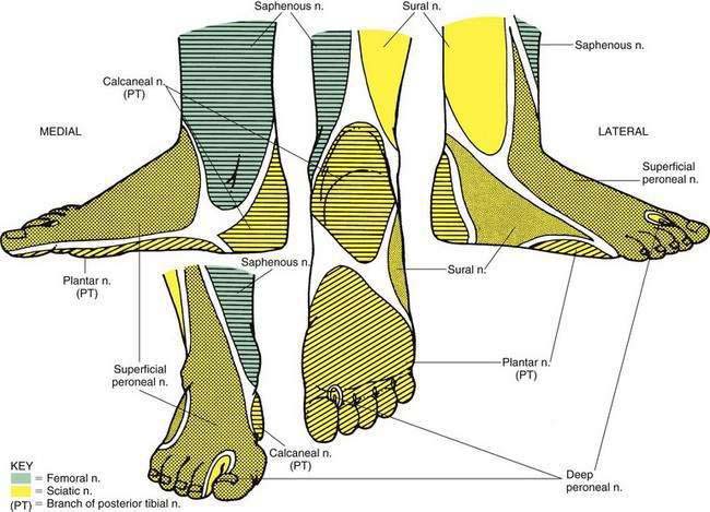
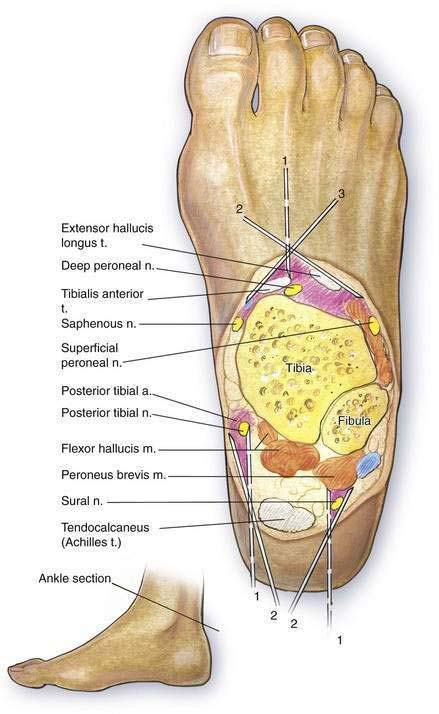
The peripheral nerves requiring block during ankle block are derived from the sciatic nerve, with the exception of a terminal branch of the femoral nerve, the saphenous nerve. The saphenous nerve is the only branch of the femoral nerve below the knee; it courses superficially anterior to the medial malleolus, providing cutaneous innervation to an area of the medial ankle and foot. The remaining nerves requiring block at the ankle are terminal branches of the sciatic nerve—the common peroneal and tibial nerves. The tibial nerve divides into the posterior tibial and sural nerves, which provide cutaneous innervation as outlined in Figure 17-1. The common peroneal nerve divides into its terminal branches, the superficial and deep peroneal nerves, in the proximal portion of the lower leg. Their cutaneous innervation is also illustrated in Figure 17-1. Figure 17-2 identifies the locations of these nerves in a cross-sectional view at the level of ankle block.
Needle Puncture: Posterior Tibial Nerve
With the patient in the prone position, the ankle to be blocked is supported on a pillow. A 22-gauge, 4-cm needle is directed anteriorly at the cephalad border of the medial malleolus, just medial to the Achilles tendon, as shown in Figure 17-2. The needle is inserted near the posterior tibial artery, and if a paresthesia is obtained, 3 to 5 mL of local anesthetic is injected. If no paresthesia is obtained, the needle is allowed to contact the medial malleolus, and 5 to 7 mL of local anesthetic is deposited near the posterior tibial artery.
Needle Puncture: Sural Nerve
The sural nerve is blocked with the patient positioned as for the posterior tibial nerve block. As illustrated in Figure 17-2, the sural nerve is blocked by inserting a 22-gauge, 4-cm needle anterolaterally immediately lateral to the Achilles tendon at the cephalad border of the lateral malleolus. If no paresthesia is obtained, the needle is allowed to contact the lateral malleolus, and 5 to 7 mL of local anesthetic is injected as the needle is withdrawn.
Needle Puncture: Deep Peroneal, Superficial Peroneal, and Saphenous Nerves
After the patient assumes the supine position, the anterior tibial artery pulsation is located at the superior level of the malleoli. A 22-gauge, 4-cm needle is advanced posteriorly and immediately lateral to this point (see Fig. 17-2). An alternative is to insert the needle between the tendons of the anterior tibial and the extensor hallucis longus muscles. Approximately 5 mL of local anesthetic is injected into this area. From this midline skin wheal, a 22-gauge, 8-cm needle is advanced subcutaneously laterally and medially to the malleoli, injecting 3 to 5 mL of local anesthetic in each direction. These lateral and medial approaches block the superficial peroneal and saphenous nerves, respectively.