Anesthesia for burn-injured patients

Published on 07/02/2015 by admin

Filed under Anesthesiology

Last modified 22/04/2025

Print this page

rate 1 star rate 2 star rate 3 star rate 4 star rate 5 star
Your rating: none, Average: 0 (0 votes)

This article have been viewed 2333 times

Anesthesia for burn-injured patients

Christopher V. Maani, MD, Peter A. DeSocio, DO and Kenneth C. Harris, MD

Patients with thermal injuries may have problems in the perioperative period that are distinct from those of other surgical populations. These issues require consideration and planning by the anesthesia provider and surgical team to obtain optimal outcomes.

Acute injury

The first 24 to 48 h after a major burn occurs is considered the resuscitation phase. Depending on the extent of injury, the patient will often require massive amounts of intravascular fluids to maintain intravascular volume, cardiac output, and urine output. Several different formulas can be used to guide therapy, but the primary goal is to administer sufficient fluid to maintain urine output between 0.5 and 1.0 mL·kg−1·h−1, not to restore intravascular euvolemia. During this phase, the patient may require escharotomy or fasciotomy to preserve blood flow to extremities or to allow for chest expansion. Less commonly, a laparotomy may be required to treat abdominal hypertension (abdominal compartment syndrome). Blood transfusion is typically not required during these initial procedures.

For patients with burns that cover less than 40% of total body surface area (TBSA), tracheal intubation is rarely required; however, almost all patients with burns that cover more than 60% of TBSA require intubation. Patients with inhalation injury may require intubation to protect their airways regardless of the size of their burn. The decision to intubate a given patient often requires considerable judgment, but, once the decision has been made, the procedure does not require any unusual considerations beyond those for any other patient with a traumatic injury. If the decision to intubate is delayed until the patient is in respiratory distress, the time remaining before complete arrest occurs may indeed be short. In this situation, because of the increased risk that these patients may develop glottis edema, an “awake” intubation may be indicated. Larger-than-normal tracheal tubes are preferred due to the likely need for bronchoscopy and suctioning of clots or mucous plugs.

General burn care

Full-thickness burns, unless very small, must be treated with excision and grafting. Partial-thickness burns may require excision and grafting depending on their depth, size, and location. Antibiotic creams and solutions—silver sulfadiazine or mafenide acetate, a carbonic anhydrase inhibitor—are the drugs most commonly used on some partial-thickness and full-thickness burns. Silver sulfadiazine is considered less painful to apply but does not penetrate intact burn eschar. Silver sulfadiazine may also cause significant leukopenia, typically in the first few days of use. Mafenide acetate cream penetrates burn eschar but can be painful to apply. In patients with large burns or renal failure, a hyperchloremic metabolic acidosis is occasionally seen that may be attributable to the application of mafenide acetate cream and may not resolve until the drug use is discontinued.

Excision and grafting

Excision of burned skin and placement of skin grafts are the primary reason that patients with burns make frequent trips to the operating room (OR). Some controversy exists over the exact timing of surgery but this approach has changed from delayed intervention to the current practice in which many burn surgeons will operate within 24 to 48 h of the patient’s admission; other surgeons will wait 48 h to ensure that the patient has been adequately resuscitated.

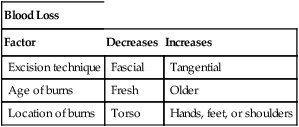
Excision may be either tangential, in which the burn is shaved off until unburned tissue is reached, or fascial, in which all skin and underlying fat is removed down to fascia, usually by using an electrocautery device. Tangential excisions generally produce a better functional and cosmetic result. Fascial excisions may be faster to perform and usually result in less blood loss, as compared with tangential excisions. Whichever method is chosen, these procedures can be quite bloody, with blood loss varying from 123 to 387 mL for each 1% of TBSA of burned tissue excised. Several factors affect the volume of blood loss (Table 233-1).

Table 233-1

Factors Related to Blood Loss in Patients with Burns

Blood Loss
Factor Decreases Increases
Excision technique Fascial Tangential
Age of burns Fresh Older
Location of burns Torso Hands, feet, or shoulders

image

The use of tourniquets and fibrin glue may substantially reduce blood loss. Harvesting of the skin graft may produce considerable blood loss itself, especially if scalp is harvested, but bleeding can be decreased with the infiltration of epinephrine solution into the area to be harvested. Pitkin solution, lactated Ringer’s solutions with 1 to 2 mg/L of epinephrine, or other combinations of vasoconstrictors in crystalloid solutions are often used to try to decrease blood loss.

Preoperative evaluation of the patient with burns

In addition to the standard preoperative evaluation, several aspects of the preoperative evaluation in patients with burns deserve special attention and will be covered in the following sections.

Care in the operating room

Setup

The OR should be heated to 90° F or as close to that temperature as possible. The patient’s entire body must often be exposed, thus limiting the usefulness of warming blankets. A rapid blood infusion system should be available. Two to six units of crossmatched packed red blood cells (pRBCs) should be immediately available, with the understanding that, for patients who will require excisions of large amounts of TBSA, 10 to 20 units may be needed, as will fresh frozen plasma and platelets. For patients with facial burns who will be intubated in the OR, the anesthesia provider should plan to secure the tracheal tube with cloth ties; suturing the tracheal tube to a tooth is another viable option, as is intubation with ties around the nasal septum.

Patient care

Consideration should be given to induction of anesthesia on the patient’s bed if movement is especially painful for the patient. Standard placement of monitors is usually routine but may be limited by injuries and dressings. Because standard electrocardiographic lead placement is rarely essential, leads are placed where space permits; in unusual situations, leads may be stapled in place after induction of anesthesia. Noninvasive blood pressure cuffs work surprisingly well over most dressings. Creativity may be needed in placing the probe for a pulse oximeter; the ears, nose, lips, forehead, and hard palate have all been used successfully. Monitoring of body temperature is always required; patients’ inability to maintain a temperature of 36°C warrants maximum effort to warm the patient. A Foley catheter should be used for most patients.

Several people, all with clearly defined roles, should be involved in transferring the patient to, or from, the operating table to minimize the risk of inadvertent removal of vascular cannulas or tubes.

Following induction of anesthesia, neuromuscular blocking agents are usually administered, either to facilitate tracheal intubation or as part of a balanced anesthetic. The use of succinylcholine is safe for the first 24 h after a burn, but, beyond that period and for up to a year afterward, can result in dramatic and fatal hyperkalemia. Nondepolarizing neuromuscular blocking agents are regularly used in patients with burns but at larger and more frequent doses than in patients without burns. The exception is mivacurium, which lasts as long or longer in patients with burns, as compared with patients without burns.

With the exception of opioids and catecholamines (e.g., phenylephrine), to which they may be relatively resistant, patients with burns typically respond normally to the usual induction agents. Following induction and intubation, the use of inhalation agents supplemented with opioids works well for most patients. Propofol may be used if the anesthesia provider wishes to administer a total intravenous anesthetic. Ketamine, a traditional agent used for many patients with burns, is also acceptable either as the primary agent or as part of a balanced anesthetic. Emergence delirium is seldom an issue with the use of ketamine because most of these patients receive a benzodiazepine.

Blood loss during the excision portion of the operation may be dramatic, with 1 to 2 L of blood not uncommonly lost in a short period of time. Young healthy adults can easily tolerate hematocrit concentrations of 20%, but once the hematocrit level drops below 18%, patients typically become hypotensive. Older patients with comorbid conditions are less tolerant of hematocrit concentrations of 24% or less. Serum ionized calcium levels should be monitored in patients receiving a large number of pRBCs over a short period of time. At many centers, the patient who has traumatic injury in addition to the thermal injury will receive a ratio of fresh frozen plasma to pRBCs of 1:1. However, the need for non-RBC products varies considerably from case to case and is usually driven by laboratory values, clinically observed bleeding, and judgment. Recombinant activated factor VIIa has been used occasionally in patients who develop coagulopathy, but, at present, there are no outcome data to support its use.

Hypotension secondary to a decrease in systemic vascular resistance from bacteremia or other factors released during excision of the wound is not uncommon but, assuming that the patient’s intravascular volume is adequate, responds well to vasopressin, norepinephrine, or phenylephrine.

After skin grafts are placed, they may be covered with a negative pressure dressing or conventional gauze dressings. For those patients who are to be extubated at the end of the operation, care should be taken to provide a smooth emergence from anesthesia to decrease the chance that patient movement (i.e., thrashing about on the bed) may damage the grafts.