ANATOMY OF JOINTS, GENERAL CONSIDERATIONS, AND PRINCIPLES OF JOINT EXAMINATION

Published on 18/03/2015 by admin

Filed under Rheumatology

Last modified 22/04/2025

Print this page

rate 1 star rate 2 star rate 3 star rate 4 star rate 5 star
Your rating: none, Average: 0 (0 votes)

This article have been viewed 3849 times

1 ANATOMY OF JOINTS, GENERAL CONSIDERATIONS, AND PRINCIPLES OF JOINT EXAMINATION

Applied Anatomy

TYPES OF JOINTS

Skeletal joints, the sites of articulation between one bone or cartilage and another, are generally of three types: fibrous joints (skull-type sutures), cartilaginous and fibrocartilaginous joints (discovertebral joints), and synovial joints (most limb joints).

Fibrous joints, or skull-type articulations, are called synarthroses: one bone is joined to another by an unossified fibrous membrane or residual plate of cartilage. Fibrous joints, such as skull sutures, allow little or no movement. Bones united by ligamentous attachments, such as the proximal and distal tibiofibular joints and the superior sacroiliac joints, are called syndesmoses.

Cartilaginous joints are amphiarthroses and are of two types: fibrocartilaginous and cartilaginous. Bony surfaces coated with hyaline cartilage and united by fibrocartilaginous disks are symphyses (fibrocartilaginous joints). Examples include discovertebral, manubriosternal, xiphisternal, and costosternal joints and symphysis pubis. United epiphyseal hyaline cartilage is an example of a cartilaginous amphiarthroidal joint. Amphiarthroses allow a limited range of movement but provide considerable stability.

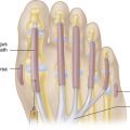
Synovial joints or diarthroses are freely movable joints. The bony surfaces are coated with hyaline cartilage and united by a fibrous articular capsule. The synovial membrane lines the inner surface of the capsule but does not cover the articular cartilage (Figure 1-1). The capsule is strengthened by collateral ligaments. Synovial joints comprise most of the joints of the extremities and are the most accessible joints to direct inspection and palpation. Synovial joints share important structural components: subchondral bone, hyaline cartilage, a joint cavity, synovial lining, articular capsule, and supporting ligaments.
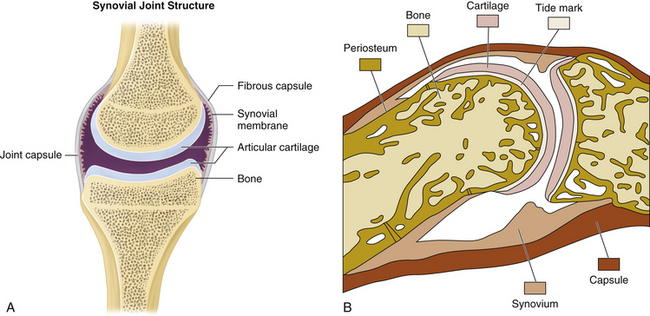
Synovial joints serve a variety of functions and differ in configuration, permitting specific movements while restricting others. Synovial joints can be subdivided into seven major types:

STRUCTURE OF SYNOVIAL JOINTS AND SUPPORTING TISSUES

The articular cartilage is an avascular, aneural, resilient, low-friction, load-bearing tissue covering the articulating bony surfaces. It is capable of absorbing impact by virtue of its compressibility, elasticity, low hydraulic permeability, and self-lubrication (squeeze-film hydrodynamic lubrication). It derives its nourishment through diffusion from the synovial fluid that bathes its surface.

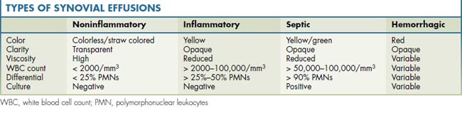
The synovial membrane provides an unobtrusive, flexible, low-friction, well-lubricated lining for diarthrodial joints, tendon sheaths, and bursae. Histologically the synovium consists of two layers: an intimal or synovial lining cell layer—made up of one to three layers of cells or synoviocytes—and a subintimal or subsynovial layer of loose, vascular, fibro-fatty connective tissue. Synovium has important phagocytic functions and produces synovial fluid. Table 1-1 shows different types of synovial effusions.

Joint capsules, ligaments, and tendons are dense fibrous tissues with a major role in musculoskeletal function. Collectively they allow and guide joint motion while resisting high tensile loads without deformation. Laxity or adaptive shortening of these structures affects joint motion and may lead to injury (see Figure 1-1).

Joint capsules attach around articular surfaces to form a continuous envelope for the joint. Capsular cells are predominantly fibroblasts with a dense fibrous matrix surrounding them. The capsule is lined with synovium that provides lubrication for the joint. Ligaments attach bone to bone and function to stabilize adjacent bones by restricting abnormal movements. Ligaments may be intracapsular, capsular, or extracapsular, and they share similar cell and matrix characteristics with the joint capsule and tendon tissues.

The anterior cruciate ligament (ACL) is an intracapsular ligament that connects anterior aspect of intercondylar eminence of tibia with medial surface of lateral femoral condyle.

The medial (tibial) collateral ligament (MCL) is a capsular ligament connecting the medial epicondyle of the femur with the medial surface of the medial tibial condyle; it is continuous with the capsule and is essentially a thickening of the capsule.

The lateral (tibial) collateral ligament (LCL) is an extracapsular ligament, not part of the fibrous capsule of the knee; it connects the lateral epicondyle of the femur with the fibular head.

Tendons vary in size and shape, but all attach muscles to bone to transmit the force of muscle contraction to bone. Maintaining tendon tension plays a role in homeostasis of the tendon. Overuse and repetitive trauma results in degenerative changes that outweigh the regenerative process and weaken the tendon.

General Considerations and History

The patient’s history is the essential first step in all musculoskeletal diagnoses, and it focuses on the physical examination. Since diagnosis of nearly all musculoskeletal problems relies upon demonstrating objective findings, the physical examination is enormously important. Despite this, the musculoskeletal examination is often poorly understood and inadequately performed by physicians at all levels of training. Without the ability to perform a proper physical examination, your use of additional diagnostic laboratory testing may be excessive, expensive, and lacking the precision that only comes from recognizing important musculoskeletal physical findings.

CATEGORIES OF MUSCULOSKELETAL PROBLEMS

Musculoskeletal problems and rheumatic diseases can be practically classified into five major categories, defined by the tissues predominantly affected: periarticular, articular, bone, nerve, and extraarticular.

At each point in your evaluation—as you gather essential historical, physical, and selected laboratory information—ask what tissues are primarily affected. Thinking of musculoskeletal problems in this way provides a useful framework for organizing clinical information. Recognizing the pattern of predominant tissue involvement then directs your attention toward well-defined members of each category, helping to organize your differential diagnosis (Table 1-2).

TABLE 1-2 MUSCULOSKELETAL PROBLEMS AND RHEUMATIC DISEASES, AFFECTED TISSUES, AND PATTERNS OF INVOLVEMENT

Periarticular (Soft-Tissue Problems)
Bursitis, tendinitis
Capsular, ligamentous sprain
Muscular strain
Articular (Synovial and Cartilaginous Joints)
Osteoarthritis
Rheumatoid arthritis
Spondyloarthritis
Crystalline arthritis
Infectious arthritis
Bone
Trauma
Osteoporosis
Avascular necrosis
Infection
Tumor
Nerve
Radiculopathy
Entrapment
Neuroarthropathy
Complex regional pain syndromesFibromyalgia (generalized musculoskeletal pain)
Extraarticular (Systemic Connective Tissue Diseases)
Systemic lupus erythematosus
Antiphospholipid (anticardiolipin) antibody syndrome
Sjögren syndrome
Polymyalgia rheumatica
Systemic vasculitis
Inflammatory myopathy
Sclerodermatous diseases (skin and fascia)

CHARACTERISTICS OF MUSCULOSKELETAL PAIN

Pain in nearly any location can be well delineated using the mnemonic OPQRST, where O = onset, P = precipitating (and ameliorating) factors, Q = quality, R = radiation, S = severity, and T = timing. This information, combined with the pattern of joint involvement, is helpful in narrowing the preliminary differential diagnosis. The number of joints involved (monoarticular, oligoarticular, or polyarticular); the presence of symmetry or asymmetry; peripheral versus axial joint involvement; small and/or large joint involvement; and a fixed, migratory, or additive pattern of evolution of arthritis are all important features of clinical pattern recognition. Careful questioning about any past joint complaints may supply missing data relating to the location of prior articular problems and the time course and pattern of evolution of previous musculoskeletal symptoms and signs.

Patterns of Involvement

A critical feature in clinical diagnosis is the distinction between arthralgia (subjective joint pain) and arthritis (objective joint swelling or impaired function). Demonstration of objective joint swelling (bony or synovial) on examination is required to make a secure diagnosis of arthritis.

The distribution of joint involvement is one of the most essential aspects of pattern recognition in patients with arthritis. Monoarthritis describes involvement of a single joint, oligoarthritis (pauciarthritis) involvement of two to four joints, and polyarthritis involvement of five or more joints. Peripheral and axial describe predominant extremity (distal) and spinal (central) joint involvement. Symmetric versus asymmetric describes whether involvement of joint pairs is bilateral or unilateral. Small joint (joints of the hands and feet) and/or large joint (shoulder, hip, knee) involvement has considerable diagnostic significance.

Although the presence of pain and tenderness may initially suggest arthritis, periarticular structures such as bursae, ligaments, and tendons may be responsible for the symptoms and are very important to correctly identify. The term enthesitis or enthesopathy describes inflammation or abnormality at the insertion site of tendons, ligaments, or joint capsules into bone. This is a particularly prominent clinical feature of spondyloarthropathies, such as ankylosing spondylitis and psoriatic arthritis.

In addition, muscles may play a critical role in musculoskeletal symptom development. Throughout day-to-day work and leisure activities, muscles must adapt to sustained positions (sitting, standing, etc.) and repetitive motion that occurs in these adapted postures. As a result, some muscles may become too short and others too long. Some muscles may become hypertrophied from repetitive use, and others become atrophied from disuse. Invariably, substitution for weak muscles by other muscles occurs, and faulty motor patterns develop that lead to compromise of normal joint mechanics. Tendinitis, bursitis, and eventually osteoarthritis may result from this deviation from optimal kinesiology. Restoration of correct muscle length, strength, and motor control is essential to prevent joint injury.

For instance, in the shoulder girdle, supraspinatus function is necessary to produce normal rotation of the humeral head on the glenoid fossa during abduction. Supraspinatus weakness results in substitution by the deltoid, a large muscle much farther away from the center of rotation of the humeral head. Preferential use of the deltoid muscle in abduction results in superior gliding of the humeral head, producing an impingement syndrome with tendinitis, bursitis, rotator cuff tears, and arthritis.

Physical Examination

ESSENTIAL CONCEPTS

A central axial spine, paired peripheral joints, and symmetric musculature provide the basis for essential side-to-side comparison during the musculoskeletal examination. Recognizing asymmetry is important and may provide an initial clue in diagnosing an abnormality.

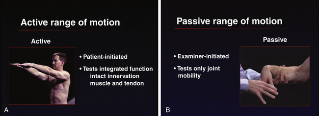
Both active and passive ranges of motion are important in assessing joint function. Active range of motion is patient-initiated movement of the joint that tests integrated function and requires intact innervation, muscle and tendon function, and joint mobility. Passive range of motion is examiner-initiated movement of a joint and tests only joint mobility. The combined use of passive as well as active range of motion minimizes the need for patient instruction and maximizes the speed and efficiency of the exam (Figure 1-2). Whenever joint movement is anticipated to be painful, it is best to first observe active range of motion (patient-initiated movement) to appreciate the degree of pain and dysfunction before gently attempting passive range of motion (examiner-initiated manipulation).

Importance of Objective Findings

Joint tenderness is at least in part a subjective reaction (joint tenderness ≠ arthritis). The location of tenderness may be helpful in distinguishing an articular problem (joint-line tenderness) from periarticular problems (bursitis, tendinitis) where more localized tenderness is found over a lesion. Tenderness must be correlated with the finding of an objective, visible, or palpable abnormality for a diagnosis of arthritis to be made.

Joint redness, or erythema, is an objective abnormality and depends on the acuteness and severity of the underlying inflammation. When present, significant erythema may suggest the possibility of infection or crystalline arthritis.

Joint warmth is also an objective finding and depends on the acuteness and severity of the underlying inflammation. When present, joint warmth can be a very helpful clinical sign of arthritis. (Clinically important chronic inflammation, however, is often cool to palpation, as frequently seen in chronic rheumatoid arthritis.)

Joint swelling is an extremely important and definitive clinical sign. Swelling due to synovial fluid (joint effusion) or swollen synovial tissue (synovial membrane inflammation = synovitis) provides the clinician with definitive evidence of the presence of arthritis. Swelling due to bony enlargement (osteophytes) is also an extremely important physical finding, indicating the presence of underlying primary or secondary osteoarthritis.

Crepitus refers to a “grating” sensation felt under the examiner’s hand during joint movements, and it may indicate roughening of the cartilaginous surface (cartilaginous crepitus) or complete loss of hyaline cartilage with bone-on-bone contact (bony crepitus).

Joint damage and deformity are important, usually permanent, signs of prior injury (ligamentous laxity, tendon rupture, flexion contracture) or arthritis (ulnar deviation, subluxation). The pattern of such damage and deformity can provide important clinical clues to the underlying process.

Manual muscle testing is an important part of the physical examination of musculoskeletal conditions. Careful evaluation of muscle length and strength provides valuable information regarding diagnosis, the focus of rehabilitation, and a basis for determining progress. The art and science of manual muscle testing involves specific positioning to isolate a muscle, test it in its shortest position, and grade its strength on a scale from 0 to 5. For instance, grade 3 strength means the patient can hold the test position against gravity but not with any other pressure applied. Manual muscle testing is the tool of choice for evaluating muscle imbalances.

EXAMINATION COMPONENTS
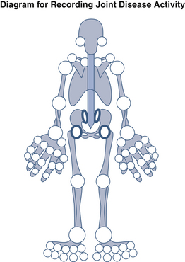
There are four essential steps in the examination of joints: inspection, palpation, active and passive range of motion, and the assessment of supporting structures and special testing. Inspect the joint for asymmetry, erythema, swelling, and deformity. Palpate for tenderness; warmth; synovial thickening; effusion (in inflammatory synovitis); bony, osteophytic swelling (in primary and secondary osteoarthritis); and crepitus. Take the joint through a combination of active (patient-initiated) and passive (examiner-initiated) range-of-motion tests appropriate to the specific joint. Finally, assess supporting structures—such as ligaments, tendons, and muscles—and perform special testing using regional evaluations specific to particular joints. Examples of special testing include manual muscle testing, impingement testing (shoulder), and testing for Tinel sign (entrapment neuropathies).