CHAPTER 7 Anatomical considerations
Applied anatomy
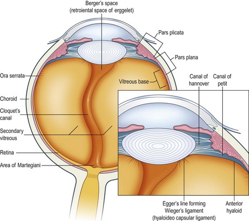
The structures in the perilenticular area, the lens capsule, nucleus and cortex, zonules, anterior vitreous, and ciliary body all interact to provide an optically clear (in health) structure which has the ability to focus, to minimize chromatic aberrations and produce the sharpest image possible on the retina for differing lighting conditions and points of focus. The ciliary muscle contracts to reduce the tension in the zonules and allow alteration in shape in the lens capsule, and therefore curvature of the lens. There is much discussion about whether it can continue to function throughout life, but it is thought the ciliary muscle/zonule/capsule interaction have relatively normal function for life1.
The anterior vitreous maintains a close relationship with the lens, is separated from it by Berger’s space, and attached to the mid-periphery of the posterior part of the lens by Weigert’s ligaments. These are all remnants from the embryological development of the lens (Fig. 7.1).
Zonular defects
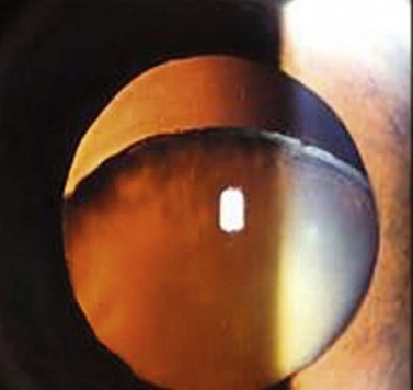
These can be congenital or acquired. The commonest congenital forms are related to Marfan syndrome, homocystinuria, Stickler/Wagner syndrome and idiopathy. Traditionally, in those patients with Marfan syndrome, the lenses are subluxed superotemporally and in homocystinuria inferotemporally (Fig. 7.2). In Marfan, the zonules are abnormal due to a defect in the fibrillin gene which accounts for the subluxation. Depending of the degree of decentration the refractive error can be relatively unchanged or higher degrees of myopia or a prismatic effect can be induced. Interference with vision occurs when the edge of the lens cuts across the visual axis.
Previous glaucoma surgery
Pseudoexfoliation
Late subluxation of the intraocular lens/capsular bag complex can occur, requiring an IOL exchange.


