Anaphylactic and anaphylactoid reactions

Published on 07/02/2015 by admin

Filed under Anesthesiology

Last modified 22/04/2025

Print this page

rate 1 star rate 2 star rate 3 star rate 4 star rate 5 star
Your rating: none, Average: 1 (1 votes)

This article have been viewed 5008 times

Anaphylactic and anaphylactoid reactions

Cornelius B. Groenewald, MB, ChB

Anaphylactic and anaphylactoid reactions, which produce an identical spectrum of clinical signs and symptoms, are acute, potentially life-threatening syndromes that occur in 1 out of every 10,000 to 20,000 anesthetics given. Anaphylactic reactions, which are associated with higher morbidity and mortality rates than anaphylactoid reactions, are type 1, typically IgE-mediated, immune reactions that result in mast cell and basophil degranulation and release of pharmacologically active mediators that affect almost all organ systems. Anaphylactoid reactions reflect nonimmune release of histamine from mast cells and basophils.

Pathophysiology

Prior sensitization or cross-sensitization to similar antigens is necessary to activate an anaphylactic reaction. Antigen-antibody interaction links two IgE molecules that, in turn, bind to and initiate mast cell and basophil degranulation and release of vasoactive mediators, including histamine, serum proteases (such as tryptase), proteoglycans, prostaglandins, and leukotrienes (Figure 246-1).

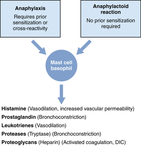
During anaphylactoid reactions, administered medications may cause nonimmunologic mast cell and basophil degranulation by activation of the complement cascade, of the coagulation system, of the kinin-generating system, or of the fibrinolytic system or by direct nonspecific mechanisms (Figure 246-1).

Clinical presentation

Target organs include the skin, upper and lower respiratory tracts, cardiovascular system, hematologic system, and the gastrointestinal tract (Box 246-1); mental status may also be altered. The signs and symptoms of anaphylaxis and anaphylactoid reactions that occur during the perioperative period depend on the types of anesthesia and monitors used. For example, early skin changes may not be appreciated in a covered patient, whereas abdominal and neurologic signs may be apparent only in awake patients.

The clinical presentation is not uniform and depends on the patient’s sensitivity and the concentration and route of administration of the antigen. For example, intravenous administration of penicillin may trigger acute cardiovascular collapse, whereas cutaneous exposure to latex may result in only urticaria.

Diagnosis

The occurrence of an anaphylactic or anaphylactoid reaction is suggested by the development of signs and symptoms soon after administration of a potential agent. To distinguish between the reactions, obtaining a serum tryptase concentration may be useful. Tryptase, a serine protease, is released from mast cells during anaphylactic reactions and may correlate with the severity of the reaction. In contrast, anaphylactoid reactions are associated with normal or only slightly elevated tryptase levels. Serum levels rise within 15 min to 2 h and return to baseline levels within 24 h after the reaction, so tryptase concentration should be tested within this time period. Histamine levels rise early during both anaphylactic and anaphylactoid reactions but return to normal within 30 min; therefore, obtaining histamine levels is rarely useful in assisting with the diagnosis. Sensitivity to a specific medication is diagnosed by skin prick, intradermal testing, and, occasionally, serologic testing.

Substances commonly implicated in anaphylactic and anaphylactoid reactions

Neuromuscular blocking agents

Neuromuscular blocking agents (NMBAs) are the most common medications involved in perioperative anaphylactic and anaphylactoid reactions, accounting for 50% to 70% of reported cases. The incidence of anaphylaxis is estimated at 1 in 6500 administrations of an NMBA. Cross-reactivity between the NMBAs may approach 60% to 70%. Succinylcholine and rocuronium account for most of the reported cases. The increased incidence of anaphylaxis occurring with the use of these drugs may be due to reporting bias; no epidemiologic studies have shown rocuronium to be more allergenic than other NMBAs. Atracurium, mivacurium, and doxacurium are the NMBAs most likely to cause anaphylactoid reactions that potentially lead to hypotension and tachycardia. However, these effects may be mitigated by slow intravenous administration of the drugs and pretreatment with antihistamines.

Other substances

Anaphylactic reactions to propofol have been reported and occur in 1 in 30,000 to 60,000 administrations. Current evidence suggests that patients who are allergic to egg lecithin (present in the yolk) are perhaps more likely to experience anaphylaxis during propofol administration, as compared with patients without egg-lecithin allergies.

Anaphylactic reactions to opioids are rare but have been reported with the use of fentanyl. Morphine and meperidine cause anaphylactoid reactions rather than anaphylaxis.

Anaphylactic reactions to methylene blue are very rare; however, the incidence approaches 1% to 2% following injection of isosulfan blue or patent blue dye.

The paraaminobenzoic acid preservative in the ester class of local anesthetic agents (procaine, chloroprocaine, tetracaine, and cocaine), rather than the local anesthetic agent itself, is the cause of most anaphylactic reactions with this type of drug; because drugs in this class are not often used, anaphylactic reactions in patients receiving local anesthetic solutions is rare. Drugs in the amide class of local anesthetics (lidocaine, bupivacaine, mepivacaine, prilocaine, and ropivacaine) are not allergenic; however, some individuals are allergic to the sodium bisulfite or metabisulfite used to stabilize epinephrine, and, for these individuals, a local anesthetic without epinephrine should be used.

Reports have implicated colloid volume expanders in 2.3% of perioperative allergic reactions; dextrans have the highest risk, and hetastarch seems to be safest. There is no cross-reactivity between different colloids.

Treatment

The first step in treating an anaphylactic reaction is to discontinue possible causative drugs, thereby decreasing the antigen load. Consideration should be given to switching to a latex-free environment. Supportive treatment is initiated with maintenance of the airway and administration of 100 % O2. All anesthetic medications should be discontinued. Inhaled anesthetics may produce myocardial depression and worsen hypotension and are not the treatment of choice for bronchospasm in this situation. Increased vascular permeability may result in rapid transit of fluid from the intravascular to the interstitial compartment and, along with vasodilation, could result in hypotension that, if severe enough, could cause hypovolemic shock. Immediate blood volume expansion with 2 to 4 L of crystalloid or colloid solution is indicated. The drug of choice for treating systemic effects is epinephrine, which increases intracellular concentrations of cyclic adenosine monophosphate and thereby stabilizes membrane permeability, resulting in decreased release of pharmacologically active mediators. Vasodilation responds to the α1-agonist effects of epinephrine on vascular smooth muscle; in addition, epinephrine, with its β2-agonist activity on bronchial smooth muscle, is the bronchodilator of choice when cardiac collapse is associated with bronchospasm. The route and dose of administration depend on the patient’s clinical condition. For reactions that are not severe, a 1:1000 solution of epinephrine may be given subcutaneously at a dose of 0.3 to 0.5 mg. Patients with hypotension should receive 0.5 to 1 mL of double dilute (1:100,000) epinephrine intravenously every 1 to 3 min until they are hemodynamically stable. In cases of life-threatening cardiac collapse, increased doses will be needed. In settings in which intravenous access has not been established, epinephrine may be given via the tracheal tube. It is important to note that epinephrine should not be given intravenously to normotensive patients.

Bronchospasm is treated with inhaled β2-agonists (such as albuterol or terbutaline).

Antihistamines and corticosteroids are often administered. However, their benefit is less clear. H1-receptor antagonists, such as diphenhydramine, may competitively bind to histamine receptors and ameliorate some of the signs and symptoms, but there is no evidence to support this use. Corticosteroids (methylprednisolone) are given to prevent delayed release of inflammatory mediators. The use of sodium bicarbonate and arginin vasopressin therapy should be considered in patients not responsive to epinephrine.

Every reaction that occurs perioperatively should be investigated immediately as well as with skin testing 6 weeks postoperatively.