Anaesthesia for Orthopaedic Surgery

Published on 27/02/2015 by admin

Filed under Anesthesiology

Last modified 22/04/2025

Print this page

rate 1 star rate 2 star rate 3 star rate 4 star rate 5 star
Your rating: none, Average: 0 (0 votes)

This article have been viewed 1785 times

Anaesthesia for Orthopaedic Surgery

One in five operations in the United Kingdom is for orthopaedic, spinal or trauma surgery. Anaesthesia for trauma surgery is discussed in Chapter 37. This chapter provides a framework for the conduct of anaesthesia for orthopaedic surgery.

THE PATIENT POPULATION

A large proportion of patients presenting for orthopaedic surgery are young and healthy. Sporting injuries and disease processes without systemic impact are common and these patients are at low risk of complications relating to anaesthesia or surgery. However, several disease processes are more common in patients presenting for orthopaedic surgery than in the general surgical population, and these are discussed below.

Comorbidities

Rheumatoid Arthritis

Rheumatoid arthritis is a chronic inflammatory disease of unknown aetiology, affecting women more often than men. Rheumatoid factor is found in 90% of affected patients and there exists a genetic predisposition with associated human leucocyte antigen HLA-DR4. It is a multisystem disease which may present the anaesthetist with problems of a difficult airway, cervical spine instability (and cervical cord vulnerability) and widespread vasculitis-induced organ dysfunction. Additionally, drug therapy for rheumatoid disease frequently produces severe and widespread side-effects. These are detailed below. The airway of the rheumatoid patient may present problems because of stiffness of the temporomandibular joint, stiffness or instability of the neck and cricoarytenoid arthritis. Radiological examination shows involvement of the cervical spine in 80% of patients, and 30% have neurological symptoms suggesting instability of the neck. Atlantoaxial subluxation, subaxial subluxation and cervical spine ankylosis are common, and should be investigated through history-taking, clinical examination and cervical X-ray. Flexion-extension views may be necessary to observe instability. Magnetic resonance imaging provides good assessment of the rheumatoid neck. Systemic disease is very common, and includes pericardial effusion, constrictive pericarditis, heart block, aortic and mitral valve disease, pleural effusion, interstitial fibrosis, anaemia, thrombocytopaenia and renal and hepatic dysfunction.

A thorough history and examination are important for patients with rheumatoid disease. Careful assessment of the airway and cervical spine should be performed. The range of neck movement should be assessed, and any associated neurological symptoms should be noted. A full blood count and serum urea and electrolyte concentrations should be measured, and an ECG and chest X-ray should be considered. Additionally, a lateral cervical X-ray, preferably with flexion-extension views, should be considered, particularly if tracheal intubation is planned. Systemic disease may indicate the need for arterial blood gas analysis, lung function tests, echocardiogram or liver function tests to be undertaken. Suspicion of cricoarytenoid involvement should prompt preoperative indirect laryngoscopic examination.

Regional anaesthesia should be used if possible. It has the advantage of avoiding airway and neck manipulation, and may be safer than general anaesthesia in patients with severe systemic disease. However, epidural and spinal anaesthesia may be very difficult because of spinal ankylosis and osteophyte formation. If general anaesthesia is used, patients with an unstable neck should be managed by an experienced anaesthetist, especially if tracheal intubation is planned. Tracheal intubation is made more difficult if movement of the temporomandibular joint is restricted. The need for tracheal intubation should be considered carefully in patients with severe disease because of the associated risks and difficulty. For many procedures, the use of a laryngeal mask airway is a suitable and a potentially less traumatic alternative. If tracheal intubation is required, intubation aids such as the intubating laryngeal mask airway or fibreoptic-guided intubation may be safer alternatives to tracheal intubation using direct laryngoscopy.

Concurrent Drug Therapy

Many young healthy patients presenting for orthopaedic surgery do not take concurrent medication. However, use of analgesics is very common in the orthopaedic population because of the painful nature of their disease process. Concurrent therapy with antihypertensive, antianginal, antidepressant or cholesterol-lowering medication is common in older patients presenting for orthopaedic surgery. These patients often present for arthroplasty and this major procedure may place significant demands upon their physiological reserves. Preparation of the patient taking these drugs is discussed in detail in Chapter 18. Patients may also be using orthopaedic disease-modifying drugs such as methotrexate, steroids and gold.

Non-Steroidal Anti-Inflammatory Drugs

Thromboxane A2 and prostaglandin endoperoxide, which are needed for the haemostatic function of platelets, are synthesized from arachidonic acid by the cyclo-oxygenase (COX) enzyme system. Non-steroidal anti-inflammatory drugs (NSAIDs) inhibit this enzyme system, impairing the formation of clots and, consequently, haemostasis. There are two COX isoforms: COX-1 synthesizes prostaglandins, which protect the gastric mucosa; COX-2 is involved with inflammatory responses. Inhibition of these systems ceases rapidly when administration of NSAIDs is stopped. However, the effects of aspirin persist for up to 10 days after treatment because of its covalent bonding with cyclo-oxygenase. Although NSAIDs taken up to the time of surgery may increase surgical blood loss, this does not imply that preoperative administration should be avoided. NSAIDs are valuable in providing analgesia pre- and postoperatively, and increased surgical blood loss is usually modest. Of more concern is gastroduodenal ulceration, of which the first symptom may be life-threatening upper gastrointestinal haemorrhage. The risk of ulceration is dose-related, commoner as age advances and even commoner if corticosteroids are also used to control inflammation. This complication stimulated the production of specific COX-2 inhibitors, but their use has been curtailed following reports of increased cardiovascular and cerebrovascular complications. NSAIDs should be avoided in patients who have a history suggestive of gastrointestinal ulceration or bleeding.

Corticosteroids

Regular medication with glucocorticoid drugs (e.g. prednisolone, hydrocortisone, dexamethasone) produces suppression of endogenous glucocorticoid production. There is an increase in glucocorticoid concentration as part of the stress response after surgery, and these patients are at risk of an Addisonian crisis, including a precipitous fall in blood pressure because they may not be able to synthesize sufficient endogenous glucocorticoid. Patients who have taken doses of steroids greater than the equivalent of prednisolone 10 mg daily during the past 3 months require replacement corticosteroid therapy. Corticosteroid therapy may cause poor wound healing and gastrointestinal ulceration; consequently, low-dose replacement therapy is currently favoured. For patients undergoing minor surgery under general anaesthesia, the usual oral dose of corticosteroid should be given on the morning of surgery or a single dose of hydrocortisone 25–50 mg intravenously at induction. For patients undergoing moderate or major surgery, the usual morning dose of corticosteroid should be given and 25–50 mg of intravenous hydrocortisone at induction followed by further doses of 25–50 mg intravenously three times daily for 24 h after moderate surgery or for 48–72 h after major surgery. The patient’s usual corticosteroid regimen is then re-established.

Other Drugs

A large variety of potentially toxic drugs is used to reduce the symptoms and retard the disease process in rheumatoid arthritis. Antimalarials, such as chloroquine, may cause retinopathy and cardiomyopathy. Gold and penicillamine cause undesirable side-effects in up to 40% of patients; these include nephrotic syndrome, thrombocytopaenia, agranulocytosis, marrow aplasia, hepatitis and pneumonitis. Sulfasalazine may cause haematological toxicity and fibrosing alveolitis. Administration of azathioprine may result in gastrointestinal side-effects, cholestatic hepatitis, leucopaenia, thrombocytopaenia and anaemia.

It should be apparent that the provision of anaesthesia for any patient with rheumatoid arthritis must be associated with a thorough search for the potentially dangerous side-effects of concurrent drug therapy.

TECHNIQUES OF ANAESTHESIA

General Anaesthesia

This is appropriate for all types of orthopaedic surgery, but regional anaesthesia may be the preferred technique for many procedures, for reasons discussed below. Patients undergoing procedures of long duration (e.g. hip revision) often require general anaesthesia because of the discomfort incurred by remaining in the same position for a prolonged period of time. In many countries, including the United Kingdom, patients often expect to receive general anaesthesia, and may not have been aware in advance of their surgery that regional anaesthesia represents a viable option. Thus, the use of general anaesthesia offers the benefit to patients of familiarity. General anaesthesia causes the greatest loss of control for the patient and many patients are pleasantly surprised to find that regional anaesthesia is an option for their operation.

Regional Anaesthesia

Central neuraxial block (spinal or epidural anaesthesia) reduces the stress response to surgery and has been shown to reduce some serious complications following many types of surgery. Benefits may include a reduction in the incidences of deep vein thrombosis, blood loss, myocardial infarction, respiratory and renal complications, and possibly pulmonary embolism. There is a high incidence of thromboembolic events in patients undergoing major lower limb arthroplasty, which makes this type of anaesthesia an attractive option.

Lower limb arthroplasty and minor lower limb procedures are frequently carried out using central neuraxial block. For longer procedures, such as hip arthroplasty, sedation or light general anaesthesia may be added. The combination of general anaesthesia with a central neuraxial block has not been shown to reduce the benefits attributable to this form of regional anaesthesia.

Following central neuraxial block, the patient is usually pain-free in the immediate postoperative period. Careful thought should be given to administration of analgesia after the nerve block has worn off (see below). There is a higher incidence of urinary retention in patients who have undergone joint arthroplasty under central neuraxial block and this leads to an increased risk of urinary tract infection. Patients may be managed by prophylactic urethral catheterization or monitoring of bladder volume postoperatively using ultrasound.

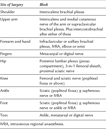
Peripheral nerve block is commonly used as a sole technique for many procedures, with the advantages of excellent pain relief, reduction of surgical stress, avoidance of complications of general anaesthesia and earlier discharge in the day-case setting. Peripheral surgery in ‘high-risk’ patients may also be carried out under peripheral nerve block to avoid the potential complications of general anaesthesia or central neuraxial block. Patients report a high degree of satisfaction following surgery carried out using this form of anaesthesia. Table 28.1 shows the sites at which surgery may be performed in association with specific nerve blocks. This form of anaesthesia requires a high level of expertise and an understanding of the issues of managing a conscious patient during surgery.

Intravenous regional anaesthesia (IVRA) is suitable for manipulation of fractures and brief operations (less than 30 min) on the forearm and lower leg. It is technically easy to perform but fatalities have occurred as a result of a large dose of local anaesthetic reaching the systemic circulation. Before performing IVRA, it is essential to understand how the risk of complications may be minimized and how they may be treated if they occur. Details of the technique and safety precautions are described in Chapter 24.

POSTOPERATIVE ANALGESIA

Oral and Intravenous Agents

Many patients are already taking regular analgesics for pre-existing bone and joint pain. Paracetamol is very useful in reducing the dose requirements of other analgesics, and may occasionally be sufficient analgesia alone. It is virtually free from side-effects in standard doses, and is contraindicated only in patients with liver dysfunction. If gastric motility is impaired, it may be administered rectally. The addition of NSAIDs, in the absence of contraindications, is usually useful and reduces the requirement for opioid analgesia.

NSAIDs inhibit the formation of prostaglandins and are widely used as analgesics in the treatment of acute bone-related pain. The newer COX-2 inhibitors potentially widened the number of patients who could benefit from these agents by reducing the potential for gastroduodenal ulceration, although as mentioned above, their use has been curtailed due to reports of increased incidences of myocardial infarction and stroke in patients taking long-term COX-2 inhibitors, leading to the withdrawal of rofecoxib in September 2004.

Prostaglandins are known to have an important role in bone repair and homeostasis. Animal studies have demonstrated that both non-specific and specific inhibitors of COX impair fracture healing. Some studies have suggested that this impairment results from COX-2 inhibition. This has raised concerns regarding the use of NSAIDs as anti-inflammatory or analgesic drugs in patients undergoing orthopaedic procedures; however, the clinical implications of this are probably minimal and NSAIDs remain extremely important analgesic agents for orthopaedic patients.

NSAIDs also affect platelet function and would therefore be expected to increase perioperative blood loss. The clinical evidence for increased blood loss in major arthroplasty surgery patients receiving NSAIDs is minimal.

Intravenous opioids are frequently used following major joint arthroplasty. Patient-controlled analgesia systems are the most commonly used delivery systems. The doses of opioid required are much reduced by the other analgesic agents prescribed, thus minimizing the risk of side-effects.

Central Neuraxial Drugs

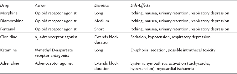
Single-dose spinal or epidural anaesthesia using local anaesthetic alone usually provides analgesia for only a relatively short period of time after operation. An adjuvant administered into the intrathecal or epidural space with the local anaesthetic improves the quality of the block and extends the duration of analgesia. Table 28.2 describes some of the more commonly administered drugs.

An epidural (or, rarely, intrathecal) infusion of local anaesthetics may be combined with an opioid to produce excellent analgesia. The combination of local anaesthetic and opioid is synergistic, reducing the side-effects of both and minimizing motor block. However, relatively high incidences of itching, nausea and urinary retention are encountered. It is routine practice in most hospitals to insert a urethral catheter in the anaesthetic room to avoid urinary retention in the postoperative period.

Epidural infusions are commonly used for up to 5 days following major orthopaedic surgery. Careful observation for signs of inadequate analgesia (often a result of catheter migration) and infection is required. The involvement of an acute pain team is very useful in this regard. Many units manage these patients in an extended recovery or high-dependency setting to increase the level of nursing care and to facilitate early detection and prompt management of complications.

Peripheral Nerve Blocks

Peripheral nerve block, with or without a central neuraxial block or general anaesthesia, often provides excellent pain relief for a number of hours postoperatively, allowing transition to oral or intravenous analgesia when required. The use of continuous 3-in-1 (femoral sheath) nerve block after knee replacement surgery results in better pain relief, faster postoperative rehabilitation and earlier discharge from hospital than opioid analgesia alone. Posterior lumbar plexus block (psoas compartment) or 3-in-1 femoral sheath block combined with proximal sciatic nerve block (e.g. Labat’s approach or a parasacral approach) can be used for hip surgery, although it is difficult to obtain analgesia of the entire surgical area with peripheral blocks alone. More recently, the introduction of enhanced recovery programmes, involving extensive local anaesthetic infiltration around the joint capsule and early mobilization following surgery, has reduced the use of formal peripheral nerve blockade following lower limb arthroplasty.

Single-dose peripheral nerve blocks using a long-acting local anaesthetic such as levobupivacaine may last for over 16 h. Additives such as clonidine may be used to prolong the duration of single-dose blocks, although few additives have been shown clearly to be effective in this regard. Alternatively, a catheter may be inserted, allowing an infusion of a low concentration of a local anaesthetic drug (e.g. 0.2% ropivacaine) to allow selective return of motor power.

Nerve injury due to peripheral nerve block is rare (see Ch 43); it occurs in 1:5000 to 1:10 000 blocks performed, and patients with concurrent comorbidity, such as diabetes or vascular disease, may have an increased risk. However, the incidence of nerve injury secondary to orthopaedic surgery (direct trauma, tourniquet or positioning) is more frequent and often occurs in the sensory distribution of the nerve block. This reduces the popularity of peripheral nerve blocks in some institutions lest the block is blamed for nerve damage.

SURGICAL CONSIDERATIONS

Positioning

Patients with arthritis frequently have restricted mobility of joints. Positioning at the extremes of the range of movement of diseased joints may cause severe postoperative pain in addition to the pain resulting from the operation. Consequently, a patient’s ability to assume the position required for operation must be assessed carefully; it is often useful to ask the patient to adopt that position before induction of anaesthesia if there is concern that mobility of joints may be an issue. Orthopaedic surgery often requires the use of unusual positions, some of which carry risks of nerve damage, soft tissue ischaemia, electrical and thermal injury, and joint pain. Care must be taken in protecting areas at risk of injury. These include bony promontories, sites of poor tissue viability and locations where nerves run close to the skin or close to the surface of a bone.

Forceful movement of the patient by the surgeon is often inevitable during orthopaedic surgery. When such movement occurs, it is advisable to re-check the patient’s position, ensuring that soft tissues, nerves, eyes and venous access sites are protected. Although some procedures may be performed under regional anaesthesia alone, long operations may result in discomfort related to posture, and when the block wears off there may be significant discomfort if positioning has been poor during the procedure.

Some positions adopted during orthopaedic surgery are associated with venous air embolism. These postures include the lateral position for hip surgery, the sitting position for shoulder surgery and the prone position for spinal surgery. Monitoring for, and treatment of, air embolism are discussed in detail in Chapter 43.

Prophylaxis Against Infection

Prophylactic intravenous antibiotics are often used during orthopaedic surgery. Infection of bone is particularly threatening to the patient and is very difficult to eradicate; consequently, prevention is a high priority. Allergic reactions to antibiotics may occur and facilities must be available to treat such a reaction when intravenous antibiotics are used.

Laminar flow is used commonly in orthopaedic theatres to provide a constant flow of microscopically filtered air over the surgical field, and to minimize the risk of wound infection by environmental pathogens. This high flow of air over the patient’s body surface greatly speeds convective heat loss, and precautions should be taken to avoid hypothermia.

Various in-theatre rituals exist for the prevention of cross-infection. These include the wearing of face masks and hats; however, the evidence supporting their use is scant.

Prophylaxis Against Hypothermia

Following induction of general or regional anaesthesia, heat is redistributed from the core to the peripheries. Following induction of general anaesthesia, there is typically a reduction in core temperature of 1°C in the first 30 min of anaesthesia. Core temperature reduces more slowly after this initial redistribution phase, typically by approximately 0.5°C per hour, although the rate of fall is heavily dependent upon ambient temperature, exposure and insulation, and the use of warming devices (see Ch 11).

Hypothermia is known to be associated with increased blood loss because of the narrow temperature range in which enzyme-dependent systems work, and perhaps because of platelet sequestration in the spleen. Hypothermia is also associated with poor postoperative wound healing and postoperative hypoxaemia.

The most effective method of reducing heat loss is forced air warming. However, warmed intravenous and surgical irrigation fluids and impermeable surgical drapes to reduce heat loss by evaporation are also useful.

Prophylaxis Against Thromboembolism

Deep venous thrombosis (DVT) may complicate any surgery, but is associated particularly with surgery involving the pelvis, hip and knee. Pulmonary embolism (PE) may be fatal and accounts for 50% of all deaths after surgery for hip replacement. Although an infusion of dextran has been shown to reduce the incidence of PE after surgery, there is a relatively high risk of anaphylaxis associated with its administration, and low-dose heparin regimens have become the norm. There is evidence that heparin reduces the incidence of fatal PE in high-risk groups, including patients who undergo surgery on the pelvis, hip or knee. Compared with unfractionated heparins (UFH), low molecular weight heparins (LMWH) inhibit the coagulation enzyme Xa and bind antithrombin-3 to a similar extent, but bind less to thrombin. The use of LMWH might be expected to result in less surgical bleeding than when UFH is used. LMWH probably protects better against DVT after hip replacement but the evidence for better prophylaxis against PE is less firm. The simplicity of once-daily administration of LMWH is an added advantage compared with UFH. Newer oral agents have the potential to provide patients with an even simpler postoperative prophylaxis treatment. This topic is discussed in detail in Chapter 13.

Dehydration and immobility increase the risk of the development of postoperative DVT. Consequently, adequate hydration and encouragement of early postoperative mobilization are advisable. Good analgesia improves mobilization and regional anaesthesia may be particularly helpful in this regard.

Epidural anaesthesia reduces fibrinolysis and activation of clotting factors, reduces the risk of DVT and may reduce the risk of PE. These advantages, and the very small risk of epidural haematoma in patients who have received heparin, must be considered in an overall risk–benefit assessment of the use of epidural anaesthesia or analgesia during and after surgery. Current practice is to wait at least 12 h after the administration of LMWH before insertion of an epidural catheter. A similar interval should be used between administration of LMWH and removal of the epidural catheter.

Correctly applied graduated stockings and intermittent calf compression devices reduce the incidence of DVT, but there may be no extra benefit for patients who receive heparin.

Arterial Tourniquets

Effective exsanguination of a limb and application of an arterial tourniquet greatly improve the visibility of the surgical field, as well as minimizing surgical blood loss. Exsanguination may be performed by elevation of the limb or by wrapping it in a rubber bandage. The tourniquet cuff should be 20% wider than the diameter of the limb; this correlates to approximately one-third of the circumference of the limb. To avoid damage by shearing and compression of skin, nerves and other tissues, the tourniquet should be lined with padding and applied over muscle bulk. To avoid injury from chemical burns, entry of spirit-based cleansing lotions under the cuff must be prevented. This is achieved usually by wrapping adhesive tape round the distal edge of the tourniquet and the adjacent skin.

The pressure in the arterial tourniquet should, in all cases, exceed arterial pressure, but, for reasons explained below, pressures are required that significantly exceed arterial pressure if arterial ooze is to be prevented. For the lower limb, this pressure is typically 300 mmHg (or 150 mmHg above systolic arterial pressure) and for the upper limb, 250 mmHg (or 100 mmHg above systolic arterial pressure). These rather wide margins are used for two reasons. First, the pressure on the measuring gauge is not the same as the effective tourniquet pressure; the narrower the cuff, the greater is the difference. Second, blood pressure commonly increases about 30 min after the tourniquet is inflated. This is not caused by the autotransfusion during exsanguination or by the increased systemic vascular resistance caused by tourniquet inflation, but results probably from activation of C-fibres by ischaemia (mediating ‘slow’ pain). This pain may be difficult to relieve, and patients whose operation is conducted under regional anaesthesia may find the pain intolerable, and may therefore require general anaesthesia. Some temporary tolerance may be achieved by administration of a short-acting opioid (e.g. alfentanil 250 μg), inhaled nitrous oxide or intravenous ketamine (e.g. 0.2 mg kg−1). Dense regional anaesthesia, whether spinal, epidural or nerve block, may prevent tourniquet pain. However, despite an apparently adequate block, occasions may arise in which the patient becomes intolerant of the tourniquet after some time. The noxious stimulation of tourniquet pain is also apparent during general anaesthesia, when the arterial pressure often increases progressively until the tourniquet is deflated.

Electromyographic and histological changes which follow prolonged application of a tourniquet reverse after deflation. The maximum period of safe ischaemia is unknown. Lasting damage is unlikely if a tourniquet time of 90–120 min is not exceeded. Current practice is that 2 h represents the absolute upper limit of tourniquet inflation time. Brief deflation followed by re-inflation of a tourniquet that has been in place for 2 h is not adequate ‘rest’ for the limb; several hours are required for restoration of metabolic normality within the limb.

When the tourniquet is deflated, the products of anaerobic metabolism in the limb are released. A bolus of cold, acidic, hypercapnic and hypoxic blood is returned to the circulation. The systemic vascular resistance suddenly decreases, and venous volume increases. This may result in transient cardiovascular changes, including cardiac arrhythmias, myocardial ischaemia and changes in arterial pressure. There may also be an increase in intracranial pressure (which is of importance in patients with reduced intracranial compliance, e.g. as a result of recent head injury). Bleeding may also occur at the operative site. Tourniquets on more than one limb should never be deflated (or inflated) simultaneously.

Tourniquets may cause damage to peripheral tissues, to the tissue underlying the cuff and to the patient as a whole due to the release of altered blood once the tourniquet is deflated. They are contraindicated to differing degrees in patients with poor peripheral circulation, crush injuries, infection and sickle cell disease or trait. The use of a tourniquet in a patient with sickle cell disease may result in within-limb sickling and subsequent ischaemia or thrombosis.

Blood Conservation

An arterial tourniquet is used during a large proportion of orthopaedic operations. Consequently, intraoperative blood loss is often slight. However, tourniquets cannot be used for some procedures, such as hip arthroplasty and shoulder surgery, which may result in significant blood loss. Spinal surgery, in particular, is frequently associated with very extensive blood loss; bleeding from epidural veins is often responsible, and the techniques of blood conservation described below have made possible several spinal surgical procedures which were previously too dangerous to contemplate. Transfusion of donated blood carries significant risks, including cross-infection, hypothermia, clotting dysfunction, electrolyte disturbances, mismatched transfusion and allergic reactions. Donor blood is also a very expensive and rapidly dwindling resource. For these reasons, it is considered appropriate to avoid blood transfusion where possible. Various techniques are in popular use.

Cell Salvage

The collection and retransfusion of blood lost during surgery has become popular in recent years. Few contraindications exist, although some of these are relevant in patients presenting for orthopaedic surgery.

Modified Transfusion Triggers

Most modern clinical practice guidelines recommend restrictive red blood cell transfusion practices with the goal of minimizing transmission of blood-borne pathogens. The haemoglobin concentration used as a trigger for transfusion has reduced progressively in recent years as awareness has increased that patients are relatively tolerant of anaemia if they do not have any organs with perfusion or oxygenation problems, and if an adequate cardiac output (and therefore an adequate circulating intravascular volume) is present. The context of the patient’s anaemia is also of importance; if the patient is still losing blood, a haemoglobin concentration of 8 g dL−1 is less tolerable than if bleeding has ceased. Independent of the patient’s coexisting pathology, anaemia is less acceptable half-way through a hip arthroplasty than it would be at the end of a knee arthroscopy. Thus, the patient’s haemoglobin concentration is relevant, but must be considered together with coexisting organ function and oxygenation (e.g. angina, renal dysfunction, transient ischaemic attacks), the nature of the operation and the timing of the measurement in relation to the progress of the procedure.

Healthy patients tolerate a haemoglobin concentration of 7 g dL−1 well if they have no additional requirements for physiological reserve. It may be safer to use a higher trigger than this for patients with known organ malperfusion. The adoption of lower transfusion triggers for patients undergoing surgery mandates that intravascular volume is maintained meticulously, because anaemia is tolerated poorly in the presence of a reduced cardiac output. It is also necessary to check the patient’s haemoglobin concentration frequently during the operation and in the early postoperative period; this is performed easily using a HemoCue® haemoglobinometer.

SPECIFIC SURGICAL PROCEDURES

Primary Hip Arthroplasty

The operation is performed in either a supine or a modified lateral position. The femoral head is removed, and the new cup and femoral components are fixed to prepared bone with polymethylmethacrylate cement. Application and hardening of the cement, particularly after its insertion into the femoral shaft, are sometimes accompanied by sudden reductions in end-tidal CO2 concentration and arterial pressure. Although attributable in part to toxic monomers released as the cement polymerizes, the high incidences of these changes reported when the technique was relatively new were probably related to a high frequency of air embolism; air was forced into the circulation as the prosthesis was pushed into the femoral shaft. Techniques such as filling the shaft with cement from the bottom upwards, or venting the shaft with a cannula, have dramatically reduced the incidence of adverse events. However, insertion of cement may still cause embolism of marrow, fat or blood clots. Embolization of air is also possible if the intramedullary pressure increases above venous pressure. Intramedullary pressure reaches its highest values when intact bone is first opened and reamed.

Regional anaesthesia is regarded by many anaesthetists as the preferred technique for hip replacement (see above) and immediate postoperative pain may be controlled by the addition of a spinal opioid to the local anaesthetic used. Blood loss is rarely large during primary replacement but vigilance is required, as assessment is made difficult by the large volumes of irrigation fluid used during the operation. Temperature homeostasis should be maintained by the use of active warming devices such as a warm air blanket.

To reduce the risk of dislocation of the new joint, the patient is placed supine in an abduction splint at the end of the procedure. This device makes it difficult to move the patient, and extra assistance is needed if the patient needs to be turned during the immediate recovery phase. After the first few postoperative hours, analgesic requirements are usually fairly low, irrespective of the anaesthetic technique employed. Newer enhanced recovery strategies involve extensive infiltration of the whole joint capsule with a large volume of dilute local anaesthetic (plus a variety of adjuvant agents), to allow early mobilization and encourage rapid recovery following surgery.

Hip Resurfacing Arthroplasty

This is a more recently developed surgical technique for primary hip arthroplasty with the advantage that only the joint surfaces are removed during surgery. Most of the normal bone is preserved, including the femoral head and neck. The medullary canal is not opened and no femoral stem prosthesis is necessary. It is an operation designed to postpone definitive joint replacement in younger patients with progressive disease. The operation is intended to interfere minimally with the normal mechanics of the joint and it is also anticipated that the longevity of the prosthesis should be greater than when a rigid stem prosthesis is placed in elastic bone. The anaesthetic management for this procedure is essentially the same as for traditional primary hip arthroplasty. The risk of embolic events is low because of the reduced bone destruction and lack of exposure of the femoral medullary canal.

Revision of Hip Replacement

Hip prostheses have a finite life, and increasing numbers of patients present for removal of the original prosthesis and insertion of a new one. This procedure is of longer duration and usually involves greater blood loss than primary hip replacement. General anaesthesia, often combined with a regional block, is used commonly. In addition to the precautions for primary hip replacement, central venous and invasive arterial pressure monitoring may be considered. A bladder catheter should be inserted to monitor urine output. Greater heat loss is experienced because of the increased length of the procedure and particular attention needs to be paid to maintenance of core temperature to reduce intraoperative coagulation abnormalities and postoperative complications. The use of blood conservation techniques such as intraoperative cell salvage should be considered. Replacement clotting factors may be required to correct abnormalities of coagulation if major blood loss occurs. Patients who have undergone revision of a hip replacement may require a period of high-dependency care postoperatively.

Knee Replacement

Spinal or general anaesthesia are appropriate techniques for this operation. Knee replacement is performed with the patient in the supine position. Pain after knee replacement is more severe than after most other major joint replacements. Administration of a spinal opioid or performance of sciatic and 3-in-1 (femoral sheath) blocks results in prolonged analgesia. Paracetamol and NSAIDs should be prescribed on a regular basis (if there are no contraindications), together with an opioid. Again, newer enhanced recovery strategies involve extensive infiltration of the whole joint capsule with a large volume of dilute local anaesthetic (plus a variety of adjuvant agents) to allow early mobilization and encourage rapid recovery following surgery.

There is less risk of thromboembolism after knee replacement than after other major joint replacements. Close observation for evidence of hypotension and cardiac arrhythmias, particularly in frail patients, is required following deflation of the tourniquet as the products of cellular metabolism are washed out of the tissues into the circulation. Significant blood loss may occur when the tourniquet is deflated and it may be necessary to reassess fluid and blood transfusion needs in the early recovery period. Specialized drains, which collect postoperative blood loss and allow immediate retransfusion, are often inserted by the surgeon.

Manipulation under anaesthesia is sometimes needed in the postoperative period. Muscle relaxants are not required. Depending on the extent of manipulation, intravenous opioid analgesia may be required to control pain, especially in the first hour after the procedure. Nerve blocks may be given to aid passive mobilization of the joint following the procedure.

Shoulder Replacement

Patients undergoing shoulder replacement are often younger than those requiring hip or knee arthroplasty. They usually mobilize more rapidly in the postoperative period and rarely require a prolonged infusion of intravenous fluids or blood transfusion.

During surgery, the patient is placed in a lateral or ‘deckchair’ position. The patient’s head is relatively inaccessible during the procedure; tracheal intubation with a reinforced tube provides a secure airway. Surgery often involves vigorous manipulation of the arm, so the head needs to be fixed firmly to the operating table. To avoid sudden hypotension, elevation to the deckchair position should be undertaken with a freely running intravenous infusion, with vasopressors available. Because the shoulder is above the heart during surgery, there is a risk of air embolism. Interscalene brachial plexus block with insertion of a catheter provides effective analgesia after surgery; indeed, it is possible to carry out the whole procedure under this block in combination with judicious sedation. Transient neuropraxia may be attributed to these blocks but, as with lower limb surgery, this is more likely to be caused by the surgical procedure. After other operations on the shoulder, when no prosthesis is inserted and the infection risk is lower, intermittent injections of local anaesthetic through a subacromial catheter may be used for pain management. More peripheral blockade of the nerve supply to the shoulder (suprascapular and axillary) may also be carried out to provide postoperative analgesia.

Spinal Surgery

Spinal surgery is a major orthopaedic subspecialty. It provides several challenges for the anaesthetist; these include massive blood loss, difficult airway management, single-lung ventilation and consideration of a variety of pathologies seldom seen outside this surgical population. Spinal surgical procedures include trauma surgery, vertebral fusion, laminectomy and correction of scoliosis. The very young and the very old may present for spinal surgery.

Active warming is required during most procedures to prevent hypothermia caused by extensive surgical exposure through a long wound, blood transfusion and laminar airflow systems.

Airway management may be difficult in patients with cervical spine instability; these patients may have external spinal fixation. Patients with a cervical spinal cord injury may develop autonomic hyperreflexia and cardiovascular instability. Succinylcholine may produce a dangerous increase in serum potassium concentration in patients who have a denervating spinal cord injury which is more than 24 h old. This is caused by a proliferation of nicotinic cholinergic receptors at the neuromuscular junction. Difficult airway management skills are often needed to achieve tracheal intubation in patients with an anatomical abnormality of the spine, e.g. ankylosing spondylitis or scoliosis.

Scoliosis is associated with neuromuscular diseases in many patients. There is some evidence that such diseases (e.g. muscular dystrophies) may be associated with an increased risk of malignant hyperthermia or a malignant hyperthermia-like syndrome of abnormal metabolism in muscles, with a rapid and progressive increase in core temperature. There may also be increased difficulty with spontaneous ventilation in the postoperative period because of muscle weakness.

Patients with scoliosis may have severely limited respiratory function (e.g. a restrictive defect due to scoliosis) and may be at risk of increased intraoperative bleeding. Single-lung ventilation is often required to achieve adequate surgical access during the correction of thoracic scoliosis.

Spinal cord function may be compromised during correction of scoliosis because of ischaemia caused by excessive straightening of the spine. Spinal cord integrity may be tested using an intraoperative wake-up test. This requires preoperative psychological preparation of the patient and a suitable anaesthetic technique. However, the wake-up test has been superseded almost entirely by advances in spinal cord monitoring techniques including somatosensory and motor evoked potential recording, which give an early warning of compromised spinal cord blood supply during surgery to correct scoliosis.

Peripheral Surgery

Most peripheral orthopaedic surgery may be carried out in the day-case setting. If general anaesthesia is required, a simple inhalational technique usually suffices. Regional techniques provide excellent analgesia postoperatively and reduce the degree of disability which the patient suffers. Regional techniques may obviate the need for general anaesthesia and may lead to earlier discharge and a high level of patient satisfaction. There is increasing interest in the use of regional techniques for both intraoperative and postoperative management; one or more catheters are inserted at the time of operation, and used to infuse a local anaesthetic. It is easy to underestimate the degree of pain and disability that the patient may experience following peripheral orthopaedic operations. Analgesia should be prescribed on a regular basis postoperatively and additional ‘as required’ analgesia should be made available. Regular paracetamol, NSAIDs and opioids, if required and not contraindicated, should be prescribed. At the end of many procedures, a plaster cast is applied. If anaesthesia ends before the plaster hardens, the patient may move, break the cast and need to be reanaesthetized.