Anaesthesia for ENT, Maxillofacial and Dental Surgery

Published on 27/02/2015 by admin

Filed under Anesthesiology

Last modified 22/04/2025

Print this page

rate 1 star rate 2 star rate 3 star rate 4 star rate 5 star
Your rating: none, Average: 0 (0 votes)

This article have been viewed 2060 times

29

Anaesthesia for ENT, Maxillofacial and Dental Surgery

Ear, nose and throat (ENT), maxillofacial and dental surgical procedures account for a significant proportion of work in most anaesthetic departments. Recent cost-benefit and evidence-based analyses have reduced the number of common procedures performed such as tonsillectomy, insertion of grommets and removal of impacted wisdom teeth. Bodies such as the National Institute for Health and Clinical Excellence (NICE) have reviewed the evidence relating to many procedures and developed rigorous guidelines for referral and intervention.

Other trends in surgical practice have offset this reduction, e.g. the prevalence of alcohol-related facial trauma and the increasing use of surgery in the treatment and palliation of cancer of the head and neck. The incidence of these cancers, particularly of the oral cavity, presents a significant and increasing global burden of disease.

The development of anaesthetic practice in these areas has therefore been concentrated on increasing the use of day-case surgery for more minor procedures and facilitating long and technically challenging operations to remove tumours and reconstruct defects. The effect of surgical pathology on the upper airway continues to require meticulous attention to airway management and has led to the proliferation of new devices and techniques to overcome difficult intubation.

ENT SURGERY

The Shared Airway

Special problems are caused when the airway is shared by both anaesthetist and surgeon (Table 29.1). If bleeding is anticipated, the airway must be protected and the oropharynx may be packed to avoid contamination of the larynx with blood, pus and other debris. If a pack is used, it should either be labelled or the tail left obviously emerging from the mouth as a reminder that it must be removed at the end of the operation. The anaesthetic circuit connections are usually hidden under the drapes and may well be ‘knocked’ by the surgeon during the procedure. Anaesthetic disconnections are, therefore, a constant threat. It is important to realize that disconnections on the machine side of the capnograph sampling tube, in a patient who is breathing spontaneously, does not lead to a loss of the capnograph trace and so careful observation of the reservoir bag is mandatory.

TABLE 29.1

Potential Problems Associated with the Shared Airway

Disconnection of tracheal tube

Dislodgement of tracheal tube

Access for surgeon or anaesthetist

Airway soiling

Tube damage, e.g. laser

Lack of visual confirmation of ventilation

Eye care

At the end of the procedure, the pack, if present, must be removed and the pharynx cleared of blood and debris before the trachea is extubated with the patient in a head-down lateral position. The fact that a pack was used and has been removed should be recorded.

Tonsillectomy

The number of tonsillectomy operations has decreased by about a third since 1996, but there are still approximately 50 000 procedures performed annually in England, just under half of which are in children. Almost all are performed under general anaesthesia, with 34% undertaken as day-case surgery.

Premedication is frequently impractical with modern admission practices but robust preoperative assessment is mandatory, in particular to obtain any history of obstructive sleep apnoea or other airway problems. Often, the patient is young and otherwise fit, and routine investigations are unnecessary.

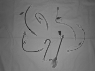
Surgical access to the pharynx requires the insertion of a Boyle Davis gag. To facilitate this, a secure airway is usually maintained with a ‘south-facing’ moulded tracheal tube (Fig. 29.1). Alternatively, a reinforced laryngeal mask airway (LMA) can be used successfully provided that the surgeon carefully avoids displacement of the LMA during the insertion and removal of the gag.

Spontaneous ventilation following the use of a short-acting muscle relaxant can be used to facilitate deep extubation in the lateral head-down position to protect the airway from soiling during emergence. Alternatively, positive pressure ventilation can be maintained throughout the procedure, with extubation fully awake in the sitting position. Various surgical techniques can be employed including cold steel dissection, electrodiathermy, laser and coblation. Blood loss can be significant and vigilance must be maintained regarding fluid replacement; however, blood transfusion is rarely necessary.

Tonsillectomy is painful and requires adequate postoperative analgesia. This frequently involves a multimodal approach with an initial dose of intravenous morphine together with paracetamol and a non-steroidal anti-inflammatory drug (NSAID). The latter can be given orally before surgery or parenterally during the procedure. Some evidence may point towards an increased risk of bleeding associated with the use of NSAIDs, but this is not clear-cut and most centres use this combination of drugs to facilitate early discharge. Multimodal antiemetic therapy should also be used because postoperative nausea and vomiting is a frequent cause of delay in discharge. There is also evidence to support the use of steroids for control of emesis and pain, usually as a single dose of dexamethasone. Some evidence supports the use of topical or locally infiltrated local anaesthetic. The early establishment of oral intake of food, fluids and analgesia encourages early discharge and should enable most operations to be performed as a day-case.

Rigid Endoscopy and Microlaryngoscopy

Rigid endoscopy is performed commonly in ENT to facilitate examination, biopsy and treatment of abnormalities of the upper aerodigestive tract.

General anaesthesia is required, usually with tracheal intubation to provide a safe airway during surgery. Provided that no difficulty with intubation is predicted, intravenous or gaseous induction is followed by the administration of a muscle relaxant dependent on the anticipated duration of the procedure. In general, a small cuffed (microlaryngoscopy) tube with internal diameter 4–6 mm is inserted into the trachea to allow the surgeon greater access to the pharynx. This should be placed in the left side of the mouth to allow passage of the rigid endoscope down the right.

Examination, with or without biopsy, is usually of a short duration and mivacurium or suxamethonium is often used. Increasingly however, the operating microscope is used to resect neoplasms of the upper airway, especially laryngeal carcinoma, allowing less invasive damage to voice function. These operations may be prolonged, requiring attention to normothermia and fluid balance. Microlaryngeal tumour resection is often carried out using a precision laser cutting tool which requires either a tube specifically designed to tolerate lasers or extreme care on the part of the surgeon to avoid the risk of damage to the tube and potential airway fire. Short-acting opioids provide balanced anaesthesia but morphine may be required for longer operations. Blood loss is not usually significant and is often controlled by the topical application of adrenaline with or without local anaesthetic. Safe extubation is normally achieved with full emergence and recovery of airway reflexes, and careful pharyngeal suction prior to the removal of the tube.

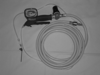
Occasionally, the surgeon requires access to the larynx without the presence of a tracheal tube. In this situation, oxygenation can be provided by jet insufflation of the lungs via a subglottic catheter or an attachment to the endoscope (Fig. 29.2). The catheter can be inserted into the trachea either down the endoscope or through the cricothyroid membrane. Anaesthesia is maintained using an intravenous agent, usually propofol.

Thyroid Surgery

Thyroid surgery is increasingly performed by specialist ENT surgeons although some general surgeons still undertake the operation.

Preoperative assessment of the anatomy of any goitre, usually by computed tomography, is vital to predict any impact on the ease of intubation of the trachea or ventilation of the lungs. Preoperative assessment also allows the surgical access route to be planned; this is usually via the neck, but may require intrathoracic access such as a sternal split if significant retrosternal extension of the tumour exists. Medical management of thyroid disease must be optimized preoperatively because abnormalities such as a thyroid storm can cause gross physiological problems intraoperatively.

For routine thyroid surgery via the neck, it is sufficient to provide balanced general anaesthesia usually, using a reinforced tracheal tube to allow the operative area to be draped safely without risking occlusion of the tube. If the goitre is causing significant subglottic stenosis without stridor, a small tube must be available which will pass easily through the stenosed area. If the stenosis extends into the chest towards and beyond the carina, specialized thoracic techniques such as bronchial intubation may be required. If any difficulty is anticipated in securing ventilation or if stridor exists, intravenous hypnotics and muscle relaxants are contraindicated until a definitive airway has been established.

Tracheostomy

Surgical tracheostomy is usually performed in a sedated or anaesthetized intubated patient. Occasionally, emergency tracheostomy is required in the unintubated patient, for example in stridor, and may even take place under local anaesthetic if general anaesthesia with a secure airway cannot be performed (see ENT emergencies below). Many procedures now take place percutaneously on the intensive care unit. If surgical tracheostomy is required, the patient is stabilized and the lungs ventilated in the operating theatre with the head and neck extended to allow access. When the surgeon has dissected down to the trachea, the lungs are ventilated with 100% oxygen and the tracheal tube is withdrawn carefully into the proximal trachea to allow the tracheal window to be excised without perforating the cuff. At this point, positive pressure ventilation of the lungs becomes impossible but in the event of surgical failure to insert the tracheostomy tube, the anaesthetic tracheal tube can be advanced back down the trachea past the defect to allow ventilation to be reinstituted. After the tracheostomy tube has been inserted, the breathing system is connected to it and ventilation confirmed with visualization, auscultation and capnography. The anaesthetic tube may be removed and discarded after the tracheostomy tube has been secured.

Nasal and Sinus Surgery

Various operations are performed on the nose and sinuses to treat and prevent epistaxis, to improve the nasal airway, to reduce the symptoms of chronic rhinosinusitis or to improve the external appearance of the nose. Nearly all can be performed as day-case procedures. Major invasive access to the nasal sinuses such as the Caldwell Luc procedure have largely been replaced by the use of endoscopic sinus surgery which is more cost-effective in terms of symptom relief.

Most nasal procedures in the UK are performed under general anaesthesia and range from simple diathermy of the inferior turbinates to prolonged cosmetic external rhinoplasty. The application of a mixture of topical local anaesthetic agents and other adjuncts (e.g. Moffat’s solution, which is a mixture of cocaine, adrenaline and bicarbonate) provides vasoconstriction before surgery. The airway must be secured to allow the delivery of oxygen and a volatile anaesthetic agent and also to protect the trachea from soiling by blood from the operative site. This can be achieved satisfactorily by the use of a reinforced LMA if there are no specific indications for tracheal intubation such as obesity or the expectation of a prolonged operation. Special attention must be paid to avoid disconnection or occlusion of the breathing system by the surgeon, or soiling of the trachea. Balanced anaesthesia is achieved using increments of a short-acting opioid or a longer-acting drug for prolonged or painful procedures. Careful pharyngeal suction is performed at the end of surgery to ensure the removal of blood and other debris which may have accumulated. The use of a pharyngeal pack is generally unnecessary but, if used, it is vital to ensure that it has been removed before emergence. Serious complications, including death, have been reported after failure to remove a throat pack. The usual principles applying to day-case anaesthesia are adhered to including preoperative assessment and postoperative care (see Ch 26).

Ear Surgery

Examination under anaesthetic, suction clearance and myringotomy with insertion of grommets are extremely common operations, particularly in children, and are performed to relieve the symptoms of chronic otitis media with effusion and to improve hearing. They may be combined with adenoidectomy and tonsillectomy for recurrent tonsillitis or chronic rhinosinusitis. Recent guidance published by NICE may reduce the prevalence of surgical management of these conditions. In general, these are quick operations requiring attention to the principles of paediatric day-case anaesthesia. Postoperative pain is usually managed with a combination of paracetamol and an NSAID to allow early discharge.

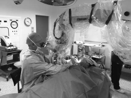
More complex procedures are performed on the structures of the ear using a microscope (Fig. 29.3), such as tympanoplasty to repair defects in the tympanic membrane, mastoidectomy to reduce the risk of abscess and infection in the mastoid air cells and stapedectomy to improve hearing in otosclerosis. These are performed under general anaesthesia, increasingly as day-case procedures. Moderate hypotension has been employed to minimize bleeding in the operative field but hypotensive agents such as β-blockers and vasodilators have largely been superseded by the use of short-acting narcotic agents given by intermittent bolus or infusion. These provide smooth anaesthesia without variations in blood pressure associated with surgical bleeding, which can obscure the surgeon’s view through the microscope. If hypotensive anaesthesia is to be employed, care should be taken to maintain vital organ perfusion and keep the mean arterial pressure above the lower limit of autoregulation of about 55 mmHg. In general, these techniques require positive pressure ventilation with an element of muscle relaxation. A ‘south-facing’ moulded tracheal tube or reinforced LMA can be used. Nitrous oxide is avoided in the gas mixture because it can increase the pressure in the middle ear, thereby increasing the risk of graft failure. Postoperative pain is not usually severe and can usually be managed using oral analgesia, often allowing same-day discharge.

ENT Emergencies

Bleeding Tonsil

Primary haemorrhage occurs in the immediate postoperative period, usually in the recovery room, whereas secondary haemorrhage occurs at home some days later and may present via the Emergency Department. Blood loss may be profound and, occasionally, death still occurs as a result of post-tonsillectomy haemorrhage. Initial treatment involves attention to the primary goals of life support with the establishment of good intravenous access and volume resuscitation as well as the administration of oxygen. A blood sample must be sent for urgent cross-match. Emphasis has been placed on restoration of blood volume and pressure before induction of anaesthesia but in the face of profuse active bleeding, urgent surgical haemostasis is of paramount importance. Most experienced practitioners advocate a rapid sequence induction of anaesthesia with cricoid pressure because the stomach may be full of blood. Preoxygenation is often difficult with the patient sitting up regularly spitting out blood, but it is vital. As soon as loss of consciousness has been achieved, the patient is placed in the supine position and the trachea is intubated as quickly as possible, with suction readily available. Alternatively, gaseous induction can take place in the left lateral head-down position and the trachea intubated under deep inhalational anaesthesia. When haemostasis has been achieved, full resuscitation takes place. Restoration of normal blood pressure must be ensured to reveal all potential bleeding points. Consideration can then be given to postoperative management, which will depend on the condition of the patient.

Epistaxis

Common causes of epistaxis include surgery, trauma and spontaneous epistaxis, the last particularly in elderly hypertensive patients. Blood loss can be profound and resuscitative measures may be necessary. General anaesthesia may be required to facilitate anterior or posterior packing of the nose, ligation of the sphenopalatine or anterior ethmoid arteries (often done endoscopically) and occasionally exploration of the neck to tie off the external carotid artery. Elderly patients are often on long-term anticoagulant therapy which may require urgent reversal using vitamin K and/or plasma derivatives, and blood samples must be sent for cross-matching. The patient has usually ingested significant amounts of blood and rapid sequence induction with tracheal intubation is mandatory. If haemorrhage is swift and ongoing, preoxygenation may take place in the sitting position with immediate transition to the supine position with the application of cricoid pressure as soon as consciousness is lost. When surgical control has been achieved, normal blood pressure must be restored to reveal any further bleeding points prior to emergence and extubation fully awake. Occasionally, if surgical control is incomplete or tenuous, it may be prudent to keep the patient sedated with the trachea intubated to allow for full correction of clotting abnormalities and for clots to form and organize before extubation.

Epiglottitis and Stridor

Epiglottitis is an acute life-threatening illness characterized by inflammation of the epiglottis and surrounding structures which can progress rapidly to complete airway obstruction. Previously, it was considered to be primarily a disease of childhood but since the introduction of routine H influenzae type B vaccination, it is now seen less frequently in children and is more common in adults. Presentation commonly involves a combination of one or more of drooling, dysphagia and distress, often with other signs of systemic illness such as fever. The onset of respiratory symptoms such as breathlessness or stridor indicates an extreme emergency and action should be taken to secure the airway. The cause is usually H influenzae type B bacterial infection (even in previously immunized individuals) and treatment involves airway support and intravenous antibiotics. If progression of airway compromise is anticipated, intubation of the trachea is indicated. Airway management may be very difficult and senior anaesthetic and ENT support must be summoned urgently. Tracheal intubation should take place if possible in an operating theatre environment with ENT surgical support and equipment immediately available, including facilities for rigid bronchoscopy and emergency tracheostomy. Fear and stimulation are known to worsen the airway compromise, and it is recommended that attempted venous access should not be undertaken in a child before induction of anaesthesia. Intubation may be difficult because of distortion of the laryngeal anatomy and most experts advocate inhalational induction with laryngoscopy under a deep plane of anaesthesia, with equipment for difficult intubation readily to hand. In the adult, fibreoptic-assisted intubation has been recommended. The trachea must remain intubated on an intensive care unit until it is confirmed that the swelling has largely subsided, usually by fibreoptic nasendoscopy. If ventilation becomes compromised before intubation, creation of an emergency surgical airway may be necessary.

Stridor, defined as a harsh high-pitched noise of breathing usually on inspiration, may also be due to other pathologies. Treatment is aimed at resolving the cause but in the face of worsening respiratory distress, emergency intubation of the trachea may be required. The anaesthetic management principles are as above; senior anaesthetic and ENT personnel must be available and intubation should take place in an operating theatre environment. In general, supraglottic stridor caused by neoplasm or abscess may cause extreme difficulty in rigid laryngoscopy and specialized methods must be available such as flexible fibreoptic endoscopy or retrograde intubation. In the event of failure, rapid surgical access to the airway may be necessary. In the presence of periglottic stridor, e.g. caused by laryngeal neoplasia, or subglottic stridor, e.g. due to thyroid disease, tracheal intubation can usually be achieved using a rigid laryngoscope under deep inhalational anaesthesia. Intravenous hypnotics or muscle relaxants must never be administered to a stridulous patient before securing the airway.

ORAL AND MAXILLOFACIAL SURGERY

Oral Surgery

The scope of modern oral surgical practice under anaesthesia continues to encompass primarily the removal of impacted teeth and treatment of associated dental pathology which cannot be dealt with under local anaesthesia by either the dentist or the oral surgeon due to the severity of the disease or the inability of the patient to tolerate dental procedures. Referral guidelines now restrict the removal of impacted third molar teeth only to patients who are symptomatic, which has reduced the incidence of those operations.

Consideration must be given to the principles of management of the shared airway and often nasotracheal intubation is required to allow a safe airway with good surgical access to the mouth. Careful attention is paid to the correct length of tube, and visualization of the reservoir bag and capnography. The muscle relaxant used is determined by the anticipated length of the procedure and whether the anaesthetist prefers to use a spontaneous breathing technique or positive pressure ventilation. In simple surgery, the anaesthetist and surgeon may elect to work around a laryngeal mask airway but vigilance is required to avoid displacement or disconnection, leading to ventilation problems. The use of a throat pack is largely historical because the surgeon should pay close attention to pharyngeal toilet at all times and the airway used should protect the trachea from soiling. Extubation may occur either under deep anaesthesia in the lateral head-down position, or fully awake. Most procedures are undertaken as day-cases and the principles of pain control involve a multimodal combination of local anaesthetic infiltrated by the surgeon, simple analgesics, and NSAIDs. Strong opioids are rarely required.

Orthognathic Surgery

Patients with severe facial architecture abnormalities or bite asymmetry problems may present for treatment requiring mandibular or maxillary osteotomy and advancement procedures. This occurs generally at a young age with minimal comorbidities but requires a general anaesthetic, usually lasting for several hours. Difficult laryngoscopy may be anticipated in patients with a severely retrognathic mandible and can require flexible fibreoptic intubation. Nasotracheal intubation is necessary and attention must be paid to fluid balance and intraoperative temperature control. Prophylactic intravenous antibiotics are given because microplates and screws are inserted to fix the skeleton into the new position. Postoperative pain and swelling can be severe and patients are often monitored on a high dependency unit and given ice packs and morphine if required. Postoperative jaw wiring is rarely indicated but multimodal antiemetic therapy is still important, including dexamethasone, which may reduce swelling as well as emesis. Paediatric orthognathic surgery, e.g. for cleft lip or palate repair, may also be performed by oromaxillofacial surgeons.

Facial Trauma and Fractures

In the field of maxillofacial trauma, the anaesthetist may be called upon to provide help in the treatment of the acutely injured patient in the Emergency Department or, more commonly, in the operating theatre for scheduled repair of facial fractures sustained previously.

Facial trauma due to road traffic accidents became less common after the implementation of legislation relating to seat belts and driving while intoxicated, but this has been more than offset in the UK by the increase in alcohol-related violent injury. The management of trauma follows the principles of resuscitation of airway, breathing and circulation and this is the primary goal particularly in victims of polytrauma. In facial trauma, airway difficulties may result from obstruction, disruption of normal anatomy, intoxication and cervical spine immobilization. Emergency tracheal intubation may be required if the clinical features of respiratory obstruction, hypoxaemia or coma are progressing and also if the anticipated course of events is likely to lead to airway compromise, e.g. facial burns. In these circumstances, rapid sequence induction of anaesthesia with rigorous preoxygenation and cricoid pressure is the technique of choice if extreme difficulty in intubation is not anticipated. Equipment must be available for difficult laryngoscopy such as the gum elastic bougie and McCoy laryngoscope, and senior anaesthetic personnel familiar with these techniques must be present. In the event of failure to intubate the trachea, a plan must be in place to maintain oxygenation if bag-mask ventilation is difficult. This may involve the laryngeal mask airway followed by cricothyroid airway access if that fails. If intubation using direct laryngoscopy is predicted to be extremely difficult or impossible, awake intubation may be necessary, or tracheostomy can be performed under local anaesthesia. Either technique can be challenging in the intoxicated and combative individual. Some advocate the ‘awake look’ which involves gentle insertion of the laryngoscope before anaesthesia to ascertain whether a good view of the posterior pharyngeal structures can be obtained. If this is satisfactory, intravenous induction of anaesthesia may be attempted safely.

Patients with facial fractures usually present for reduction and fixation 24–48 h after injury, when swelling has subsided, intoxication is no longer present and the presence of a significant head injury has been excluded. Depending on the mechanism of injury, the fracture may be mandibular, mid-face or isolated fractures of the zygoma or orbit. General anaesthesia is required and tracheal intubation is necessary in all but the simplest zygomatic elevation in a starved patient. Attention must be paid to the principles of the shared airway and consideration given to the optimal route of access to allow surgical intervention to proceed. Rarely, difficult intubation may be encountered because of anatomical disruption or residual swelling. Reduced mouth opening is usually caused by pain and stiffness. Most repairs of the mandible or mid-face involve intraoperative intermaxillary fixation to optimize postoperative function and nasotracheal intubation is required. Isolated orbital or zygomatic repairs can be managed with a ‘south-facing’ oral tube. Occasionally, nasotracheal intubation is not possible if complex repairs involving the naso-ethmoidal bony skeleton are also to be undertaken, or if a fracture of the base of the skull is suspected (which may occur in Le Fort III fractures of the mid-face). In these circumstances, tracheostomy may be required or, alternatively, submental intubation in which the tube is passed via an incision in the floor of the mouth through the oral cavity and into the trachea.

When the operation has been completed, the tracheal tube can be removed safely and the patient should be nursed in an environment in which postoperative pain and swelling can be monitored and treated.

ANAESTHESIA FOR HEAD & NECK CANCER SURGERY

Tumours of the head and neck can arise from the lips, oral cavity, salivary glands, nose or nasal sinuses, oropharynx, hypopharynx or larynx. Worldwide, cancer of the mouth and oropharynx is the tenth most commonly occurring form of cancer. The tumours are most commonly squamous cell carcinomas which metastasize to lymph nodes in the neck. Neck disease may present with an unknown primary which can often be identified by examination or radiological scanning. Squamous cell carcinomas are known to be associated with alcohol and tobacco use, and increase in incidence with age. However in the UK and other developed countries, for reasons which are unclear, there has been a recent increase in the incidence of oral cancer in the age group 40 to 65 years, particularly in men (Fig. 29.4). Surgery, radiotherapy or chemotherapy may be the primary treatment modality depending on patient factors and the staging of the disease. Combinations are often used. Surgical resection may be performed by ENT surgeons or maxillofacial surgeons depending on the site of the tumour, and the expertise of plastic surgeons may be used for the reconstruction of defects. Anaesthesia must create conditions which allow surgery to take place safely, ensuring physiological support of all systems, lack of awareness, pain control and excellent surgical access.
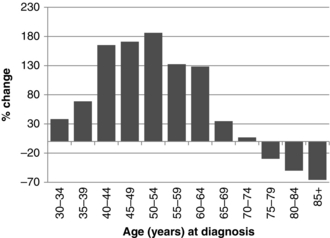
Laryngectomy

Small superficial tumours of the larynx are treated surgically by laser microlaryngoscopy (see above) or by partial or hemilaryngectomy. Total laryngectomy is performed for more advanced disease, usually without neck dissection because tumours of the glottis rarely metastasize to the neck. Preoperative assessment of patients for laryngectomy must establish whether there is any evidence of respiratory compromise, which may be due to airway obstruction by the tumour or concurrent smoking-related illness. Flexible endoscopic examination of the glottis and CT staging of the neck can be used to predict difficulty in direct laryngoscopy. Tumours at the glottic level rarely present difficulty in intubation and, if no respiratory obstruction exists, intravenous induction followed by tracheal intubation can be performed. A smaller non-cuffed tube may be required if there is significant glottic or subglottic stenosis. If respiratory obstruction with stridor is present, inhalational induction and laryngoscopy under deep inhalational anaesthesia should be performed. Fibreoptic intubation may be necessary if difficult laryngoscopy is encountered or predicted, e.g. because of radiation-induced scarring of the floor of the mouth. During surgery, an end-tracheostomy is fashioned and a tracheostomy tube is inserted after the trachea has been divided below the larynx. To facilitate the division, the oral tracheal tube must first be withdrawn proximally and the lungs ventilated with 100 % oxygen. The operation usually lasts for several hours and attention must be paid to intraoperative warming, monitoring and fluid balance.

Neck Dissection

This may be performed as a curative procedure or for local control of disease. It is usually combined with excision of the primary lesion, which may be oropharyngeal or nasal, but it is performed occasionally as a sole procedure if the primary is unknown or has been treated using another modality such as radiation. Cervical lymph tissue is dissected out, preserving blood vessels and nerves if possible. The tissue can then be examined microscopically to stage the disease and guide further treatment and prognosis. The neck dissection is often followed by creation of a free tissue graft pedicle with anastomoses to the blood vessels in the neck. General anaesthesia with tracheal intubation is required, using a reinforced tube facing away from the neck to allow access. A long procedure should be anticipated. In radical neck dissection, the internal jugular vein may be ligated; if this is performed bilaterally, severe oedema of the face and neck may develop and tracheostomy may be necessary to protect the airway.

Surgery for Oral, Nasal and Oropharyngeal Cancer

If surgery is used, it generally consists of wide excision of the primary tumour, dissection of the cervical lymph nodes (depending on the staging of the disease) and reconstruction of the resulting defect. Procedures include glossectomy, pharyngectomy, maxillectomy and mandibular resection. Operations can be extremely prolonged (up to 15 h), particularly if microvascular tissue transfer is performed.

Preoperative assessment may reveal significant co-morbidities, particularly in the elderly, and these may influence the anaesthetic technique and postoperative care. Supraglottic tumours can cause difficult intubation and this should be anticipated by careful examination of the patient and radiological investigations. Techniques such as awake fibreoptic intubation or the use of other difficult airway devices may be required.

The functions of airway control and ingestion are usually secured by insertion of a tracheostomy and gastrostomy feeding tube which can be removed after bleeding and swelling have resolved and healing has produced a stable and secure tract. Prolonged operations require careful attention to fluid balance and warming, and blood loss may be significant.

Reconstruction of defects is an important element of surgery. This commonly involves the insertion of a tissue graft into the defect. The graft may be swung on a vascular pedicle (e.g. pectoralis major flap) or a free tissue graft may be anastomosed with the blood vessels of the neck. A free flap tends to give a more favourable cosmetic and functional result but is extremely time-consuming. Grafts may be soft tissue only (e.g. radial forearm skin) or composite for bony defects (e.g. fibula). A free jejunal graft or a pull-up of the stomach from the abdomen may be required to restore integrity of the gastrointestinal tract if total pharyngectomy has been performed. Both will require laparotomy. Consideration should be given to allowing adequate rest breaks for the entire theatre team, including the anaesthetist. Postoperatively, the patient should be nursed on a high dependency unit where monitoring can continue and pain can be controlled effectively.

DENTAL ANAESTHESIA

Anaesthesia and dentistry have a strong historical association. The development of both disciplines and the increasing awareness of the benefits of dental care and oral hygiene resulted in the uncontrolled proliferation of the use of anaesthesia in dental practice, often by dentists themselves, who had had minimal training in anaesthesia, sedation and resuscitation. At its peak in the 1950s, over 2 million outpatient dental anaesthetics were given annually in the UK. Disquiet relating to the possible risks of death associated with the use of anaesthesia in dentistry resulted in the Department of Health commissioning a report in 1990 which recommended that general anaesthesia should be avoided if possible and that pain and anxiety associated with dental procedures should be ameliorated by local anaesthesia and conscious sedation. Further reports and guidelines since then have reinforced this viewpoint and general anaesthesia is now reserved almost exclusively for small children, and adults with learning difficulties. General anaesthesia for dental procedures must be administered in a hospital setting with full theatre resources and anaesthetic support. Conscious sedation can be used in the dental surgery if anxiolysis is required. This is administered by an anaesthetist or trained sedationist. The traditional dental chair anaesthetic has ceased to exist.

General Anaesthesia

In paediatric practice, the vast majority of procedures are for the extraction of carious teeth. There is a low incidence of systemic disease but upper respiratory tract infections are common. Premedication may consist of topical local anaesthetic cream together with an oral analgesic and, if necessary, a sedative such as oral midazolam. Psychological preparation is an important element and is best carried out in a specialized paediatric environment. The majority of procedures are extremely short. Induction may be intravenous or inhalational, and airway support is commonly provided using a face mask, nasal mask or laryngeal mask airway delivering a combination of oxygen, nitrous oxide and a volatile anaesthetic agent. The principles of care of the shared airway must be observed and good communication between the anaesthetist and dentist is essential. Full anaesthetic monitoring is required, although it may be necessary to wait until after induction of anaesthesia in unco-operative patients. A combination of simple analgesics and NSAIDs is generally used for pain control and allows early discharge.

In the adult, general anaesthesia for simple dentistry is indicated only for patients who are unable to tolerate local anaesthesia with sedation for psychological reasons or those unable to co-operate, e.g. because of severe learning difficulties. The vast majority of dental procedures under general anaesthesia in adults are performed when it is technically difficult to achieve the result without general anaesthesia, e.g. severely impacted third molar teeth or roots, or if poorly controlled carious disease has resulted in significant infection or anatomical derangement. These operations take place on oral surgery lists, commonly as day-case procedures. Nasotracheal intubation may be required for more difficult operations (see above). Patients are generally young and fit but concurrent disease may be present in patients undergoing a total dental clearance before treatment for head and neck cancer or cardiac valve surgery.

Sedation

The greatly reduced use of general anaesthesia in dental surgery has resulted in an increase in the use of sedative techniques to allow surgery to take place comfortably in anxious patients or when complex dental work is undertaken. Sedation is defined as the use of a drug or drugs to render a state of reduced consciousness to allow treatment to be carried out but in which verbal contact is maintained with the patient throughout the period of sedation. The drugs and techniques used should carry a margin of safety large enough to make loss of consciousness unlikely. In the UK, intercollegiate working parties from dentistry and anaesthesia have developed guidance relating to the safe conduct of sedation in dental surgery, stressing the need for adequate equipment, training, and documentation. It is recognized that potentially life-threatening complications may occur rarely.

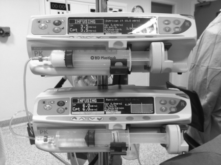
Common techniques involve the administration of an intravenous benzodiazepine or the use of nitrous oxide. Sedation may be administered by the operator before surgery or there may be a dedicated sedationist. Anaesthetists are often asked to take on this role and may use more complex techniques such as a continuous infusion of propofol using a target-controlled infusion device (Fig. 29.5).

FURTHER READING

Chesshire, N.J., Knight, D.J.W. The anaesthetic management of facial trauma and fractures. BJA CEPD Reviews. 2001;1:108–112.

Chester, A.C., Antisdel, J.L., Sindwani, R. Symptom-specific outcomes of endoscopic sinus surgery: a systematic review. Otolaryngol. Head Neck Surg. 2009;140:633–639.

Goldman, A.C., Govindaraj, S., Rosenfeld, R.M. A meta-analysis of dexamethasone use with tonsillectomy. Otolaryngol. Head Neck Surg. 2000;123:682–686.

Grainger, J., Saravanappa, N. Local anaesthetic for post tonsillectomy pain: a systematic review and meta-analysis. Clin. Otolaryngol. 2008;33:411–419.

http://eng.mapofmedicine/evidence/map/epiglottitis_adult_2.html

http://guidance.nice.org.uk/CG60

http://guidance.nice.org.uk/TA1

http://rcoa.ac.uk/docs/SCSDAT.pdf

http://www.hesonline.nhs.uk

http://www.rcoa.ac.uk/docs/GPAS-headneck.pdf

Marret, E., Flahault, A., Samama, C.M., et al. Effects of postoperative, nonsteroidal, anti-inflammatory drugs on bleeding risk after tonsillectomy: meta-analysis of randomized, controlled trials. Anesthesiology. 2003;98:1497–1502.

Mehanna, H., Paleri, V., West, C.M., Nutting, C. Head and neck cancer – Part 1: epidemiology, presentation, and prevention. Br. Med. J. 2010;341:663–666.

Murphy, M.F., Walls, R.M. Identification of the difficult and failed airway. In: Walls R.M., ed. Emergency airway management. second ed. USA: Lippincott Williams & Wilkins; 2004:70–82.

Ravi, R., Howell, T. Anaesthesia for paediatric ear, nose, and throat surgery. Continuing Education in Anaesthesia Critical Care and Pain. 2007;7:33–37.

Somerville, N., Fenlon, S. Anaesthesia for cleft lip and palate surgery. Continuing Education in Anaesthesia Critical Care and Pain. 2005;5:76–79.

Van den Aardweg, M.T., Schilder, A.G., Herkert, E., Boonacker, C.W., Rovers, M.M. Adenoidectomy for otitis media in children. Cochrane Database Syst. Rev. 2010;20(1):CD008282. Jan