Anaesthesia for Cardiac Surgery

Published on 27/02/2015 by admin

Filed under Anesthesiology

Last modified 22/04/2025

Print this page

rate 1 star rate 2 star rate 3 star rate 4 star rate 5 star
Your rating: none, Average: 0 (0 votes)

This article have been viewed 1538 times

Anaesthesia for Cardiac Surgery

In the United Kingdom and much of the developed world, more than half of all cardiac surgical procedures are undertaken to revascularize ischaemic myocardium. Of the remainder, surgery for acquired valvular disease, congenital anomalies and disorders of the great vessels comprise the majority. Impaired ventricular function is not uncommon in this group of patients, the severity of which may greatly affect the conduct of anaesthesia and surgery as well as outcome. The combination of underlying cardiac pathology, comorbid conditions and concomitant medications – such as β-blockers and angiotensin converting enzyme (ACE) inhibitors – make many patients with cardiac disease susceptible to the adverse haemodynamic effects of anaesthetic agents, particularly peripheral vasodilatation. Regardless of the disease process or state, all efforts should be made to maintain haemodynamic stability and promote a positive myocardial oxygen balance during anaesthesia and throughout the postoperative period.

Undoubtedly, there is more equipment and technology on show in the cardiac surgical theatre than in other operating theatres, and the number of staff present is often large. This makes familiarity with equipment and multidisciplinary team working imperative, as well as a specialist knowledge of cardiovascular and respiratory physiology. The replacement of the functions of the heart and lungs by cardiopulmonary bypass (CPB) is often required, although some coronary surgery can be performed on the beating heart (off pump). Indeed, novel ‘minimally invasive’ methods may allow repair of various structures within the heart and even valve replacement or repair without CPB, and this is an exciting area of development.

TRENDS IN SURGICAL PRACTICE

The 6th National Adult Cardiac Surgical Database Report, published in 2009 by the Society for Cardiothoracic Surgery (SCTS) in Great Britain and Ireland, provides detailed information about trends in UK cardiac surgical practice. Although the total number of cardiac surgical procedures carried out in the UK in the period 2001–2008 increased year-on-year, coronary artery bypass graft (CABG) surgery plateaued at just under 23 000 operations per year, possibly due to advances in percutaneous intervention. Coincidentally, the number of elderly patients undergoing cardiac surgery of all types increased such that patients over the age of 75 years now make up more than 20% of the cardiac surgical population, and over 5% are over 80 years of age. Despite this, crude mortality rates decreased significantly between 2001 and 2008: 2.3% to 1.5% for isolated CABG; 5.2% to 3.5% for isolated valve surgery and 8.3% to 6.1% for combined procedures.

Ischaemic Heart Disease

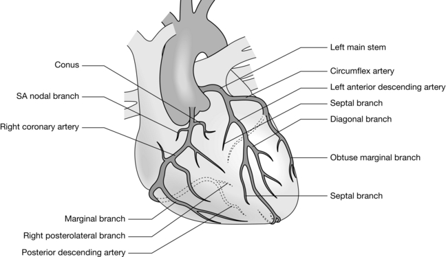
The concept of aorto-coronary bypass grafting for the relief of coronary ischaemia was conceived and performed in animals in the early 1900s. It was not until the 1960s, following development of the heart-lung machine and the chance discovery of coronary angiography, that direct revascularization of the ischaemic myocardium using the autologous saphenous vein (Fig. 34.1) replaced indirect therapies such as sympathectomy, thyroidectomy and pericardial poudrage.

Since being popularized in the late 1960s, coronary artery bypass grafting (CABG) has become the most commonly performed cardiac operation. The internal mammary artery is used routinely as a graft conduit, and there is good evidence that this provides good survival benefit. Complete arterial revascularization is possible using arteries such as the radial and epigastric arteries. Improved surgical techniques have increased the popularity of off-pump coronary artery surgery but its precise role remains uncertain and any advantages over surgery using CPB have not yet been proven. Technological advances in coronary stent technology, especially coated (drug eluting) stents have led to a huge expansion in the use of percutaneous coronary intervention (PCI; angioplasty, atherectomy and stenting) in the cardiac catheter laboratory. These procedures are typically performed under sedation, and length of stay in hospital and return to normal activities is undoubtedly markedly improved. However, the long-term efficacy of stenting has recently been called into question, and traditional CABG, once thought to be in terminal decline, remains a popular procedure.

Valve Disease

Stenosis or incompetence (regurgitation or insufficiency) most commonly involves the mitral and aortic valves. The most common diseases are calcific degeneration (causing aortic stenosis, with or without regurgitation), chronic rheumatic disease (affecting mitral and aortic valves) and myxomatous disease (most often causing mitral regurgitation). It should be borne in mind that valve dysfunction may occur as the result of systemic disease (e.g. carcinoid syndrome, infective endocarditis) and disruption of nearby anatomical structures (e.g. aortic regurgitation in acute dissection of the ascending aorta and mitral regurgitation following papillary muscle rupture).

Surgery usually entails repair or prosthetic replacement, guided by intraoperative transoesophageal echocardiography (TOE). The use of bioprosthetic or ‘tissue’ (porcine, bovine, cadaveric homograft) valves obviates the necessity for, and risks associated with, life-long anticoagulation but exposes the patient to the prospect of reoperation within 15–20 years. In contrast, mechanical (tilting disc) valves tend to last longer than bioprostheses and are therefore better suited to younger patients and those already anticoagulated for other reasons (e.g. chronic atrial fibrillation). Improvements in technology have led to some prostheses lasting more than 20 years, especially in patients aged > 70 years at the time of surgery. Minimally invasive transcatheter aortic valve replacement (TAVR) is now possible, making ‘redo’ sternotomy unnecessary in case of valve failure, by inserting a new tissue valve within the old prosthesis.

Cardiopulmonary Bypass

The essential components of a cardiopulmonary bypasshe essential components of a cardiopulmonary bypass (CPB) circuit (Fig. 34.2) are:
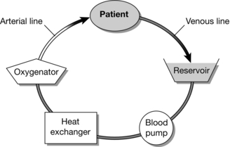
Full anticoagulation of the patient, typically with unfractionated heparin, is required to prevent coagulation in the CPB circuit caused by contact between the blood and the plastic components, which would otherwise lead to potentially lethal CPB/oxygenator blockage and failure. Despite anticoagulation, blood/plastic contact leads to the release of a number of active substances which cause vasodilatation, consumption of clotting factors and fibrinolysis. These include cytokines, thromboxane A2 and leukotrienes, and they are responsible for the hypotension and increased bleeding associated with CPB.

Blood from the venous side of the circulation, the venae cavae or right atrium, is drained by gravity to a venous reservoir, from where it is pumped into a gas exchange unit (oxygenator) where oxygen is delivered to, and carbon dioxide removed from, the blood. The blood can also be cooled or warmed efficiently at this point, using water pumped through a countercurrent heat exchanger located within the oxygenator. Oxygenated or ‘arterialized’ blood is then delivered into the systemic circulation, usually via a cannula in the ascending aorta. The heart and lungs are thus ‘bypassed’ or isolated and their function maintained temporarily by mechanical equipment remote from the body. Any blood in or around the bypassed heart (whether spilt or drained) may be drained and returned to the venous (cardiotomy) reservoir for filtration, oxygenation and subsequent return to the circulation.

Pumps

Roller pumps displace blood around the circuit by intermittent, semi-occlusive compression of the circuit tubing during each rotation. Intermittent acceleration of the roller head can be used to produce a ‘pulsatile’ pressure waveform although there is little evidence that a more physiological flow pattern improves outcome. Alternatively, a centrifugal pump may be used. Movement of a disc at very rapid speeds (> 3000 revolutions per minute) leads to exertion of gravitational force on blood and results in propulsion at a flow which is dependent on the resistance (afterload) offered by the arterial tubing and the patient’s systemic vascular resistance. There is some evidence that centrifugal pumps cause less blood component damage and activation, but this has not translated into improved outcome, and their use is usually confined to prolonged or complex surgery. Unlike roller pumps, which impede all flow when stopped, centrifugal pumps permit passive retrograde blood flow when switched off.

Fluid Prime

The CPB circuit must be primed with fluid (de-aired) prior to use. When CPB is commenced and the patient’s blood is mixed with the clear fluids which prime the bypass circuit, the haematocrit decreases by approximately 20–25%. Although oxygen content is reduced, oxygen availability may be increased by improved organ blood flow resulting from reduced blood viscosity. In some patients (low body weight, children or preoperative anaemia, when dilution would reduce the haematocrit to below 20%), blood may be added to the prime. In the normal adult, ‘clear’ primes are used almost exclusively (usually a crystalloid/colloid mixture). Most units have individual recipes for addition to the prime (e.g. mannitol, sodium bicarbonate and potassium) to achieve an isosmolar solution at physiological pH.

PREOPERATIVE ASSESSMENT

In recent years, there has been a trend towards the assessment of elective patients in pre-admission clinics, typically one to two weeks before surgery. This allows routine paperwork, laboratory tests and radiological imaging to be completed before admission, which may not be until the day of surgery. Despite undergoing an extensive array of specialized investigations to diagnose and quantify cardiac disease, there is evidence that a significant number of cardiac surgical patients have additional and hitherto undocumented pathology. Thorough preoperative evaluation by the anaesthetist remains an essential component of perioperative care. This should, at the very least, include confirmation of the documented history and symptoms, documentation of current drug therapy, a review of the results of diagnostic investigations and a physical examination focused on the cardiovascular and respiratory systems.

Cardiac Catheterization

Left heart catheterization typically comprises coronary angiography, aortography, left ventriculography and manometry. This provides the following information (Table 34.1):

TABLE 34.1

Cardiac Catheterization

Technique Procedure Parameter
Manometry Pressure measurement with catheter in aortic root and LV Aortic valve gradient
LV end-diastolic pressure
Angiography Coronary arteries selectively cannulated, contrast injected Coronary anatomy
Ventriculogram Catheter in LV, contrast injected very rapidly LV size and function
Ejection fraction
Severity of mitral regurgitation
Aortogram Catheter in aortic root, contrast injected Severity of aortic regurgitation

LV, left ventricle.

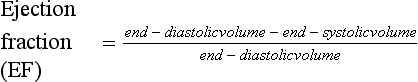
The efficiency of ventricular contraction (ejection fraction) can be estimated using the formula:

image

Right heart catheterization allows measurement of right heart and pulmonary artery pressures. When combined with measurements of cardiac output, these can be used to determine the pulmonary and systemic vascular resistances (Table 34.2).

TABLE 34.2

Measurements Obtained During Cardiac Catheterization

Parameter Normal Values
Left heart Systemic arterial/aortic pressure < 140/90 (mean 105) mmHg
LV pressure < 140/12 mmHg
Right heart RA pressure < 6 (mean) mmHg
RV pressure < 25/5 mmHg
PA pressure 25/12 (mean 22) mmHg
PAWP 12 mmHg
Cardiac index 2.5–4.2 l min– 1 m– 2
PVR 100 dyne s cm– 5
SVR 800–1200 dyne s cm– 5

LV, left ventricle; RA, right atrium; RV, right ventricle; PA, pulmonary artery; PAWP, pulmonary artery wedge pressure; PVR, pulmonary vascular resistance; SVR, systemic vascular resistance.

Echocardiography

Transthoracic echocardiography (TTE) is used frequently to define cardiac anatomy and assess ventricular and valvular function. TTE is non-invasive, and can be performed at intervals to monitor disease progression and to optimize the timing of surgical intervention before irreversible ventricular damage has occurred. It may also assist planning of the type of intervention required. Doppler techniques allow recognition of the direction and velocity of blood flow and are valuable in the diagnosis of valvular disease.

Unfortunately, TTE is of limited use in obese patients and patients with chronic lung disease (because of poor ultrasound windows caused by tissue or air). In addition, certain parts of the heart may not be visualized adequately because of their distance from the probe (such as the left atrium and interatrial septum). Therefore, transoesophageal echocardiography (TOE) may be required preoperatively (usually performed under sedation). TOE may also be indicated in mitral valve pathology to aid surgical decision-making between valve replacement and repair.

Preoperative Drug Therapy

Cardiac surgical patients typically take five or more prescription medicines for the control of symptoms (e.g. nitrates), cardiac risk modification (e.g. vasodilators, β-blockers) and the management of related conditions (e.g. hypoglycaemic agents, statins, bronchodilators). Care is required to balance the risks of discontinuation in the perioperative against the risk of major adverse cardiovascular events, e.g. withholding antiplatelet agents such as aspirin and clopidogrel.

β-Blocking agents. Continued administration of these drugs up to the time of surgery is desirable because discontinuation may increase the risk of perioperative myocardial infarction.

Calcium antagonists have a negative inotropic effect but, as with β-blockers, it is preferable to continue therapy throughout the perioperative period.

Nitrates should be continued to prevent rebound angina.

Digitalis. In most centres, digoxin is discontinued 24–48 h before surgery to diminish digoxin-associated arrhythmias after surgery.

Diuretics should be continued until the day before surgery.

Anticoagulants, including aspirin and clopidogrel, are usually stopped up to 1 week before surgery to permit platelet function to return towards normal. However, there is recent evidence that stopping aspirin is associated with increased morbidity, and it is continued throughout the perioperative period in many centres.

Angiotensin-converting enzyme (ACE) inhibitors are prescribed for hypertension and cardiac failure. They may produce significant vasodilatation and hypotension intraoperatively and postoperatively. Perioperative use varies from unit to unit; they may be stopped up to 1 week before surgery or continued until the day of operation.

Angiotensin receptor blockers (ARBs) are used for similar indications to ACE inhibitors and managed similarly.

Potassium-channel activators may be continued up to the day of operation.

Investigations

Blood count. Chronic anaemia (Hb < 120 g L− 1) should be investigated because it is associated with blood transfusion and worsened outcome. Anaemia may predispose to excessive haemodilution during CPB. Iron deficiency or any other cause should be investigated and treated before surgery if detected in advance. Quantitative platelet or leucocyte abnormalities should also be excluded.

Coagulation. Clotting studies should be performed before surgery. In the absence of anticoagulant administration, the finding of a seemingly trivial prolongation of the activated partial thromboplastin time (APTT) should prompt further investigation because it may indicate the presence of a coagulopathy (e.g. factor IX or XI deficiency).

Electrolytes. Serum potassium concentration should be within normal limits; hypokalaemia is invariably associated with hypomagnesaemia. Chronic diuretic therapy may produce total body sodium depletion and uraemia. Raised serum concentrations of urea and creatinine may indicate chronic renal insufficiency and an increased risk of postoperative renal failure.

Liver function tests. Abnormal values may indicate congestive cardiac failure, alcohol abuse or metastatic malignancy.

RISK ASSESSMENT

Outcome from cardiac surgery has been the subject of intense scrutiny for the last two decades. Despite advances in surgical techniques, anaesthesia and critical care, cardiac surgery still carries a finite risk of death and serious complications. While this risk has decreased steadily over the last 10 years (< 1% for isolated CABG or aortic valve replacement in young male patients), outcomes vary from centre to centre, and from surgeon to surgeon. Although helping the patient to understand the benefits (symptomatic and prognostic) and risks of surgery during the consent process is the responsibility of the surgeon, it is essential that the anaesthetist understands how risk is assessed so that the patient is not given contradictory information.

In the late 1980s, Parsonnet and colleagues identified 14 independent risk factors for death after cardiac surgery. The so-called Parsonnet score was adopted by many centres worldwide and is still in use today. However, most present day cardiac surgeons ‘out-perform’ Parsonnet, reducing the usefulness of the scoring system as a measure of both risk and surgical performance. The European System for Cardiac Operative Risk Evaluation (EuroSCORE), developed in the late 1990s, provides a more robust risk assessment, which, like its predecessor, can be calculated easily at the bedside (Table 34.3). The EuroSCORE has been validated in the UK, Europe and North America and has been shown to be predictive of major complications, duration of critical care stay and resource utilization.

TABLE 34.3

EuroSCORE—The European System for Cardiac Operative Risk Evaluation additive risk stratification model. http://www.euroscore.org.

Factor Points
Age Per 5 years or part thereof > 60 1
Gender Female 1
Chronic lung disease Bronchodilators or steroids 1
Extra cardiac arteriopathy Claudication, carotid stenosis > 50%, abdominal aortic, limb artery or carotid surgery planned or undertaken 2
Neurological dysfunction Severe effect on ambulation or function 2
Previous cardiac surgery Pericardium opened 3
Serum creatinine > 200 μmol L– 1 before surgery 2
Active endocarditis On antibiotics 3
Critical preoperative state VT, VF, cardiac massage, invasive ventilation, inotropic support, IABP 2
Unstable angina Angina at rest requiring intravenous nitrates 2
LV dysfunction Moderate (LV EF 30–50%)
Poor (LV EF < 30%)
1
3
Recent myocardial infarct < 90 days 2
Pulmonary hypertension PA systolic > 60 mmHg 2
Emergency operation Carried out on referral, before next working day 2
Other than isolated CABG 2
Surgery on thoracic aorta 3
Post infarct septal rupture 4

VT, ventricular tachycardia; VF, ventricular fibrillation; IABP, intra-aortic balloon pump; CABG, coronary artery bypass graft; EF, ejection fraction; PA, pulmonary artery; LV, left ventricle.

For higher risk patients, calculation of the logistic EuroSCORE provides a more accurate prediction than the simple additive score. For example, a fit 60-year-old man with asymptomatic critical left main stem coronary artery stenosis and normal LV function undergoing elective CABG surgery has a EuroSCORE predicted mortality of 1% (0.94% logistic). In contrast, a 75-year-old woman with poor LV function, chronic pulmonary disease and class IV angina undergoing emergency CABG surgery has a predicted mortality of 13% (38.74% logistic).

MONITORING

Extensive and accurate physiological monitoring is essential throughout the perioperative period for the safe practice of cardiac surgery. Instrumental monitoring should be considered an adjunct to, rather than a replacement for, routine clinical observation of the patient.

Electrocardiograph

The ECG should be monitored throughout the perioperative period. The ideal system is one which allows simultaneous multiple-lead monitoring or at least switching between leads II and V5, for accurate identification of ischaemia. Rate and rhythm should also be observed.

Echocardiography

Transoesophageal echocardiography (TOE) is indicated whenever valve surgery is undertaken, in cases of impaired left ventricular function or when the underlying diagnosis is uncertain. Performed immediately before surgery, it may be used to help guide the surgeon in the choice of procedure (e.g. valve repair). In addition, it may identify lesions which have not been detected or diagnosed correctly during preoperative evaluation, and may change surgical practice in up to 15% of cases. Immediately after CPB, the adequacy of surgery can be judged and the need for re-operation in case of failure identified. Abnormal motion of the ventricular wall (dyskinesia or akinesia) detected at this time but not present before surgery may indicate myocardial ischaemia, prompting further surgical revascularization or inotropic support.

Cerebral Monitoring

Despite being available for many years, monitors of cerebral function and cerebral substrate (oxygen) delivery are rarely used during routine cardiac surgery. In recent years, however cerebral near-infrared spectroscopy (NIRS) has gained popularity. This non-invasive technique allows measurement of capillary oxygen saturation in the frontal cortex throughout the perioperative period, and may prompt titration of haemodynamic and respiratory indices to improve cerebral oxygen delivery or reduce consumption (cooling). The effect of such measures on outcomes remains unproven and the subject of ongoing research.

PATHOPHYSIOLOGY

The anaesthetist should have a clear understanding of the fundamental principles of cardiac and cardiovascular physiology. Accurate monitoring reveals alterations in cardiac function and permits the anaesthetist to manipulate factors which ensure adequate cardiac output and myocardial blood supply.

Preload and contractility determine the work performed by the heart (Fig. 34.3). In the failing heart, afterload determines the work expended in overcoming aortic pressure compared with that used to provide forward flow. Thus, cardiac output may be increased by increasing preload or contractility, or by reducing afterload. Any increases in heart rate, contractility, preload or afterload result in increased myocardial oxygen consumption. For this reason, augmentation of cardiac output by increasing preload or contractility may have a detrimental effect on oxygen balance. Reducing afterload may increase cardiac output while simultaneously reducing oxygen demand.
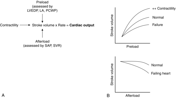
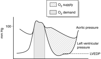
Adequate coronary perfusion demands the maintenance of an adequate diastolic aortic pressure. Oxygen supply to the myocardium occurs predominantly during diastole and is dependent on the gradient between diastolic aortic pressure and intraventricular pressure, and on the duration of diastole. The portion of myocardium most at risk of developing ischaemia is the left ventricular endocardium. Figure 34.4 illustrates how these variables affect oxygen supply and demand in the myocardium and how a satisfactory supply/demand ratio may be preserved.

Care of the patient with valvular heart disease depends on the type, severity and consequences of the valvular lesion. In general, patients with valvular heart disease are intolerant of extremes in heart rate. In patients with valvular regurgitation, reducing afterload tends to reduce the regurgitant fraction and increases forward flow. In contrast, patients with valvular stenosis often require increased preload and are intolerant of acute reductions in peripheral resistance (afterload). This is particularly true of patients with aortic stenosis, when most of the afterload to left ventricular ejection is caused by the stenosed valve itself. This afterload is fixed and cannot be reduced simply by lowering peripheral resistance. Vasodilatation in these patients produces marked hypotension and results in failure of perfusion of the hypertrophied myocardium with no increase in forward flow through the stenosed valve. Furthermore, diastolic dysfunction secondary to left ventricular hypertrophy dictates that the majority of ventricular filling occurs later in diastole as a consequence of atrial systole. For this reason, atrial fibrillation and nodal (junctional) rhythms may be tolerated poorly.

ANAESTHETIC TECHNIQUE

There is no single preferred anaesthetic technique for cardiac surgery. The choice of a specific drugs is less important than the care with which they are administered and their effects monitored. The techniques described here are suitable for standard CABG with cardiopulmonary bypass. Anaesthesia for off-pump coronary surgery may be complicated by significant haemodynamic disturbances while the heart is positioned by the surgeon and by intraoperative myocardial ischaemia when coronary arteries are cross-clamped during anastomosis of the grafts. The reader is directed to more specialized texts for details of management.

Induction of Anaesthesia

All drugs and equipment should be ready and the theatre and bypass circuit available for immediate use before the patient arrives in the anaesthetic room. Cross-matched blood should also be available immediately in case of rapid deterioration or surgical misadventure.

Before induction, ECG electrodes should be applied and the ECG trace displayed. Arterial and large-gauge venous cannulae should be inserted under local anaesthesia along with adequate sedation to reduce stress (e.g. midazolam 1–2 mg). The lungs should be preoxygenated.

Induction may be achieved in a variety of ways. Most often, a large dose of an opioid analgesic (e.g. fentanyl 5–15 μg kg–1) is administered with a benzodiazepine (e.g. midazolam 0.05–0.1 mg kg–1) to obtain unconsciousness. Alternatively, a small dose of propofol (0.5–2 mg kg–1) together with opioid or benzodiazepine may be used; consciousness is obtunded by an opioid in moderate dose and hypnosis is then produced by a small dose of an intravenous induction agent. An alternative is a target-controlled infusion of propofol with the target concentration increased in small steps, accompanied by an infusion of a short-acting opioid such as alfentanil or remifentanil.

As consciousness is lost, a neuromuscular blocking drug is administered and ventilation supported when necessary. Almost all currently available relaxants have been used during cardiac surgery, although pancuronium remains a popular choice. The objective is to undertake tracheal intubation without cardiovascular stimulation and thus adequate analgesia and anaesthesia are required. A tracheal tube with a low-pressure, high-volume cuff should be used. Positive pressure ventilation is continued, usually with an oxygen/air mixture. Nitrous oxide, which may depress myocardial function and increase the volume of gaseous emboli, tends to be avoided in cardiac anaesthesia.

Percutaneous cannulation of a subclavian or internal jugular vein is performed using a multilumen catheter to allow monitoring and intravenous infusions. Ultrasound assistance should be used to facilitate central vein access, and reduces complications associated with this procedure. The nasopharyngeal temperature probe and a urinary catheter are inserted. Mechanical ventilation is continued with a breathing system containing a humidifier and bacterial filter.

Previously identified ‘high-risk’ patients, such as those with poor ventricular function or severe pulmonary hypertension, may require more extensive monitoring, e.g. a PA catheter. In the critical or emergency situation, the central venous catheter can be inserted under local anaesthesia, and induction of anaesthesia can be undertaken in theatre with the full team ready for immediate surgery.

Anaesthesia Pre-CPB

After the patient is prepared and draped, the sternum is opened and, if required, the left internal thoracic (mammary) artery is harvested. Many surgeons request discontinuation of mechanical ventilation during sternotomy to reduce the risk of direct injury to the lungs. The pericardium is then opened and the heart inspected. Heparin is administered (usually 300 IU kg–1), to prolong the activated clotting time (ACT). The target ACT depends on local protocols, but is typically > 400 s. The surgeon then places cannulae in the aorta and the vena cava. Cannulation is commonly undertaken via the right atrium into the inferior vena cava using a two-stage cannula, allowing drainage of both lower body (IVC) and upper body (right atrium). Alternatively, if the heart chambers are to be opened (for example when mitral valve surgery is scheduled), the cavae are cannulated separately.

The goals of haemodynamic management are to maintain a stable heart rate and arterial pressure during this period, particularly at moments of profound stimulation, notably skin incision, sternotomy and sternal retraction. If a technique based on opioids or volatile anaesthetics has been chosen, additional analgesia or inhalational anaesthesia should be given before stimulation. Alternatively, the opioid infusion rate can be increased temporarily as necessary, perhaps accompanied by increased target concentrations of propofol. The tendency of isoflurane to produce a ‘coronary steal’ (the diversion of blood away from ischaemic muscle) is considered not to be of clinical importance. There is evidence that volatile anaesthetic agents increase myocardial tolerance to ischaemia by a mechanism known as preconditioning thought to be mediated via ATP-dependent potassium channels.

If arterial pressure decreases during the period before institution of CPB, small doses of a vasoconstrictor (e.g. metaraminol or phenylephrine 250–500 μg) may be administered. A mean arterial pressure (MAP) sufficient to allow vital organ perfusion should be maintained. This will obviously depend on the individual patient, but in most patients MAP > 70 mmHg is sufficient. During aortic cannulation, hypertension should be avoided to reduce the risk of aortic dissection. Many surgeons request a maximum MAP at this delicate point in the procedure (typically MAP < 70 mmHg). Manipulation of the arterial pressure is required at frequent intervals to achieve a number of different goals.

Cardiopulmonary Bypass

Two factors complicate the provision of anaesthesia during CPB. First is the impact of haemodilution, hypotension, non-pulsatile blood flow and hypothermia on the pharmacokinetics of anaesthetic agents. Second is the inability to administer volatile inhalational agents via the lungs – mechanical ventilation is discontinued when full pump flow is reached and ventricular ejection ceases. Temporary loss of the lungs as a route for drug administration can be circumvented by use of total intravenous anaesthesia or the addition of a volatile agent to the CPB oxygenator ‘sweep’ gas.

Surgery is usually preceded by placing an aortic cross clamp proximal to the arterial cannula to isolate the coronary circulation. During CABG surgery, the distal anastomoses are usually fashioned first and the cross-clamp then removed to permit restoration of myocardial perfusion. Application of a side-biting aortic clamp then allows the proximal (aortic) anastomoses to be fashioned without interfering with perfusion of the native coronary arteries.

Myocardial Preservation

Most surgical techniques require that the heart be immobile. During CPB, the aorta is cross-clamped between the aortic cannula and the aortic valve, thus isolating the heart from the flow of oxygenated blood. Ischaemic damage to the myocardium can be reduced by hypothermia and the institution of diastolic cardiac arrest. The latter is typically achieved by instilling 500–1000 mL of crystalloid cardioplegic solution, often mixed with the patient’s blood, into the coronary arteries. Many cardioplegic solutions are available; the majority contain potassium and a membrane-stabilizing agent, such as procaine.

Myocardial cooling is achieved by using ice-cold cardioplegia and by pouring cold saline (4°C) into the pericardial sac. Depending on surgical preference, the patient may also be cooled systemically. This is most often carried out when more complex or prolonged surgery is proposed, to allow better organ preservation due to reduced metabolic rate. Administration of cardioplegia is usually repeated at regular intervals, for example every 20–30 min.

Perfusion on Bypass

At normothermia, a pump flow of 2.4 L min–1 m–2 of body surface area is required to prevent inadequate perfusion of the tissues. Mean systemic (arterial) pressure is dependent on pump flow and systemic vascular resistance. Controversy exists regarding the optimum perfusion pressure because essential organs, particularly the brain, may be damaged if mean arterial pressure is < 45 mmHg. Unfortunately, perfusion is difficult to assess clinically, especially in the hypothermic patient.

Following the onset of CPB, haemodilution causes marked decreases in peripheral resistance and arterial pressure, which in most instances resolve spontaneously in 5–10 min. If this does not occur, arterial pressure may be increased by raising systemic resistance with a vasoconstrictor. Frequently, peripheral resistance and arterial pressure increase during hypothermic CPB as a result of increasing concentrations of catecholamines, and then decrease during active rewarming because of profound vasodilatation.

Acid–Base Balance

The development of metabolic acidosis suggests that perfusion is inadequate and, if necessary (base deficit > 6–8 mmol L–1), sodium bicarbonate may be administered.

When systemic hypothermia is used during CPB, consideration needs to be given to the effect of hypothermia on the solubility of gases in blood, and how these affect values obtained from arterial blood gas analysis. As temperature decreases, the solubility of gases in liquids increases, and the proportion of gas in equilibrium with the gas phase (partial pressure) decreases, although the total content of each gas remains the same. The net result of this phenomenon is a metabolic acidosis secondary to reduced PaCO2. There are two strategies for dealing with this issue. Not correcting arterial blood gas measurements for temperature allows a normal pH to be maintained. This is known as alpha-stat, because this maintains the degree of ionization of alpha-histidine. Alternatively, adding additional CO2 to maintain a normal pH on the basis of corrected blood gas measurements is known as pH stat. In most centres, alpha-stat is used, but pH-stat offers a number of theoretical benefits in patients undergoing procedures requiring deep hypothermia.

Weaning from CPB

Following removal of the aortic cross-clamp, oxygenated blood flows into the coronary arteries again, washing out cardioplegia and repaying the oxygen debt. In many cases, the heart regains activity spontaneously. In a minority of patients, it starts to beat in sinus rhythm but reverts usually to ventricular fibrillation; internal defibrillation is required to convert fibrillation to sinus rhythm and is successful only if pH, serum potassium concentration, oxygenation and temperature are approaching normal values. The heat exchanger in the oxygenator is used to increase the temperature of blood, but peripheral temperature is often depressed for some time. If a spontaneous heartbeat cannot be maintained, external pacing via epicardial wires should be started.

When core body temperature exceeds 36°C, metabolic indices are normal and a regular heartbeat is present, the establishment of spontaneous cardiac output is attempted. By gradually restricting venous drainage to the venous reservoir, venous blood is diverted to the right atrium. When the pulmonary circulation has been restored, mechanical ventilation is restarted; 100% oxygen is given because the efficiency of pulmonary gas exchange is unknown at this stage and any gas bubbles which have not been vented may enlarge in volume if nitrous oxide or nitrogen (air) is introduced.

Left ventricular ejection produces an upward deflection on the arterial pressure trace after a QRS complex. If the myocardium is contracting satisfactorily, pump flow is reduced cautiously and the heart, now receiving all the venous return, achieves normal output.

Although arterial pressure is the most easily measured index of successful termination of CPB, it is merely the product of cardiac output and peripheral resistance. If there is doubt about efficiency of the heart, cardiac output should be measured and peripheral resistance derived.

Following successful termination of CPB, preload can be adjusted by retransfusing any blood left in the CPB circuit, by altering the patient’s posture and by administering a vasodilator or vasoconstrictor.

Low Cardiac Output State

Failure to wean a patient from extracorporeal support should prompt the resumption of CPB while the cause is identified and supportive treatment initiated. TOE may be particularly helpful in this situation, permitting continuous assessment of preload, ventricular wall motion and valvular function. If causes such as failing to ventilate the lungs or an appropriate vaporizer setting have been excluded, attention should be focused on cardiac contractility and afterload.

Coronary aeroembolism, which may occur during and after separation from CPB, causes myocardial ischaemia manifest as a regional ventricular wall motion abnormality and rhythm disturbance. The problem usually resolves when arterial pressure is elevated and the heart permitted to eject on CPB.

The choice of inotrope in this setting is largely a matter of personal preference; there is little evidence to suggest that one drug is superior to another. Despite the theoretical risk of worsened myocardial reperfusion injury, calcium salts (e.g. CaCl2 250–1000 mg) are commonly used first. Other drugs commonly used – either alone or in combination – include adrenaline (0.05–0.2 μg kg–1 min–1), dobutamine (2–20 μg kg–1 min–1) and dopamine (2–20 μg kg–1 min–1) by infusion; all increase myocardial oxygen demand and tend to precipitate tachyarrhythmias. Both adrenaline and dopamine cause vasoconstriction at high doses.

Phosphodiesterase III inhibitors, such as milrinone and enoximone, may be a suitable alternative or adjunct to conventional inotropes, particularly in patients with right ventricular dysfunction and pulmonary hypertension. By inhibiting the breakdown of cytosolic cyclic adenosine monophosphate, they improve myocardial performance and dilate both arterioles and veins. By reducing afterload, they reduce myocardial oxygen demand and augment ventricular ejection. Arterial hypotension, more commonly seen with milrinone, can be treated with an infusion of either noradrenaline or vasopressin.

Failure to achieve an adequate spontaneous circulation by pharmacological means alone is an indication for mechanical support such as intra-aortic balloon counterpulsation.

Bleeding

Following decannulation of the heart, it is necessary to restore normal coagulation and achieve haemostasis. In the case of heparin anticoagulation, protamine sulphate (~ 1 mg for each 100 units of heparin given) is administered cautiously. Protamine typically produces a transient and occasionally profound fall in arterial pressure because of peripheral vasodilatation. Rarely, protamine may cause acute pulmonary vasoconstriction. In excessive dosage, protamine may itself act as an anticoagulant.

Excessive bleeding after cardiac surgery is associated with increased resource utilization, morbidity and mortality. Coagulopathic bleeding may be caused by residual anticoagulation or the consumption of clotting factors and platelets during CPB. Failure to achieve adequate haemostasis within 45 min of separation from CPB should prompt a full blood count, coagulation screen and, where available, thromboelastography. The results should then be used to guide the selection of blood component therapy.

While assisting the surgeon to achieve haemostasis, the anaesthetist must maintain adequate anaesthesia, maintain haemodynamic stability and correct any metabolic or biochemical abnormalities.

HAEMODYNAMICS AFTER CPB

Myocardial injury and a degree of cardiac dysfunction are inevitable consequences of cardiac surgery and CPB. Virtually all patients exhibit brief increases in serum troponin and cardiac enzyme concentrations in the early postoperative period. The net result is reduced contractility (depressed Frank–Starling curve), reduced sensitivity to adrenergic agonists (endogenous and exogenous) and increased sensitivity to myocardial depressants (e.g. β blockers). In the normal course of events, myocardial contractility tends to improve in the week after surgery.

Peripheral vasoconstriction may persist for several hours after CPB. This may lead to hypertension in patients with preserved ventricular function (particularly after surgery for aortic stenosis) or a low cardiac output state in patients with impaired ventricular function. The treatment of hypokalaemia and hypomagnesaemia, and administration of a vasodilator such as sodium nitroprusside, decrease myocardial oxygen demand, improve peripheral perfusion and may increase cardiac output. In addition, blood pressure control reduces the stress placed on vascular anastomoses.

In some patients, however, a profound systemic inflammatory response produces excessive vasodilatation and hypotension. Having excluded hypotension secondary to low cardiac output, it may be necessary to initiate treatment with phenylephrine, noradrenaline or vasopressin.

Other Aspects

In addition to maintaining cardiac function and oxygen supply to the tissues during this period, the anaesthetist should ensure that normality is regained as soon as possible, and maintained, in respect of the following.

Cardiac Rhythm

Transfer to Postoperative Care Unit

In most centres, intraoperative support and monitoring are extended into the postoperative period. The duration of this care depends on institutional practice and the patient’s speed of recovery. In some centres, patients are routinely cared for in the intensive care unit (ICU). However, there may be some advantages to managing these patients on separate extended recovery units. In such instances, the interval between admission and weaning from mechanical ventilation can usually be considerably shortened. This so-called ‘fast-track’ approach may be associated with earlier discharge and reduced morbidity from unnecessarily prolonged sedation and mechanical ventilation of the lungs.

Transferring patients from the operating theatre is not without risk. Care must be taken to prevent inadvertent injury and avulsion of indwelling tubes, catheters and cannulae. Mechanical ventilation, drug therapy and haemodynamic monitoring should be continued throughout transfer.

POSTOPERATIVE CARE

Regardless of location, there should be a well-practised routine for the care of patients after surgery. Usually, ventilation of the lungs and full cardiovascular monitoring are recommenced immediately. The principles of care in this phase are similar to those described for the period of anaesthesia after termination of bypass.

The principles underpinning the management of patients in the first few hours after cardiac surgery are the maintenance of haemodynamic stability, adequate pulmonary gas exchange, normal acid-base homeostasis, haemostasis and renal function.

In the uncomplicated patient, sedation can be discontinued 3–4 h after admission, and the patient weaned from mechanical ventilation. In a significant minority of patients however, haemodynamic instability, poor gas exchange, bleeding, hypothermia or agitation may necessitate prolonged sedation.

Bleeding

Bleeding after cardiac surgery is normal and to be expected. However, excessive bleeding (> 150 mL h–1) should be considered abnormal and prompt further assessment. Coagulopathic bleeding may be due to thrombocytopenia, clotting factor deficiency and residual effects of heparin, and should be treated actively on the basis of laboratory and point-of-care investigations. Temperature and acid–base balance should be normalized. In contrast, bleeding in the setting of normal coagulation should prompt consideration of early surgical re-exploration. However, it should be borne in mind that both resternotomy and massive transfusion are associated with significantly increased morbidity and mortality.

In some instances in which chest tube drainage is inadequate, the accumulation of blood within the chest may lead to haemodynamic collapse secondary to cardiac tamponade. Falling arterial blood pressure and rising central venous pressure should be considered to be due to tamponade until proved otherwise. If deterioration occurs rapidly, resternotomy must be undertaken in the ITU.