Chapter 4 AMENORRHEA: PRIMARY
Primary amenorrhea is defined as the absence of menses by 16 years of age in the presence of normal growth and secondary sexual characteristics or lack of menses by 14 years of age in the absence of secondary sexual characteristics. In the classification of primary amenorrhea, hypogonadism refers to gonads that are not functioning, and this condition is associated with a hypoestrogenic state; eugonadism refers to gonads that maintain normal steroidogenesis, and this condition is associated with a well-estrogenized state. An evaluation of breast development can be used to determine a patient’s estrogen status. The pelvic examination then further narrows the potential causes by determining the presence or absence of a normal mullerian system.
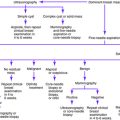
The first step in the evaluation of primary amenorrhea (Fig. 4-1) is documentation of the history and a physical examination. If secondary sexual characteristics are not present, follicle-stimulating hormone (FSH) and luteinizing hormone (LH) levels should be measured. FSH and LH levels lower than 5 IU/L indicate hypogonadotropic hypogonadism. If the FSH level exceeds 20 IU/L and the LH level exceeds 40 IU/L, hypergonadotropic hypogonadism is present; in that case, karyotype analysis is indicated.

Figure 4-1. Evaluation of primary amenorrhea.
(From Master-Hunter T, Heiman DL: Amenorrhea: evaluation and treatment. Am Fam Physician 2006; 73:1374-1382.)
Causes of Primary Amenorrhea
Key Historical Features
Key Physical Findings
✓ Height and weight compared against normative data
✓ Pubertal development according to Tanner staging
✓ Signs of an eating disorder, such as parotid gland enlargement, Russell’s sign, or dental erosions
✓ Abdominal examination for masses
✓ Pelvic examination for imperforate hymen, transverse vaginal septum, clitoral hypertrophy, or undescended testes
✓ Rectal examination for skin tags, fissures, or occult blood that may indicate inflammatory bowel disease
✓ Evaluation for striae, buffalo hump, central obesity, or proximal muscle weakness
Suggested Work-Up
| Pregnancy test | To rule out pregnancy |
| Serum FSH and LH measurement | Should be undertaken if secondary sexual characteristics are not present |
| FSH level higher than 30 and up to 40 IU/L is suggestive of premature ovarian failure; LH level is more suppressed than FSH level when amenorrhea is caused by suppression of the hypothalamic-pituitary-ovarian axis | |
| Prolactin level measurement | To evaluate for hyperprolactinemia |
| Ultrasonography of the uterus | Should be performed if primary amenorrhea is present and secondary sexual characteristics are also present |
| If the uterus is absent or abnormal, karyotype analysis should be performed; if the uterus is present and normal, the patient should be evaluated for evidence of outflow obstruction |
Additional Work-Up
| Karyotype analysis | If FSH level is persistently elevated, karyotype analysis is necessary to evaluate for a chromosomal abnormality |
| Complete blood cell count (CBC), erythrocyte sedimentation rate (ESR) measurement, thyroid-stimulating hormone (TSH) measurement, liver function tests, electrolyte measurements, blood urea nitrogen (BUN) measurement, creatinine measurement, blood glucose measurement, and urinalysis | If pubertal delay is present or systemic illness is suspected |
| Serum estradiol measurement | To confirm hypoestrogenism if premature ovarian failure is suspected |
| Serum testosterone and dehydroepiandrosterone sulfate (DHEAS) measurement | To evaluate for hyperandrogenism if signs of androgen excess are present |
| Magnetic resonance imaging (MRI) of the sella turcica | If pituitary tumor is suspected (prolactin level > 100 ng/mL) |
| Radiography of the hand and wrist | If short stature is present, to clarify skeletal maturation for chronologic age |
Adams-Hillard PJ, Deitch HR. Menstrual disorders in the college age female. Pediatr Clin North Am. 2005;52:179-197.
Kazis K, Iglesias E. The female athlete triad. Adolesc Med. 2003;14:87-95.
Master-Hunter T, Heiman DL. Amenorrhea: evaluation and treatment. Am Fam Physician. 2006;73:1374-1382.
Pletcher JR, Slap GB. Menstrual disorders: amenorrhea. Pediatr Clin North Am. 1999;46:505-518.
Timmreck LS, Reindollar RH. Contemporary issues in primary amenorrhea. Obstet Gynecol Clin. 2003;30:287-302.
Warren MP. Evaluation of secondary amenorrhea. J Clin Endocrinol Metab. 1996;81:437-442.