Allergy and the Immunologic Basis of Atopic Disease

Published on 25/03/2015 by admin

Filed under Pediatrics

Last modified 22/04/2025

Print this page

rate 1 star rate 2 star rate 3 star rate 4 star rate 5 star
Your rating: none, Average: 0 (0 votes)

This article have been viewed 3050 times

Chapter 134 Allergy and the Immunologic Basis of Atopic Disease

Allergic or atopic patients have an altered state of reactivity to common environmental and food antigens that do not cause clinical reactions in most people. Patients with clinical allergy usually produce immunoglobulin (Ig) E antibodies to the antigens that trigger their illness. The term allergy represents the clinical expression of IgE-mediated allergic diseases that have a familial predisposition and that manifest as hyperresponsiveness in target organs such as the lung, skin, gastrointestinal tract, and nose. There has been a significant increase in the prevalence of allergic diseases during the last few decades. This increase is attributed to changes in environmental factors (exposure to tobacco smoke, air pollution, indoor and outdoor allergens, respiratory viruses, obesity).

Key Elements of Allergic Diseases

Allergens

Allergens are almost always proteins, but not all proteins are allergens. For a protein antigen to display allergenic activity, it must induce IgE production, which must lead to a type 1 hypersensitivity response upon subsequent exposure to the same protein. Biochemical properties of the allergen, stimulating factors of the innate immune response around the allergen substances at the time of exposure, stability of the allergen in the tissues, digestive system, skin, or mucosa, and the dose and time of stay in lymphatic organs during the interaction with the immune system are all factors hat may cause an antigen to become an allergen. This is distinguished from general antigen responses, which induce a state of immune responsiveness without associated IgE production.

Most allergens are usually proteins of 10-70 kd molecular weight; molecules < 10 kd do not bridge adjacent IgE antibody molecules on the surfaces of mast cells or basophils; most molecules > 70 kd do not pass through mucosal surfaces, a feature needed to reach antigen-presenting cells (APCs) for stimulation of the immune system. Allergens frequently contain proteases, which promote barrier dysfunction and increase allergen penetration into host tissues. Low molecular weight moieties such as drugs can become allergens by reacting with serum proteins or cell membrane proteins to be recognized by the immune system. Carbohydrate structures have also been identified as allergens. This finding is most relevant with the increasing use of biologics in clinical practice; patients with cetuximab-induced anaphylaxis have IgE antibodies in specific for galactose-α-l,3-galactose (Chapter 146).

T Cells

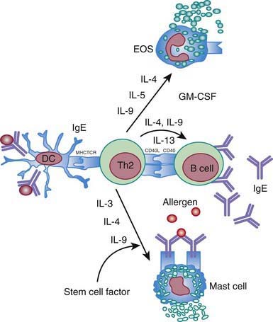
Everyone is exposed to potential allergens. Atopic individuals respond to allergen exposure with rapid expansion of T helper type 2 (Th2) cells that secrete cytokines, such as interleukins IL-4, IL-5, and IL-13, favoring IgE synthesis and eosinophilia. Allergen-specific IgE antibodies associated with atopic response are detectable by serum testing or positive immediate reactions to allergen extracts on prick skin testing (Chapter 135). The Th2 cytokines IL-4 and IL-13 play a key role in immunoglobulin isotype switching to IgE (Fig. 134-1). IL-5 and IL-9 are important in differentiation and development of eosinophils. The combination of IL-3, IL-4, and IL-9 contributes to mast cell activation. Th2 cytokines are important effector molecules in the pathogenesis of asthma and allergic diseases; acute allergic reactions are characterized by infiltration of Th2 cells into affected tissues. In addition, IL-25 and IL-33 contribute to Th2 response and eosinophilia.

A fraction of the immune response to allergen results in proliferation of T helper type 1 (Th1) cells. Th1 cells are typically involved in the eradication of intracellular organisms, such as mycobacteria, because of the ability of Th1 cytokines to activate phagocytes and promote the production of opsonizing and complement-fixing antibodies. The Th1 component of allergen-specific immune response contributes to chronicity and the effector phase in allergic disease. Activation and apoptosis of epithelial cells induced by Th1 cell–secreted interferon-γ (IFN-γ), tumor necrosis factor-α (TNF-α), and Fas-ligand constitute an essential pathogenetic event for the formation of eczematous lesions in atopic dermatitis and bronchial epithelial cell shedding in asthma.

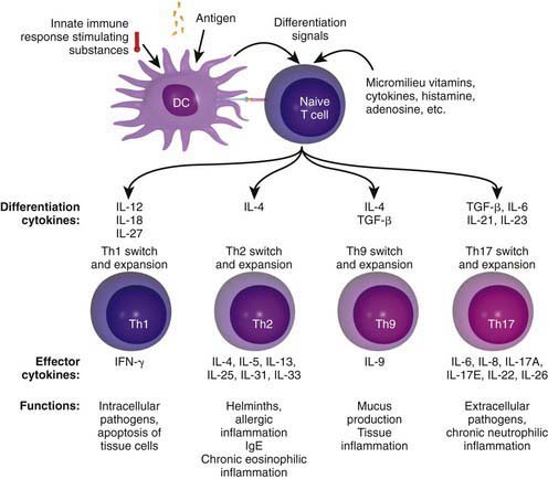
Chronic allergic reactions are characterized by infiltration of Th1 and Th17 cells. This is important because Th1 cytokines such as IFN-γ can potentiate the function of allergic inflammatory effector cells such as eosinophils and thereby contribute to disease severity. Cytokines in the IL-17 family act on multiple cell types, including epithelial cells and APCs, to cause the release of chemokines, antimicrobial peptides, and pro-inflammatory cytokines to enhance inflammation and antimicrobial responses. In addition, recently identified Th9 cells produce IL-9 but not other typical Th1, Th2 and Th17 cytokines and constitute a distinct population of effector T cells that promotes tissue inflammation. Figure 134-2 depicts the complex cytokine cascades involving Th1, Th2, Th9, and Th17 cells.

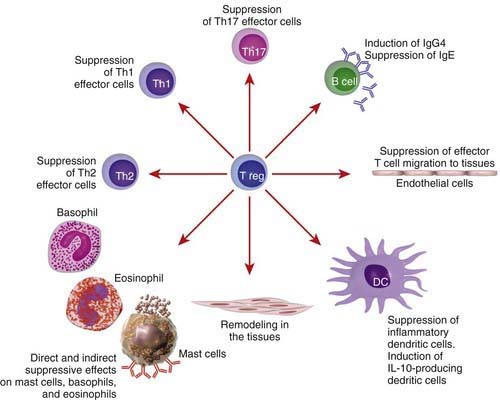
T regulatory (Treg) cells are a subset of T cells thought to play a critical role in expression of allergic and autoimmune diseases. These cells have the ability to suppress effector T cells of either the Th1 or Th2 phenotypes (Fig. 134-3). Treg cells express CD4+CD25+ surface molecules and immunosuppressive cytokines such as IL-10 and transforming growth factor-β (TGF-β1). The forkhead box/winged-helix transcription factor gene FOXP3 is expressed specifically by CD4+CD25+ Treg cells and programs their development and function. Adoptive transfer of Treg cells inhibits the development of airway eosinophilia and protects against airway hyperreactivity in animal models of asthma. T cell response to allergens in healthy individuals shows a wide range, from no detectable response to involvement of active peripheral tolerance mechanisms mediated by different subsets of Treg cells. Individuals who are not allergic even though they are exposed to high doses of allergens, such as beekeepers and cat owners, show a detectable allergen-specific IgG4 response accompanied by IL-10–producing Treg cells. It is thought that CD4+CD25+ Treg cells play an important role in mitigating the allergic immune response and that the lack of such cells may predispose to the development of allergic diseases. Patients with mutations in the human FOXP3 gene lack CD4+CD25+ Treg cells and develop severe immune dysregulation, with polyendocrinopathy, food allergy, and high serum IgE levels (XLAAD/IPEX disease) (Chapter 120).

Antigen-Presenting Cells

Dendritic cells, Langerhans cells, monocytes, and macrophages have the ability to present allergens to T cells and thereby modulate allergic inflammation by controlling the type of T-cell development. Antigen-presenting cells are a heterogeneous group of cells that share the property of antigen presentation in the context of the major histocompatibility complex (MHC) and are found primarily in lymphoid organs and the skin. Dendritic cells (DCs) and Langerhans cells are unique in their ability to prime naïve T cells and are responsible for the primary immune response, or the sensitization phase of allergy. Monocytes and macrophages are thought to contribute to activating memory T-cell responses upon re-exposure to allergen, which characterizes the elicitation phase of allergy.

Peripheral DCs residing in sites such as the skin, intestinal lamina propria, and lung are relatively immature. These immature DCs take up antigens in tissues and then migrate to the T-cell areas in locally draining lymph nodes. The DCs undergo phenotypic and functional changes during migration, characterized by increased expression of MHC class I, MHC class II, and co-stimulatory molecules that react with CD28 expressed on T cells. In the lymph nodes, they directly present processed antigens to resting T cells to induce their proliferation and differentiation.

Mature DCs have been designated as DC1 or DC2 on the basis of their ability to favor Th1 or Th2 differentiation, respectively. The critical factor for polarization to Th1 cells is the level of IL-12 produced by DC1 cells. By contrast, DC2 dendritic cells have low levels of IL-12, resulting in Th2 cell predominance. Local histamine and prostaglandin (PG) E2 inhibit IL-12 synthesis and thereby promote the development of DC2 phenotype. There is considerable interest in the role of thymic stromal lymphopoietin (TSLP), which is overexpressed in the mucosal surfaces and skin of atopic individuals. TSLP enhances Th2 cell differentiation by inducing expression of OX40L on immature myeloid DCs in the absence of IL-12 production.

Presence of allergen-specific IgE on the cell surfaces of APCs is a unique feature of atopy. Importantly, the formation of high-affinity IgE receptor I (FcεRI)/IgE/allergen complexes on APC cell surfaces markedly facilitates allergen uptake and presentation. The clinical importance of this phenomenon is supported by the observation that FcεRI-positive Langerhans cells bearing IgE molecules are a prerequisite for skin-applied, aeroallergen provocation of eczematous lesions in patients with atopic dermatitis. The role of the low-affinity IgE receptor II (FcεRII, CD23) on monocytes-macrophages is less clear, although it appears that under certain conditions it can also facilitate antigen capture. Cross linking of FcεRII as well as FcεRI on monocytes-macrophages leads to the release of inflammatory mediators. There is a critical role for DCs in induction of oral tolerance; tolerogenic DCs are compartmentalized within the mucosa and present antigen through a mechanism designed to produce a Th1/Treg–suppressive response that ablates allergen-specific T cells.

Immunoglobulin E and its Receptors

The acute allergic response depends on IgE and its ability to bind selectively to the α chain of the high-affinity FcεRI or the low-affinity FcεRII (CD23). Cross linking of receptor-bound IgE molecules by allergen initiates a complex intracellular signaling cascade followed by the release of various mediators of allergic inflammation from mast cells and basophils. The FcεRI molecule is also found on the surface of antigen-presenting DCs (e.g., Langerhans cells), but differs from the structure found on mast cells/basophils in that the FcεRI molecule found on DCs lacks the β chain. CD23 is found on B cells, eosinophils, platelets, and dendritic cells. Cross linking and FcεRI aggregation on mast cells and basophils can also lead to anaphylaxis (Chapter 143). Differential expression of tyrosine kinases responsible for positive and negative regulation of mast cell/basophil degranulation are thought to be responsible for this aberrant allergic response.

The induction of IgE synthesis requires two major signals. The first signal (signal 1) initiates IL-4 or IL-13 activation of germline transcription at the ε immunoglobulin locus, which dictates isotype specificity. The second signal (signal 2) involves the engagement of CD40 on B cells by CD40 ligand expressed on T cells. This engagement results in activation of the recombination machinery, resulting in DNA switch recombination. Interactions between several co-stimulatory molecule pairs (CD28 and B7; lymphocyte function–associated antigen-1 [LFA-1] and intercellular adhesion molecule-1 [ICAM-1]; CD2 and CD58) can further amplify signal 1 and signal 2 to enhance IgE synthesis. Factors that inhibit IgE synthesis include Th1-type cytokines (IL-12, IFN-α, IFN-γ) and microbial DNA containing CpG (cytosine-phosphate-guanine) repeats.

Eosinophils

Allergic diseases are characterized by peripheral blood and tissue eosinophilia. Eosinophils participate in both innate and adaptive immune responses and, like mast cells, contain dense intracellular granules that are sources of inflammatory proteins. These granule proteins include major basic protein, eosinophil-derived neurotoxin, peroxidase, and cationic protein. Eosinophil granule proteins damage epithelial cells, induce airway hyperresponsiveness, and cause degranulation of basophils and mast cells. Major basic protein released from eosinophils can bind to an acidic moiety on the M2 muscarinic receptor and block its function, thereby leading to increased acetylcholine levels and the development of increased airway hyperreactivity. Eosinophils are also a rich source of prostaglandins and leukotrienes; in particular, cysteinyl leukotriene C4 contracts airway smooth muscle and increases vascular permeability. Other secretory products of eosinophils include cytokines (IL-4, IL-5, TNF-α), proteolytic enzymes, and reactive oxygen intermediates, all of which significantly enhance allergic tissue inflammation.

Several cytokines regulate the function of eosinophils in allergic disease. Eosinophils develop and mature in the bone marrow from myeloid precursor cells activated by IL-3, IL-5, and granulocyte-macrophage colony-stimulating factor (GM-CSF). Allergen exposure of allergic patients causes resident hematopoietic CD34 cells to express the IL-5 receptor. The IL5 receptor activation induces eosinophil maturation, causing eosinophils to synthesize granule proteins, prolonging their survival, potentiating degranulation of eosinophils, and stimulating release of eosinophils from the bone marrow. GM-CSF also enhances proliferation, cell survival, cytokine production, and degranulation of eosinophils. Certain chemokines, such as RANTES (regulated upon activation, normal T cell expressed and secreted), macrophage inflammatory protein-1α (MIP-1α), and eotaxins, are important for recruiting eosinophils into local allergic tissue inflammatory reactions. Eotaxins mobilize IL-5–dependent eosinophil colony–forming progenitor cells from the bone marrow. These progenitors are rapidly cleared from the blood and either return to the bone marrow or are recruited to inflamed tissue sites.

Mast Cells

Mast cells are derived from CD34 hematopoietic progenitor cells that arise in bone marrow. Upon entering the circulation, they travel to peripheral tissue, where they undergo tissue-specific maturation. Mast cell development and survival relies on interactions between the tyrosine kinase receptor c-kit expressed on the surface of mast cells and the fibroblast-derived c-kit ligand stem cell factor (SCF). Unlike mature basophils, mature mast cells do not typically circulate in the blood. They are, instead, widely distributed throughout connective tissues, where they often lie adjacent to blood vessels and beneath epithelial surfaces that are exposed to the external environment, such as the respiratory tract, gastrointestinal tract, and skin. So placed, mast cells are positioned anatomically to participate in allergic reactions. At least two subpopulations of human mast cells are recognized: mast cells with tryptase and mast cells with both tryptase and chymase. Mast cells with tryptase are the predominant type found in the lung and small intestinal mucosa, whereas mast cells with both tryptase and chymase are the predominant type found in skin, the gastrointestinal submucosa, and blood vessels.

Mast cells contain, or produce on appropriate stimulation, a diverse array of mediators that have different effects on allergic inflammation and organ function. They include preformed granule-associated mediators (histamine, serine proteases, proteoglycans) and membrane-derived lipid, cytokine, and chemokine mediators arising from de novo synthesis and release. The most important mast cell-derived lipid mediators are the cyclo-oxygenase and lipoxygenase metabolites of arachidonic acid, which have potent inflammatory activities. The major cyclo-oxygenase product of mast cells is PG D2, and the major lipoxygenase products are the sulfidopeptide leukotrienes (LTs): LTC4 and its peptidolytic derivatives LTD4 and LTE4. Mast cells also can produce cytokines that promote Th2-type responses (IL-4, IL-13, GM-CSF) and inflammation (TNF-α, IL-6) and regulate tissue remodeling (TGF, vascular endothelial cell growth factor). Immunologic activation of mast cells and basophils typically begins with cross linkage of IgE bound to the FcεRI with multivalent allergen. Mast cell surface FcεRI is increased by IL-4 and IgE. Surface levels of FcεRI decrease in subjects receiving treatment with anti-IgE antibody that lowers serum IgE, which is of potential therapeutic interest.

Mechanisms of Allergic Tissue Inflammation

IgE-mediated immune responses can be classified chronologically according to 3 reaction patterns. The early-phase response is the immediate response after allergen is introduced into target organs. This response is characterized by mast cell degranulation and release of preformed mediators, occurring within 10 min after allergen exposure and resolving within 1-3 hr. Acute reactions are associated with increased local vascular permeability, which leads to leakage of plasma proteins, tissue swelling, and increased blood flow, as well as itching, sneezing, wheezing, and acute abdominal cramps in the skin, nose, lung, and gastrointestinal tract, respectively, depending on the targeted organ.

A second, late-phase response can occur within hours of allergen exposure, reaching a maximum at 6-12 hr and resolving by 24 hr. Late-phase responses are characterized in the skin by edema, redness, and induration; in the nose by sustained nasal blockage; and in the lung by airway obstruction and persistent wheezing. In general, late-phase responses are associated with early infiltration of neutrophils and eosinophils followed by basophils, monocytes, macrophages, and Th2-type cells. Recruitment of inflammatory cells from the circulation requires increased expression of adhesion molecules on their cell surfaces and expression of their ligand on endothelial cells, which are under the control of cytokines. Several hours after allergen exposure, TNF-α released by activated mast cells induces the vascular endothelial expression of cell adhesion molecules, and this change leads to transendothelial migration of various inflammatory cells. Preferential accumulation of eosinophils occurs through interactions between selective adhesion molecules on the eosinophil cell surface (e.g., α4β1 integrin or very late antigen-4 [VLA-4]); VCAM-1 (vascular cell adhesion molecule-1) surface expression can be enhanced by IL-4 and IL-13 on endothelial cells.

Chemokines are chemotactic cytokines that play a central role in tissue-directed migration of inflammatory cells. RANTES, MIP-1α, monocyte chemotactic protein (MCP)-3, and MCP-4 are chemoattractants for eosinophils and mononuclear cells, whereas eotaxins are relatively selective for eosinophils. These chemoattractants have been detected in epithelium, macrophages, lymphocytes, and eosinophils at sites of late-phase responses and allergic tissue inflammation. Blockade of these chemokines leads to significant reduction in tissue-directed migration of allergic effector cells.

In the third reaction pattern, chronic allergic disease, tissue inflammation can persist for days to years. Several factors contribute to persistent tissue inflammation, including recurrent exposure to allergens and microbial agents. The repeated stimulation of allergic effector cells such as mast cells, basophils, eosinophils, and Th2 cells contributes to unresolved inflammatory conditions. Additionally, Th2-type cytokines (IL-3, IL-5, GM-CSF) secreted during allergic reactions can prolong survival of allergic effector cells by delaying apoptosis. Local differentiation of tissue-infiltrating eosinophil precursors induced by IL-5 results in self-generation of eosinophils, further sustaining damage of local tissue. Tissue remodeling leading to irreversible changes in target organs is also a feature of chronic allergic disease. In asthma, remodeling involves thickening of the airway walls and submucosal tissue, as well as smooth muscle hypertrophy and hyperplasia, which are associated with a decline in lung function. This is an unexpected role for eosinophils in airway remodeling as well as chronic inflammation. In atopic dermatitis, lichenification is an obvious manifestation of skin remodeling.

Th2 cytokines can not only maintain allergic inflammation but also influence tissue remodeling by activating resident cells in target organs; IL-4, IL-9, and IL-13 induce mucus hypersecretion and metaplasia of mucus cells; IL-4 and IL-13 stimulate fibroblast growth and synthesis of extracellular matrix proteins; and IL-5 and IL-9 increase subepithelial fibrosis. TGF-β produced by eosinophils and fibroblasts can enhance subepithelial fibrosis. IL-11 expressed by eosinophils and epithelial cells also contributes to subepithelial fibrosis, in addition to enhancing deposition of collagen and the accumulation of fibroblasts. The resulting tissue injury amplifies further epithelial injury through proinflammatory cytokine release, extracellular matrix deposition in target organs, and angiogenesis. Genetic predisposition to aberrant injury-repair responses may contribute to chronicity of illness. Once the allergic immune response is established, it can be self-perpetuating and can lead to chronic disease in genetically predisposed individuals. The subsequent infiltration of Th1 cells and Th17 cells enhances the inflammatory potential of allergic effector cells and contributes to chronic tissue inflammatory responses through the release of proinflammatory cytokines and chemokines.

Genetic Basis of Atopy

Allergic diseases are complex genetic conditions susceptible to environmental triggers. Several major groups of genes are associated with allergic diseases: genes that regulate systemic expression of atopy (increased IgE synthesis, eosinophilia, mast cell responses) that are commonly expressed among various allergic diseases, genes that control barrier function in specific target organs (e.g., the skin in atopic dermatitis, the lung in asthma, the gastrointestinal tract in food allergy), and genes encoding pattern-recognition receptors of the innate immune system that engage microbial pathogens and influence adaptive immune responses. Once allergic responses have been initiated, a genetic predisposition to chronic allergic inflammation and aberrant injury-repair responses contribute to tissue remodeling and persistent disease.

Atopic diseases have a strong familial predisposition, with approximately 60% heritability found in twin studies of asthma and atopic dermatitis. The 5q23-35 region comprises several genes implicated in allergic disease pathogenesis, including genes coding for Th2 cytokines (IL-3, IL-4, IL-5, IL-9, IL-13, GM-CSF). Among these, IL4 is a well-studied potential candidate gene. A nucleotide change at position 589 of the IL4 promoter region is associated with the formation of a unique binding site for NF-AT (nuclear factor for activated T cells) transcription factor, increased IL-4 gene transcription, higher NF-AT binding affinity, and increased IgE production. Similarly, IL13 coding region variants have been associated with asthma and atopic dermatitis. An association between atopy and a gain-of-function polymorphism on chromosome 16, which codes for the α subunit of the IL-4R, has been found. This finding is consistent with the important role of IL-4, IL-13, and their receptors in the immunopathogenesis of allergic diseases.

Several genome-wide searches have also linked atopy to chromosome region 11q13. The gene encoding the β subunit of FcεRI-β has been proposed to be the candidate gene in this region. The β subunit gene modifies the FcεRI activity on mast cells, and several genetic variants of FcεRI-β have been associated with asthma and atopic dermatitis. Chromosome 6 contains genes coding for human leukocyte antigen (HLA) class I and class II molecules, which regulate the specificity and intensity of the immune responses to specific allergens. IgE responses to specific allergens, such as ragweed antigen Amb a V and mite allergen Der p I, have been linked to specific MHC class II loci. TNF-α, a key cytokine that contributes to the influx of inflammatory cells, is also located on chromosome 6. TNF-α polymorphisms have been associated with asthma.

Barrier dysfunction has a key role in the pathogenesis of allergic diseases. Genetic linkage studies of atopic dermatitis have demonstrated the importance of chromosome 1q21, which contains a cluster of genes involved in epidermal differentiation. This has been best demonstrated in studies involving the gene that encodes filaggrin, a protein essential in the formation of the stratum corneum, where it has been repeatedly found that null mutations of the filaggrin gene are strongly associated with early onset and severe atopic dermatitis. Mutations in the gene encoding the serine protease inhibitor SPINK5 has been shown to cause Netherton disease, a single-gene disorder associated with erythroderma, food allergy, and high serum IgE levels. A common polymorphism in SPINK5 (in particular, Glu420Lys) increases the risk of developing atopic dermatitis and asthma. SPINK5 is expressed in the outer epidermis and is thought to be critical to neutralizing the proteolytic activity of Staphylococcus aureus and common allergens such as Der p I, which use these proteases to penetrate the skin to induce allergic responses. Barrier dysfunction is involved in other allergic diseases, such as asthma and rhinosinusitis, but likely involves other barrier genes, such as those encoding gap junctions.

Candidate genes associated with asthma susceptibility have been identified by positional cloning: GPRA (G-protein coupled receptor for asthma susceptibility on chromosome 7p14), ADAM-33 (A disintegrin and metalloproteinase 33 on chromosome 20p), and DPP10 (dipeptidyl peptidase 10 on chromosome 2q14). The functions of these genes do not fit into classical pathways of atopy and therefore provide new insights into asthma pathogenesis. GPRA encodes a G-protein coupled receptor, with isoforms expressed in bronchial epithelial cells and smooth muscle in asthmatic persons, suggesting an important role for these tissues in asthma. ADAM-33 is expressed in bronchial smooth muscle and has been linked to bronchial hyperresponsiveness. DPP10 encodes a dipeptidyl dipeptidase that can remove the terminal two peptides from certain proinflammatory chemokines, a change that may modulate allergic inflammation.

Pattern-recognition receptors of the innate immune system, which are expressed by epithelial cells and dendritic cells, have been associated with disease susceptibility. These receptors recognize specific microbial components. Polymorphisms in CD14 (engages endotoxin), Toll-like receptor 2 (which engages S. aureus), and T-cell immunoglobulin domain and mucin domain (which engage hepatitis A virus) have been correlated with asthma and/or atopic dermatitis susceptibility. Dysregulation of these frontline immune defense systems would permit abnormal response to common environmental allergens.