Acute Respiratory Distress Syndrome

Published on 01/06/2015 by admin

Filed under Pulmolory and Respiratory

Last modified 22/04/2025

Print this page

rate 1 star rate 2 star rate 3 star rate 4 star rate 5 star
Your rating: none, Average: 0 (0 votes)

This article have been viewed 1936 times

Chapter 36 Acute Respiratory Distress Syndrome

The acute respiratory distress syndrome (ARDS) is an inflammatory disease process of the lungs that is a response to both direct and indirect insults, characterized clinically by severe hypoxemia, reduced lung compliance, and bilateral radiographic infiltrates. Although much has evolved in the current understanding of its pathogenesis and factors affecting patient outcome, there is still no specific treatment for ARDS.

In 1967, Ashbaugh and colleagues published an article in The Lancet in which they described for the first time a clinical syndrome that they termed the “adult respiratory distress syndrome” for its similarity to the well-recognized infant respiratory distress syndrome. From a cohort of 272 patients who were receiving respiratory support, these investigators identified 12 patients with a constellation of specific pulmonary findings: The respiratory distress in these patients was defined as sudden and catastrophic and often associated with a multiorgan system insult that led to tachypnea, hypoxemia, decreased lung compliance, and bilateral pulmonary infiltrates on the chest radiograph, in the absence of cardiogenic pulmonary edema. In this study, respiratory support consisted of oxygen therapy delivered by nasal prongs or face mask and mechanical ventilation. The mortality rate was 58%, and on histopathologic examination, the lungs of the nonsurvivors were heavier than normal and exhibited atelectasis with interstitial and alveolar edema and hyaline membranes. Since that time, the hallmarks of this syndrome, now known as ARDS, have been recognized to consist of (1) a risk factor for the development of acute respiratory distress (e.g., sepsis, trauma, pneumonia, aspiration, pancreatitis), (2) severe hypoxemia despite a relatively high fraction of inspired oxygen (FIO2), (3) decreased lung compliance, (4) bilateral pulmonary infiltrates, and (5) lack of clinical evidence of cardiogenic pulmonary edema. Acute lung injury (ALI) resulting in ARDS also can occur in the setting of left ventricular failure, but this is very difficult to diagnose without careful serial measurements of pulmonary artery and capillary (wedge) pressure. Although ALI/ARDS had been recognized for more than a century and was referenced in published data from the Second World War, it was not until the landmark paper by Ashbaugh’s group that broad clinical interest in ARDS began to emerge. In the subsequent years, very few acronyms have received as much attention in critical care medicine.

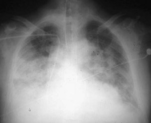
ARDS usually occurs in previously healthy people. Characteristically, there is a latent period between the insult and the development of the full-blown clinical syndrome, which usually is 18 to 24 hours in duration. After this interval, tachypnea, labored breathing, and cyanosis are observed, and arterial blood gas analysis confirms hypoxemia. The abnormalities in lung mechanics and oxygenation are assessed once the patient is intubated and receiving mechanical ventilation. The chest radiograph classically shows diffuse, bilateral, interstitial alveolar infiltrates (Figure 36-1). Resolution of the infiltrates, if it occurs at all, is much slower than with cardiogenic pulmonary edema.

Pathophysiology, Histopathology, and Etiology

ARDS is caused by an insult to the alveolar-capillary membrane that results in increased permeability and subsequent interstitial and alveolar edema. The mechanisms whereby a wide variety of insults can lead to this syndrome are not clear. ALI includes injury to both the pulmonary capillary endothelium and the alveolar epithelium. In the ARDS lung, an influx of protein-rich edema fluid into the air spaces occurs as a consequence of increased permeability of the alveolar-capillary barrier. The degree of alveolar epithelial injury is an important determinant and predictor of outcome. The normal alveolar epithelium has two types of cells. The type I cells make up 90% of the alveolar surface area and are easily injured. The type II cells make up the remaining 10% of the alveolar surface area and are more resistant to injury; their functions include surfactant production, ion transport, and proliferation and differentiation to type I cells after injury.

The loss of epithelial integrity in ARDS has several consequences. First, under normal conditions, the epithelial barrier is much less permeable than the endothelial barrier; thus epithelial injury can contribute to alveolar flooding. Second, the loss of epithelial integrity and injury to type II cells serve to disrupt normal epithelial fluid transport, impairing the removal of edema fluid from the alveolar space. Third, injury to type II cells reduces the production and turnover of surfactant. Fourth, loss of epithelial barrier can lead to sepsis in patients with bacterial pneumonia. Finally, in severe alveolar epithelium injury, pulmonary fibrosis can develop. Independent of the clinical disorders associated with ARDS (Box 36-1), it is useful to think of the pathogenesis of ARDS as a result of two different pathways: a direct insult to lung cells and an indirect insult occurring as a result of an acute systemic inflammatory response. The host’s inflammatory response to the initial direct (pulmonary) or indirect (nonpulmonary) insult is a key factor in determining the development and progression of the acute injury to the lung. Despite ongoing elucidation of the role of cellular and humoral components of the inflammatory responses in the lung, the precise sequence of events leading to lung damage is still unknown. As with any form of inflammation, ALI during ARDS represents a complex process in which multiple cellular signaling pathways can propagate or inhibit damage to the lung.

The typical histopathologic features of ARDS are collectively known as diffuse alveolar damage. The early phase of ALI—the exudative phase—is characterized by leakage of protein-rich edema fluid into the lung and inflammatory cellular alveolar infiltrates. During this phase, a cytokine storm and an array of inflammatory mediators are released into the interstitium and alveolar space, perpetuating inflammation and promoting the development of atelectasis and structural damage to the lung architecture. In addition, damage to the alveolar-capillary barrier enhances the difficulty in removing the excess of extravascular lung fluid. An important source of these inflammatory mediators is neutrophils, which play a key role in the pathogenesis and progression of ALI. Human and animal studies have demonstrated migration and activation of neutrophils in the lungs, where they cause cell damage through the production of free radicals, inflammatory cytokines, and proteases. It is well accepted, however, that a single mediator does not predominate, and that several parallel and simultaneously interacting mechanisms may be involved. Clinically, this initial phase is manifested as marked hypoxemia and reduced lung compliance. Eventually, these changes evolve to a fibroproliferative phase in which capillary thrombosis, lung fibrosis, and neovascularization take place. Most nonsurvivors of ARDS die during this phase, despite aggressive ventilatory support with high inspiratory concentrations of oxygen and positive end-expiratory pressure (PEEP). However, only a small proportion of patients with ARDS die of hypoxemia. Rather, lung injury appears to predispose patients to the development of a systemic inflammatory response that culminates in multiple system organ dysfunction. A plethora of evidence suggests that the development of multiple extrapulmonary organ dysfunction is due to alveolar epithelial-endothelial barrier disruption and the migration of cytokines produced in the lungs into the systemic circulation.

More than 50 specific conditions associated with the development of this syndrome are recognized. The risk for development of ARDS depends on the predisposing clinical condition (i.e., some events are more likely than others to progress to ARDS) but also increases with the number of predisposing factors. Sepsis, bacterial pneumonia, multiple trauma, and aspiration pneumonia are the most common predisposing factors, accounting all together for more than 70% of cases; infection is the most frequent cause. Many invading organisms can trigger host innate and acquired immune systems to initiate the inflammatory cascade. The risk for development of ARDS also depends on patient characteristics. For example, alcoholism is a predisposing factor, and new data suggest the possibility of a genetic predisposition. Overall mortality from ARDS has not decreased substantially since the publication of the 1967 report, and the current survival rate approximates to 45% in all major epidemiologic series. Sepsis-related ARDS is characterized by a higher overall disease severity, poorer recovery from lung injury, and higher mortality than non–sepsis-related ARDS. Among patients with ARDS associated with combined pulmonary and nonpulmonary sources of infection, mortality is even higher. Approximately 80% of all deaths in patients with ARDS occur within 2 to 3 weeks after the onset of the syndrome. Death traditionally has been attributed to the underlying disease, the presence of sepsis, and the failure of vital organ systems other than the lung.

Definition, Incidence, and Severity

Because it is difficult to measure changes in capillary and alveolar permeability at the bedside, diagnosis of ARDS is based on a combination of clinical, oxygenation, hemodynamic, and radiographic criteria. These criteria allow the inclusion of a highly heterogeneous group of critically ill patients, because various types of lung injury can lead to a similar pulmonary response. Despite general agreement on the overall criteria on which to base a definition of ARDS (i.e., severe hypoxemia, marked decreased of lung compliance), the specific values of these variables and the preferred conditions of measurement vary greatly among clinicians and scientists. Thus, the original description of ARDS has proved to be incapable of identifying a uniform group of patients. Several of the patients in the original report of Ashbaugh and co-workers would not be classified as having ARDS today, because fluid overload was an important etiologic factor in those cases. Some investigators have questioned whether ARDS is a distinct entity. Others have suggested that ARDS should not be considered a separate syndrome but should be seen as part of the multiple system organ dysfunction syndrome. From a clinical perspective, a strict definition of ARDS may not be required, because current management is supportive. From a therapeutic standpoint, however, a more precise definition probably is necessary, because the effects on outcome of certain ventilatory and adjunctive techniques may presumably vary depending on the degree of lung injury. In terms of prognosis, a number of investigators have examined whether various parameters of oxygenation and lung mechanics would be useful in predicting outcome. In the context of research on ARDS, a very strong argument can be made for a universal definition: It would help standardize experimental and clinical studies evaluating the natural history, incidence, pathophysiology, treatment, and outcome of ARDS. It also would help in the comparison of data among various clinical studies and centers.

A good example of the problems inherent in formulation of a definition for ARDS is the wide disparity in the literature on the incidence. Reported data in the United States suggest an occurrence rate greatly in excess of that expected from current clinical experience in Europe. The most common figure cited for the annual incidence of ARDS is 75 cases per 100,000 population. This is based on an American Lung Program Task Force of the National Heart and Lung Institute in 1972. This internal report suggested that there were about 150,000 cases per year of ARDS in the United States, a value similar to the number of all new cases of cancer. In 1988, Webster and colleagues in England estimated an incidence of 4.5 cases per 100,000 population, and in 1989, Villar and associates in Spain calculated the incidence as 3.5 new cases per 100,000 population per year. Most epidemiologic studies report an ARDS incidence ranging from 4 to 8 cases per 100,000 population per year.

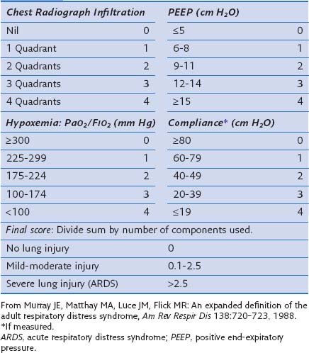
In an attempt to overcome some of these problems, Murray and colleagues proposed an expanded definition of ARDS that takes into account various pathophysiologic features of the syndrome. Their definition uses a “lung injury score” (LIS) to characterize the acute pulmonary damage by considering four components: assessment of the chest radiograph, degree of hypoxemia (determined as the ratio of arterial partial pressure of oxygen to the fraction of inspired oxygen, PaO2/FIO2), level of PEEP, and the value of lung compliance, when available (Table 36-1). The final injury score is obtained by dividing the total score by the number of components that were used. A score of 0 indicates no lung injury, a score of 1 to 2.5 indicates mild to moderate lung injury, and a score greater than 2.5 indicates severe lung injury or ARDS. The LIS is not specific for ARDS, however, and has not been validated, because it is not clear whether patients with identical LIS have similar degrees of lung injury. Furthermore, patients with a major component of cardiogenic edema may be mislabeled as having ARDS, and a postoperative patient with moderate atelectasis and mild fluid overload may fit the LIS criteria for ARDS.

Table 36-1 Lung Injury Scoring System

image

Because severe hypoxemia is the hallmark of ARDS, it should be crucial to the assessment of the severity of ARDS, for predicting the development and evolution in any given patient, and for assessing the response to treatment. To better characterize the severity of lung damage, in 1994 an American-European Consensus Conference (AECC) defined ALI and ARDS as follows:

According to these guidelines, ALI exists when the PaO2/FIO2 ratio is 300 mm Hg or less regardless of the level of PEEP and FIO2, and ARDS is present when the PaO2/FIO2 ratio is 200 mm Hg or less regardless of the PEEP setting and FIO2 (Box 36-2). Although this definition formalized the criteria for the diagnosis of ARDS and is simple to apply in the clinical setting, it has been challenged over the years in several studies. Such definitions have limitations: The physiologic thresholds do not require standardized ventilatory support, and the use of PEEP can improve oxygenation indices sufficiently to convert the patient’s status from meeting the definition of ARDS to not meeting the ARDS definition and also can change the physiology in the lung such that the patient does not meet the criteria for ARDS. Therefore, the ARDS criteria may be met when the PaO2 is measured with zero PEEP but not when measured at a PEEP of 5 or 10 cm H2O, making patient comparisons difficult. Furthermore, most of the randomized controlled studies did not use the same definition for ARDS, nor did they evaluate the same ventilatory approaches. Diversity among ARDS definitions is apparent in a large number of studies (Table 36-2).

Table 36-2 Definitions of Acute Respiratory Distress Syndrome in Several Published Reports

Published Study Criteria
Montgomery et al: Am Rev Respir Dis 132:485–489, 1985 PaO2/FIO2 <150 mm Hg
PCP <18 mm Hg
Villar et al: Am Rev Respir Dis 140:814–816, 1989 PaO2 ≤75 mm Hg on FIO2 ≥0.5
PCP <18 mm Hg
Bone et al: Chest 96:849–851, 1989 PaO2/FIO2 ≤150 mm Hg (with ZEEP)
OR PaO2/FIO2 ≤250 mm Hg with PEEP
PCP ≤18 mm Hg
Amato et al: N Engl J Med 338:347–354, 1998 Lung injury score ≥2.5 and PCP <16 mm Hg
Stewart et al: N Engl J Med 338:355–361, 1998 PaO2/FIO2 <250 mm Hg on PEEP of 5 cm H2O
Brochard et al: Am J Respir Crit Care Med 158:1831–1838, 1998 Lung injury score >2.5
Villar et al: Intensive Care Med 25:930–935, 1999 PaO2/FIO2 ≤150 mm Hg on PEEP ≥5 cm H2O
ARDSNet: N Engl J Med 342:1301–1308, 2000 AECC
Gattinoni et al: N Engl J Med 345:568–573, 2001 PAO2/FIO2 ≤200 mm Hg on PEEP ≥5 cm H2O
PCP ≤18 mm Hg
Villar et al: Crit Care Med 34:1311–1318, 2006 PAO2/FIO2 ≤200 mm Hg on PEEP ≥5 cm H2O and FIO2 ≥0.5
Meade et al: JAMA 299:637–645, 2008 PAO2/FIO2 <250 mm Hg
Mercat et al: JAMA 299:646–655, 2008 PAO2/FIO2 ≤200 mm Hg
PCP ≤18 mm Hg

AECC, American-European Consensus Conference; PCP, pulmonary capillary pressure; PEEP, positive end-expiratory pressure; ZEEP, zero PEEP.

Genetics

Critical care physicians have long recognized that some patients progress despite therapy, whereas others do better than predicted. It is now well accepted that these responses may be related to variations in the genome. Little is known, however, about the genes that are responsible for susceptibility to and outcome of ARDS. The search for genetic variants determining susceptibility and predicting outcome is still a developing field. The identification of important associations between genotype and clinical outcomes will have an impact on the development of more efficient genotype- or phenotype-guided therapies for patients with ALI or ARDS. The current understanding is that the pathogenesis of ARDS has a fundamental inflammatory component eliciting a response similar to that observed against any pathogen. In addition, common genetic risk factors with modest effects may be associated with disease susceptibility. Many studies have searched for genetic variations underlying ARDS susceptibility. Owing to the impracticality of classical genetic approximation in ARDS, association studies comparing unrelated ARDS cases with controls for genetic variants at specified locations of the human genome represent the prevailing study design for detecting such loci. The genetic variants explored usually are single base changes in the DNA, known as single-nucleotide polymorphisms (SNPs), because they are the most common variants across the genome. The genetic studies of ARDS have focused largely on candidate genes involved in the response to external stimulus and cell signal transduction, because those genes are assumed to be important in the immune response.

Extensive cross-species gene expression pattern comparisons in experimental models of ALI/ARDS have revealed that IL-6, an acute-phase response cytokine with pleiotropic effects, is highly upregulated. This finding is consistent with clinical studies indicating that IL-6 and other cytokines are released from the lungs in patients with ARDS; increased IL-6 concentrations are found in the bronchoalveolar lavage fluid and serum of these patients. IL-6 levels have been correlated with clinical outcome and implicated in the development of multiple system organ failure. A G/C SNP located at position −174 of the promoter region of the IL-6 gene has been shown to functionally affect the activity of the IL-6 gene promoter in vitro. To date, assessing the SNP variation for virtually all common variants of the gene has allowed subsequent studies to reveal a consistent picture for the association of the IL-6 gene with ALI and ARDS.

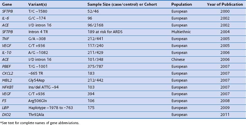
In case-control studies of patients with severe sepsis and ARDS, several investigators have explored variants of the gene encoding the lipopolysaccharide-binding protein (LBP) and serial measurements of the LBP in serum to relate them with risk gene variants. It has been reported that (1) a four-SNP risk haplotype of the LBP gene is associated with mean serum LBP concentrations within the first week of the disease process; (2) LBP levels at 48 hours are much higher in patients with ARDS than in those with ALI; and (3) a subsequent increase of LBP levels at 48 hours is associated with a four-fold increase in mortality rate. A positive association with ARDS susceptibility and/or outcome has been reported for several other genes, including surfactant pulmonary-associated protein B (SFTPB), angiotensin-converting enzyme (ACE), tumor necrosis factor (TNF), vascular endothelial growth factor (VEGF), IL-10, pre-B cell–enhancing factor (PBEF), chemokine CXC motif ligand 2 (CXCL2), mannose-binding lectin-2 (MBL2), myosin light chain kinase (MLCK), nuclear factor κ light polypeptide gene enhancer in B cells (NFKB1), coagulation factor V (F5), and type 2 deiodinase (DIO2) (Table 36-3). These genes are involved mainly in the response to external stimulus and cell signal transduction.

Altogether, significant progress has been made in the studies of genetic associations for ARDS. Because all studied candidate genes await repetitive validation in independent studies using larger samples, the search for genetic variants determining susceptibility and outcome in ARDS still needs to grow, to identify associations between genotype and clinical outcomes. The identification of genetic risk factors might allow the development of a new classification of patients and a more accurate determination of patient outcome.

Ventilator-Induced Lung Injury

Unequivocal evidence from both experimental and clinical research shows that mechanical ventilation can damage the lungs and initiate an inflammatory response, possibly contributing to extrapulmonary organ dysfunction. This type of injury, referred to as ventilator-induced lung injury (VILI), resembles the syndromes of ALI and ARDS. VILI can trigger a complex array of inflammatory mediators, resulting in a local and systemic inflammatory response. Substances produced in the lungs can be translocated into the systemic circulation as a result of injury to the pulmonary epithelium and to the capillary endothelium. This type of injury forms the basis for the use of low tidal volumes (in the range of 4 to 8 mL/kg of predicted body weight) during mechanical ventilation of patients with ALI or ARDS. The recognition of VILI has prompted a number of investigators to suggest that ALI and ARDS may in part be a product of efforts to mechanically ventilate patients, rather than representing progression of the underlying disease. On the other hand, current scientific evidence supports a link between VILI and the development of extrapulmonary organ dysfunction, in a manner similar to that in which severe cases of sepsis manifest clinically. In addition, functional genomic approaches using gene array methodology to measure lung gene expression have identified patterns of genes differentially expressed in animal models of VILI, similar to those gene pathways activated during experimental and clinical sepsis.

Ventilators are intended to deliver air or oxygen at tidal volumes and frequencies sufficient to provide adequate alveolar ventilation, to reduce the work of breathing, and to enhance oxygenation (see also Chapter 32). However, mechanical ventilation is a nonphysiologic process, and complications are associated with its application, including increased risk for pneumonia, impaired cardiac performance, and lung injury. During mechanical ventilation, pressures, gas volumes, ventilatory rates, and concentrations of inspired oxygen often are applied at levels that exceed those normally experienced by healthy lungs during spontaneous breathing. VILI is not a new concept—it is the designation historically applied to macroscopic injuries associated with alveolar rupture due to overdistention resulting from application of high inspiratory pressures. Clinical manifestations include interstitial emphysema, pneumothorax, pneumomediastinum, and pneumoperitoneum. The concept of VILI has shifted somewhat from pressure-induced (really volume-induced) injury to increased vascular permeability, accumulation of lung fluid, “atelectrauma,” and inflammation induced by mechanical ventilation.

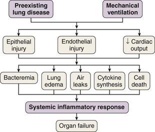
In 1998 Tremblay and Slutsky coined the term biotrauma to describe the pulmonary and systemic inflammatory response triggered by lung cell distention, alveolar disruption, and/or necrosis after the application of mechanical ventilation. Although a Consensus Conference in 1994 recommended that plateau pressure generally should be limited to 35 cm H2O, little change in ventilator practice occurred until publication of an ARDS Network study demonstrating that a lung-protective strategy using a tidal volume of 6 mL/kg of predicted body weight and moderate levels of PEEP decreased mortality in patients with ALI. This study confirmed that VILI was not just an interesting experimental entity but also was an important clinical entity. This recognition led to the widespread, albeit not universal, use of lung-protective strategies in patients with ALI. Unequivocal evidence from both experimental and clinical data has proved that mechanical ventilation can cause or aggravate ALI. Many of the pathophysiologic consequences of VILI mimic those of ARDS. A number of specific forms of injury caused by the trauma of mechanical ventilation have been identified: barotrauma, volutrauma, atelectrauma, and biotrauma. Current experimental and clinical evidence supports a link between VILI and the development of extrapulmonary organ dysfunction, by mechanisms similar to those whereby most severe cases of sepsis are clinically manifested (Figure 36-2).

Pathophysiology of Ventilator-Induced Lung Injury

Lung volumes for all mammals scale with a common function based on body mass. In spontaneously breathing mammals, tidal volumes are approximately 6 to 7 mL/kg of body weight (Figure 36-3), yet historically, tidal volumes of 12 to 15 mL/kg were used in mechanically ventilated patients with acute respiratory failure, and peak alveolar pressures were allowed to increase above 40 cm H2O. This “one tidal volume fits all” approach was formulated in the 1960s, because anesthesiologists and critical care pioneers had demonstrated that ventilation with small tidal volume resulted in a gradual loss of lung volume (i.e., atelectasis) and hypoxemia. Large-tidal-volume ventilation was useful to prevent this negative physiologic consequence. Since the mid-1970s, experimental studies have described the onset of pulmonary edema in rats and larger mammals ventilated with high peak alveolar pressures.

image

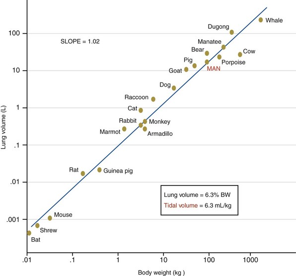
Figure 36-3 Scaling of the lung in mammals. BW, body weight.

(From Villar J, Kacmarek RM, Hedenstierna G: From ventilator-induced lung injury to physician-induced lung injury: why the reluctance to use small tidal volumes? Acta Anaesth Scand 48:267–271, 2004.)

Experimental evidence and advances in lung imaging techniques and bedside ventilator waveform analysis are providing support for the concept that any tidal volume, regardless of how small, has the potential to damage the preinjured lung. Limitation of tidal volume to 6 to 7 mL/kg and plateau pressure to a maximum of 30 cm H2O represents the standard for mechanical ventilation in patients with ALI or ARDS. However, tidal hyperinflation may occur in some patients despite limiting tidal volume to 6 mL/kg and plateau pressure to 30 cm H2O. Current evidence suggests that lung-protective ventilation strategies (in the absence of a priming pulmonary insult) cause significant gene expression in the lung. These changes can be detected even after 90 minutes of mechanical ventilation. In patients with ARDS, a further reduction of tidal volume to levels lower than 6 mL/kg in some instances minimizes tidal hyperinflation and attenuated pulmonary inflammation, as evaluated by pulmonary cytokines and computed tomography.

VILI is a dynamic process that is hard to capture at a single point in time. The damage observed in VILI reflects the primary injurious stimuli and the secondary complex interactions of inflammatory mediators on alveolar epithelial and capillary endothelial cells. Alveolar overinflation elicits a well-coordinated response that contributes to cellular proliferation and inflammation. As the epithelium is progressively stretched, a nonreversible opening of water-filled channels between alveolar cells occurs, resulting in free diffusion of small solutes and albumin across the epithelial barrier. The pulmonary endothelium is a metabolically active surface that provides a regulatory interface for the continual processing of blood-borne vasoactive molecules; it plays an active role in homeostasis and immunologic and inflammatory events, regulates vascular tone, and interacts with inflammatory cells and neighboring vascular cells. Mechanical forces originating within the alveolus can cause endothelial disruption and hemorrhagic injury, even in the absence of preexisting inflammation. Cell deformation by mechanical forces directly causes conformational changes in molecules within the cell membrane, leading to activation of downstream messenger systems. Mechanical ventilation can trigger a complex array of pro- and antiinflammatory mediators that may lead to either greater injury or enhanced lung healing and quicker restoration of pulmonary function. Some ventilatory strategies, however, can cause an acute imbalance in this normal stress response, altering lung cellular function and producing a local and a systemic response. Two mechanisms are believed to be responsible for this mechanical ventilation–induced cytokine release (biotrauma). The first is direct trauma to the cell with disruption of the membranes, resulting in translocation of cytokines into both the alveolar space and the systemic circulation. The second mechanism has been termed mechanotransduction. In vitro studies have shown that most pulmonary cells can produce cytokines in response to cyclic stretch. A large number of genes differentially expressed in the lung by mechanical ventilation have been identified in in vivo animal models of VILI, including genes involved in immunity and inflammation, stress response, metabolism, and transcription processes. However, the sensing mechanism of these physical forces and the translation into intracellular signals is largely unknown.

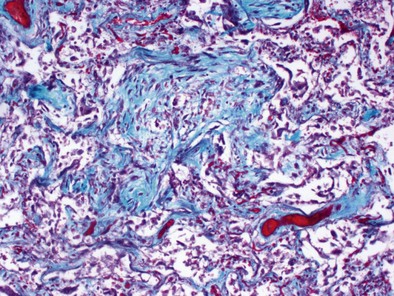
Mechanical ventilation with large tidal volumes results in extracellular matrix remodeling in patients. This effect suggests a potential contributory role of mechanical ventilation in causing lung fibrosis (Figure 36-4). In elective surgical patients, mechanical ventilation with a tidal volume of 12 mL/kg and no PEEP promote procoagulant changes, leading to fibrin deposition within airways; such changes can be prevented by use of low VT and high-level PEEP.

Mechanical Ventilation–induced Sepsis

Sepsis originating from pulmonary and nonpulmonary infections is the most frequent cause and complication of ARDS. First described by Hippocrates in 400 BCE, sepsis is a controversial topic eliciting various hypotheses for its immunopathophysiology. The controversy surrounding sepsis is a result of the way it is defined, because its etiology and pathogenesis remain unclear, and because it is defined by inflammation, regardless of the source.

Sepsis describes a complex clinical syndrome occurring as a result of a systemic inflammatory response to live bacteria or bacterial products. Sepsis develops when the initial, appropriate host response to an infection becomes amplified and is then deregulated. According to the current definition, sepsis represents a disease continuum that proceeds from infection to a systemic inflammatory response syndrome and finally to severe sepsis. The clinical management of sepsis remains very complicated because of the nonhomogeneous nature of patient populations and because of the difficulties in precise clinical classification of patients with the disorder. Whether the sepsis is treated or not, potential outcomes include multiple system organ dysfunction, shock, and death.

Because an all-encompassing single mechanism for sepsis remains elusive, mechanical ventilation–induced inflammation may be a root cause, by amplifying the sepsis cascade of microbial or nonmicrobial origin. Most pulmonary cells express a large repertoire of genes under transcriptional control that are modulated by biomechanical forces and bacterial infections. The highly integrated pulmonary defense system is capable of initiating rapid and intense immune responses to invading microbes or cell debris, resulting in profound local and systemic inflammation. Healthy experimental animal and human lungs ventilated with small, moderate, and large tidal volumes evoke early inflammatory responses similar to those evoked by endotoxin or those seen during infections (activation of nuclear factor κB [NFκB] and cytokine release). Gene expression profiles obtained from microarrays across different experimental models of VILI also suggest that the response triggered by alveolar overdistention might mimic an innate immune inflammatory response against pathogens. Mechanical ventilation enhances lipopolysaccharide (LPS)-induced expression of TNF-α. When used with moderate tidal volumes, it augments the innate immune response to bacterial products in the lung and may play a role in the perpetuation of a septic state. Ventilator-associated pneumonia is a frequent complication of mechanical ventilation and an important contributor to morbidity and mortality in critically ill patients. Endotracheal intubation may be the most important factor leading to pneumonia, because the endotracheal tube directly interferes with the barrier between the oropharynx and the lower respiratory tract, by preventing glottic closure, leading to an ineffective cough mechanism. It is not surprising that pulmonary bacterial colonization and infection are associated with a higher risk for development of ARDS, because the lung, with the largest epithelial surface area of the body, is repeatedly exposed to air-borne particles and microorganisms.

As with the association of bacteria and their products translocating from the gut and leading to sepsis, injurious mechanical ventilation may be responsible for translocation of bacteria and their products from the alveoli into the bloodstream and be a major factor in the development of systemic infection and multiple organ dysfunction. In the absence of a pulmonary infection, however, mechanical ventilation–induced epithelial and endothelial damage could be responsible for the translocation of soluble mediators derived from pathogens or tissue other than bacteria or endotoxin. In some cases, those pathogen-associated molecules could come from pathogens that are colonizing rather than infecting the airway.

VILI and sepsis involve more mediators than was previously thought. Essential components of the innate immune system are the Toll-like receptors, which recognize not only microbial products but also degradation products released from damaged tissue, providing signals that initiate inflammatory responses. Toll-like receptor agonists circulate in the bloodstream both attached to microorganisms and also separately. Because the pulmonary epithelial and endothelial barriers are damaged during ALI, lung cytokines and bacterial products can be trafficked from the alveoli and the interstitium into the systemic circulation, leading to inter- or intracellular signaling impairments in the functional metabolic properties of those populations of cells. It has been found that mechanical ventilation, in the absence of infection, can induce upregulation of the Toll-like receptor-4 signaling pathway (a receptor related to endotoxin signal transduction), resulting in an increase in proinflammatory cytokine levels in the lungs and in the systemic circulation.

Multiple System Organ Dysfunction: A Common End for Ventilator-Induced Lung Injury and Sepsis

Approximately 80% of all deaths in patients suffering from ARDS occur within 2 to 3 weeks after its onset. The exact cause of death remains elusive; no autopsy studies have revealed why patients with ARDS die. Death has traditionally been attributed to the underlying disease, the presence of sepsis, and/or the failure of vital organ systems other than the lung. The association of ARDS with multiple system organ dysfunction is not inevitable, but it certainly is common.

The relationship of ARDS with multiple system organ dysfunction is complex, with at least three possible pathophysiologic models: (1) Although sepsis may cause similar local injury (e.g., increased capillary leak), the functional implications of this injury in the lung are greater because of the exquisite susceptibility of the gas exchange apparatus of the lung to edema; (2) local injury to the lungs (trauma, aspiration, gas inhalation) may set up a secondary diffuse inflammatory response resulting in damage to other organs; or (3) infection and sepsis set off the chain of events leading to ARDS and multiple system organ dysfunction. Among those three models, the classic accepted scenario is that ARDS is complicated by sepsis, which results in multiple system organ dysfunction. A fourth possibility has emerged in light of current evidence on VILI: Ventilator-induced inflammatory response may alter cellular pathways that are important for the normal function of tissues and organs.

Multiple system organ dysfunction is a cumulative sequence of organ dysfunctions in patients suffering from the same diseases often found in patients with ARDS. Still to be resolved is why in most studies on sepsis, at least a third of the patients do not have a truly identified source of the septic process. Among several possible reasons, one of the most frequently offered is that antibiotics, administered early, have suppressed the growth of bacteria, as borne out by results of blood culture. It is plausible that mechanical ventilation could be partially responsible for the development of sepsis or a sepsis-like syndrome in critically ill patients, even if blood cultures are negative. Several investigators have suggested that the mechanical ventilation–induced inflammatory response may be a contributing factor in multiple system organ dysfunction by initiating or propagating a malignant, systemic inflammatory response (see Figure 36-2). ARDS and acute kidney injury are frequent complications in critically ill patients with multiple system organ dysfunction. Although the functional changes seen in septic patients are known to affect primarily the lungs, the cardiovascular system, and the kidneys, the gut has long been established as an important component of the immune barrier. An emerging consensus implicates the gut flora in the pathogenesis of sepsis. The gut is a reservoir of pathogens and pathogen-associated molecular patterns from microbes, and translocation of these molecular patterns and toxins other than endotoxin also induces a systemic inflammatory response (probably through Toll-like receptors and NFκB signaling pathways), contributing to the development of a systemic infection and multiple organ dysfunction in critically ill patients.

Implications of the Sepsis–ventilator-Induced Lung Injury Connection

What conclusions can be drawn from the VILI story? First, the available evidence strongly indicates that patients with ARDS must be ventilated with a tidal volume of 4 to 8 mL/kg of predicted body weight. Second, although data are too limited to support the conclusion that all critically ill patients must be ventilated with a tidal volume of 4 to 8 mL/kg, using small tidal volumes in patients without ARDS may be a reasonable strategy, with little risk for harm, if clinical issues related to maintenance of sufficient PEEP, and possibly the respiratory acidosis that may arise, are addressed appropriately. Third, in view of the relationship between mechanical ventilation and lung injury, an increasingly pertinent consideration is that ARDS may be a consequence of clinical efforts to ventilate the patient, rather than of progression of the underlying disease. Injurious ventilatory strategies have been shown to increase alveolar-capillary leak, to worsen oxygenation, to cause pulmonary infiltrates, to decrease lung compliance, and to cause an increase in lavage fluid and systemic cytokines—all hallmarks of ALI and ARDS. In the context of increased alveolar-capillary leak, use of excessive intravenous fluids—often administered to treat shock in patients at risk for ALI—can cause increased lung water and likewise impair pulmonary mechanics and gas exchange, leading to worse clinical outcomes. It may not be a coincidence that ARDS was first described in the late 1960s, at the time of the Vietnam conflict, when it was called “Da Nang lung” or “shock lung,” in the setting of aggressive resuscitation in the battlefield. Finally, endotracheal intubation affects host defense and can lead to development of colonization or pneumonia, a predisposing factor for ALI and ARDS. Thus, ALI-ARDS may be largely a “man-made” syndrome, arising as a consequence of the aggressive regimens adopted to treat acutely treat patients. If this is so, an inflection point is needed in which ALI-ARDS is no longer a syndrome that must be treated but an iatrogenic illness that should be prevented.

Ventilatory Management for Acute Respiratory Distress Syndrome

Ventilation is an essential function of life and also one of the first to be replicated by artificial means. Mechanical ventilation is the second most frequently performed therapeutic intervention after treatment of cardiac arrhythmias in the intensive care unit (ICU), and it is the most important aspect of the supportive care of patients with respiratory failure. As part of the therapy for the underlying disease, patients with ARDS invariably require endotracheal intubation and mechanical ventilation to decrease the work of breathing and to improve oxygen transport. An improvement in oxygenation can be obtained in many patients with ARDS by an increase in PEEP. To date, the only proven, widely accepted method of mechanical ventilation for ARDS is protective lung ventilation using a low-tidal-volume strategy (4 to 8 mL/kg of predicted body weight) and medium to high PEEP to keep alveoli open throughout the ventilator (respiratory) cycle.

Early interest in low-tidal-volume ventilation was prompted by animal studies in the 1970s showing that mechanical ventilation with large tidal volumes and high peak alveolar pressures resulted in the development of ALI characterized by formation of hyaline membranes and inflammatory infiltrates. Although tidal volumes of 10 to 15 mL/kg had traditionally been used in a majority of patients with respiratory failure, it was recognized in the mid-1980s that ARDS resulted in a significant reduction in the amount of normally aerated lung tissue. The ARDS “baby lung” concept—in which only a small portion of the lung is actually available for ventilation—describes marked overdistention by high tidal volumes.

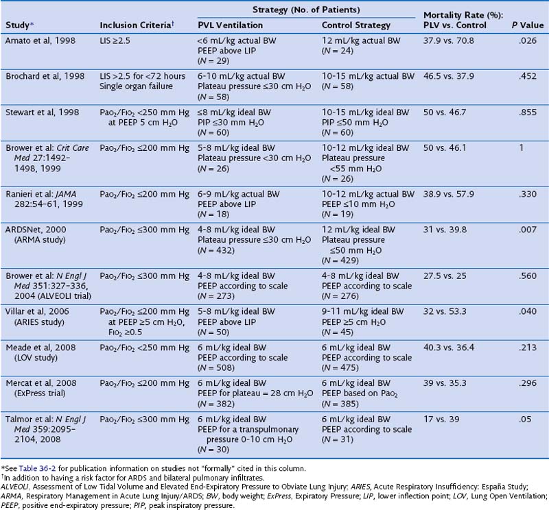
The effect of different lung-protective ventilatory strategies in patients with ALI or ARDS has been investigated in randomized controlled trials. In the late 1990s, four such trials were conducted to evaluate the benefit of low tidal ventilation in ARDS patients compared with traditional tidal volume ventilation (Table 36-4). Only one of these trials, performed in Brazil by Amato and colleagues, showed a significant reduction in mortality in the experimental treatment group. Patients randomized to receive tidal volumes of 6 mL/kg of actual body weight or lower and driving pressures of less than 20 cm H2O were significantly less likely to die during the 28-day study period than were patients randomized to receive the traditional 12 mL/kg of actual body weight tidal volumes and unlimited driving pressures. Because all four studies had limited statistical power owing to small sample sizes, a large trial was needed to definitively determine the effect of low-tidal-volume ventilation in patients with ARDS.

In response to this need, the National Heart, Lung, and Blood Institute ARDS Network enrolled 861 patients at 10 institutions between 1996 and 1999 in a randomized controlled trial known as the Respiratory Management in Acute Lung Injury/ARDS (ARMA) trial. This study compared a ventilatory protocol using tidal volumes of 4 to 8 mL/kg of predicted body weight and maintaining plateau pressures of 30 cm H2O or less with conventional mechanical ventilation using 12 mL/kg of predicted body weight tidal volumes. The lower tidal volume protocol in the ARMA study achieved more pronounced differences between the intervention and control groups in tidal volume (6.2 versus 11.8 mL/kg of predicted body weight) and plateau pressure (25 versus 33 cm H2O) than those achieved in previous studies. The hospital mortality rate was significantly reduced in the low-tidal-volume group, with a nearly 9% reduction in the absolute risk of death from that the control group (31% versus 39.8%; P = .007). Although it has been postulated that the mortality benefit demonstrated in the ARMA study was attributable to a high mortality rate in the control group resulting from tidal volumes that were higher than the standard of care, several observational studies performed later indicated that no uniform standard of care existed at the time of the ARMA trial.

Therefore, it is recommended that critical care physicians utilize the ventilation protocol outlined by the Acute Respiratory Distress Syndrome Clinical Network (ARDSNet) investigators. This protocol involves the following principles:

Since the publication of ARMA, however, despite the statistically significant decrease in mortality rates, widespread use of this therapy remains elusive. Significant barriers to the implementation of low-tidal-volume mechanical ventilation include unwillingness to accept or lack of knowledge of the published data, failure to recognize patients as having ARDS, perceived contraindications to low-tidal-volume ventilation, concerns regarding patient discomfort, and presence of hypercapnia or acidosis.

The Use of Positive End-Expiratory Pressure in Acute Respiratory Distress Syndrome

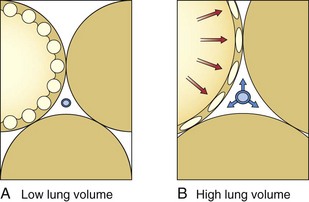
PEEP has become an essential component of the care of many critically ill patients who require ventilatory support. The rationale for the use of PEEP in ALI coincides with the theoretical basis for loss of lung volume and compliance in patients with ARDS. With the application of PEEP, the baseline end-expiratory pressure in mechanically ventilated patients is elevated above atmospheric pressure. The application of PEEP is expected to improve lung mechanics and gas exchange, because it maintains recruited lung volume open. PEEP should be utilized in patients with ARDS to decrease the proportion of nonaerated lung, resulting in improved oxygenation. PEEP prevents complete alveolar collapse and improves oxygenation by increasing functional residual capacity, probably by preventing airway closure and keeping open the previously recruited unventilated alveoli. Conversely, the increase in functional residual capacity also may increase lung compliance. In general, PEEP is applied to improve oxygenation, which usually is not observed except with a concomitant increase in functional residual capacity. The application of PEEP is expected to increase PaO2 and to decrease intrapulmonary shunt, alveolar-arterial O2 pressure difference, and arteriovenous O2 content difference because greater lung volumes are recruited. Four mechanisms have been proposed to explain the improved pulmonary function and gas exchange with PEEP: (1) increased functional residual capacity; (2) alveolar recruitment; (3) redistribution of extravascular lung water; and (4) improved ventilation-perfusion matching. The increase in lung volume is the result of three separate effects. First, PEEP increases lung volume as a result of distention of already patent airways and alveoli by an amount dependent on system compliance. Therefore, the stiffer the system, the smaller the volume change. Second, application of PEEP prevents alveolar collapse during expiration. Dependent, small airways tend to collapse at low lung volumes. Third, PEEP levels above 10 cm H2O recruit collapsed alveoli in the acutely injured lung. Alveolar recruitment describes reinflation of previously collapsed alveoli. The application of PEEP allows recruitment of flooded alveoli and improves oxygenation without diminishing lung water content, supporting the conclusion that PEEP redistributes lung water from alveoli to the perivascular space. In general, by increasing intraalveolar pressure, PEEP moves fluid from the interstitial space of alveolar vessels to the interstitial space around extraalveolar vessels. In addition, PEEP increases the transmural pressure across corner (in the peribronchial area) and extraalveolar vessels, thereby increasing flux into the interstitial space (Figure 36-5).

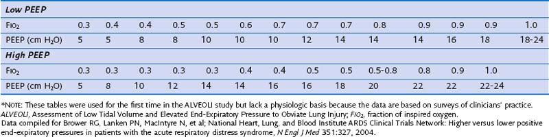
Although PEEP is well recognized to improve oxygenation in selected patients, its beneficial effects on morbidity and mortality have not been conclusively demonstrated. Early observations that PEEP greatly improves oxygenation in patients with ARDS led to its widespread use in such patients, but the level of PEEP needed to achieve maximum benefit with minimal complications has never been established. Traditionally, PEEP values of 5 to 12 cm H2O have been used in the ventilation of patients with ARDS. In the ARMA trial, PEEP levels in both the experimental and control groups were adjusted according to the required FIO2 and ranged between 5 cm H2O (when FIO2 was 0.3) and 18 to 24 cm H2O (when FIO2 was 1.0) (Table 36-5). On average, patients in the low-tidal-volume ventilation group were treated with levels of PEEP that were no different than those utilized in the high-tidal-volume group. However, it remains unclear whether these values are ideal because randomized controlled trials have not clearly shown that higher levels of PEEP lead to a reduction in mortality rate.

Several randomized controlled trials have evaluated the efficacy of high levels of PEEP in the treatment of ARDS. Thus far, a total of seven published randomized controlled trials have examined the effects of higher levels of PEEP in patients with ALI or ARDS, or both. Those trials have tested higher versus lower PEEP strategies during low-tidal-volume ventilation and lower tidal volume and PEEP titrated to above the lower inflection point of the individual pressure volume curve versus higher-tidal-volume ventilation and lower PEEP (see Table 36-4). At first look, the pooled results of a metaanalysis of these trials suggest that the application of either low or high PEEP levels in patients with ALI or ARDS does not influence outcome. However, this is not what the data show. First, because the ALI-ARDS population was not homogeneous in all the trials, the benefit of higher levels of PEEP could not be appropriately evaluated. Second, in some trials, a PEEP level of 5 cm H2O was a permissible setting in the higher PEEP group. Therefore, a patient with a PaO2 of 60 mm Hg on a FIO2 of 0.3 and 5 cm H2O of PEEP satisfied the AECC inclusion criteria. It is difficult to accept that in these patients there is a need to test the effects of high levels of PEEP. Finally, some trials (e.g., the Assessment of Low Tidal Volume and Elevated End-Expiratory Pressure to Obviate Lung Injury [ALVEOLI] and Expiratory Pressure [ExPress] trials) (see Table 36-4) included patients with ALI (without ARDS), who did not benefit and more often experienced adverse effects from higher levels of PEEP, suggesting that both trials failed to focus on the patients at highest risk. If the subjects in a trial have a very low risk for the condition that the intervention is hypothesized to prevent, the trial—regardless of sample size—will not verify the value of the experimental intervention under study. In the study of a protective ventilation strategy by Amato and Villar and their respective colleagues, PEEP was significantly higher in the intervention group than in the control group (13 to 14 cm H2O versus 9 cm H2O, respectively). Although both trials reported a significantly lower mortality in the intervention group, these findings cannot be solely attributed to higher levels of PEEP, because the intervention strategies in these trials also included both low tidal volumes and high levels of PEEP.

To determine the isolated benefit of high levels of PEEP in patients with ARDS, the ARDSNet conducted a large randomized controlled trial known as the ALVEOLI trial. Patients ventilated with low tidal volumes (4 to 8 mL/kg of predicted body weight) were randomized to a ventilator protocol utilizing high levels (12 to 24 cm H2O) or low levels of PEEP (5 to 24 cm H2O). In the first 7 days of ventilator support after initiation of treatment, patients in the high PEEP group received 13 to 15 cm H2O of PEEP and patients in the low PEEP group, 8 to 9 cm H2O of PEEP. Although patients in the high PEEP group experienced better improvement in oxygenation, the duration of mechanical ventilation and hospital mortality were similar in the two groups.

The speculation of many investigators regarding the lack of expected benefit from higher PEEP in the ALVEOLI, Lung Open Ventilation (LOV), and ExPress trials (see Table 36-4) is that in a substantial proportion of patients in those trials, the severity of lung injury was modest. In two other trials, Amato and colleagues and Villar and associates enrolled only patients with severe, established ARDS, and both groups found that the application of higher levels of PEEP was associated with a better outcome. By contrast, in the rest of the trials, the investigators studied an unselected, mixed population of ALI and ARDS and, as a result, missed the opportunity to test whether the use of higher levels of PEEP is beneficial in patients with persistent ARDS. Patients in the ALVEOLI, LOV, and ExPress trials had similar PaO2/FIO2 ratios at study entry (about 140 ± 50 mm Hg). It is conceivable, however, that a disproportionate number of patients meeting ALI criteria on standard ventilatory settings ended up in the control group, negating the beneficial effect of the treatment because of a lower mortality rate (20% or less in most series). A critical review of the two most recently published trials on the effects of PEEP in ARDS leads to an alternative interpretation. In both trials (the LOV study and the ExPress trial), patients who received higher PEEP were less likely to require rescue therapy. In addition, a lower PEEP was associated with significantly fewer ventilator-free and organ failure-free days, which may be the reason for a 4% mortality difference in favor of the high PEEP groups (P = .10).

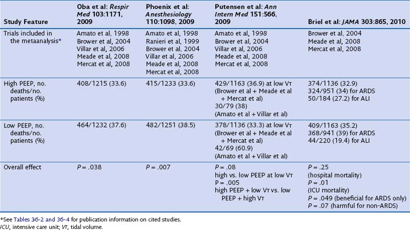
Of note, current published metaanalyses including some or all of the available trials reported conflicting results and are biased because they do not deal with all comparisons between tidal volume and PEEP (Table 36-6). The first metaanalysis from Oba and colleagues, which considered five trials (reported by Amato, Brower, Meade, Mercat, and Villar and their co-workers), showed a decrease in in-hospital mortality (P = .03) and a trend toward significance for the 28-day mortality (P = .06) with higher PEEP and without an increase in barotrauma. A second meta-analysis, by Phoenix and colleagues, included an additional study (by Ranieri and co-workers) and showed a reduction in mortality (pooling together the in-hospital and the 28-day mortality rates, P = .007) with higher PEEP. In a subsequent metaanalysis, Putensen and colleagues analyzed the five trials included in the Oba metaanalysis. Data from three of these trials—those reported by Brower, Meade, and Mercat and their co-workers—were pooled together for comparison of lower versus higher PEEP at low tidal volume ventilation. The remaining two studies (by Amato and Villar and their co-workers) were used for the comparison of higher tidal volume and lower PEEP versus lower tidal volume and higher PEEP. Despite opposite results from the two previous metaanalyses, the comparison between lower and higher levels of PEEP at low tidal volume ventilation showed similar hospital mortality rates (P = .08). However, the risk of in-hospital mortality was reduced only with the combination of a lower tidal volume and higher PEEP, compared with higher tidal volume and lower PEEP (P = .005). Thus, the beneficial effect of PEEP may have been due to a simultaneous reduction in the tidal volume and an increase in the level of PEEP. Finally, a fourth metaanalysis from Briel and colleagues (see Table 36-6) was performed on the individual data for 1136 patients (with higher PEEP) and 1163 patients (with lower PEEP) from trials that compared lower and higher PEEP values at a low-tidal-volume ventilation. The individual data analysis showed that a nonsignificant difference was found in hospital mortality rates between the higher and lower PEEP groups (32.9% versus 35.2%; P = .25). In conclusion, these metaanalyses of individual patient data suggest that higher levels of PEEP may be associated with lower mortality in patients who meet ARDS criteria, whereas such a benefit is unlikely in patients who have less severe lung injury.

Rescue Strategies for Refractory Hypoxemia

A number of alternative techniques (currently available worldwide or under evaluation) can be used to improve oxygenation and ventilation in patients with ALI or ARDS who have refractory hypoxemia. Today, refractory hypoxemia is rare and an infrequent cause of death (accounting for less than 15% of ARDS-related deaths). There is no standard definition for refractory hypoxemia in terms of a predetermined PaO2 value under a specific oxygen concentration (FIO2) and applied PEEP level for a specific period of time. In most reports, it has been defined as having a PaO2 below 60 mm Hg on an FIO2 0.8 to 1.0 and PEEP 10 to 20 cm H2O for more than 12 to 24 hours.

RecruitMent Maneuvers

As described previously, ARDS is characterized by collapsed and consolidated alveoli. Recruitment maneuvers are intended to reopen these alveoli and to attenuate the injurious effects of the repetitive opening and closing of the alveolar unit. In general, a recruitment maneuver is defined as applying a pressure higher than that applied during a normal breath either intermittently (for 2 to 3 minutes) or sustained for a short period of time (up to about 40 seconds). A recruitment maneuver usually improves oxygenation and may influence ventilation by reducing PaCO2. Recruitment occurs during the whole period of inspiration, and the amount of recruited lung area correlates with the inspiratory pressure applied. The amount of potentially recruitable lung tissue seems to correlate well with the severity of ARDS. With respect to hemodynamics, the acute and substantial increase in intrathoracic pressure during the recruitment maneuver often induces a decline in cardiac output and tissue oxygenation. Therefore, it is essential to stabilize the patient hemodynamically before a recruitment maneuver procedure. Although the potential for barotrauma is a concern during a recruitment maneuver, when recruiting pressures are maintained at or below 50 cm H2O peak alveolar pressure, barotrauma has been rarely reported.

The ALVEOLI trial also evaluated the safety and efficacy of recruitment maneuvers in the first 80 patients randomized to receive high-level PEEP. Continuous positive airway pressure (CPAP) of 35 to 40 cm H2O was applied for 30 seconds, and the results were compared with those of a sham recruitment maneuver. Because the interventions resulted in only small and transient increases in oxygenation, they were discontinued. Such maneuvers have been associated with transient hypotension and hypoxemia, and their long-term benefit remains unproved. In a metaanalysis of seven clinical trials involving 1170 patients with ALI/ARDS, there was no significant difference in survival between groups receiving an “open lung” ventilatory strategy that included recruitment maneuvers and groups given standard ventilatory care. The main limitation of that systematic review, however, was the design of the trials, in that they either did not isolate recruitment maneuvers from other variables or assessed only short-term outcomes, and few of these trials determined the patient-specific PEEP level (by decremental trial) after the recruitment maneuvers—a key to the successful use of such maneuvers. Most recent data suggest that a recruitment maneuver performed in the early phase of ARDS might be more effective than in late-stage ARDS.

Extracorporeal Membrane Oxygenation

Extracorporeal membrane oxygenation (ECMO) is a technique that originally was applied in patients with acute respiratory failure of such severity that it was impossible to provide adequate oxygenation by conventional mechanical ventilation. To supplement gas exchange, a portion of the cardiac output must go through the ECMO circuit. During ECMO, CO2 is removed by the extracorporeal circuit, but this technique usually is supplemented with conventional mechanical ventilation at low ventilatory rates, high PEEP levels, and tidal volumes adequate to maintain a plateau pressure below 30 cm H2O. Most long-term adult ECMO procedures are performed using the venovenous approach. Access for both blood removal and return is by way of the femoral, saphenous, or jugular vein. Despite the excitement generated by earlier reports of success, the results of ECMO trials led to a loss of enthusiasm for its use in acute respiratory failure. Some investigators, however, believe that there exists a role for ECMO in young adult patients with single organ system failure who are deemed to have potentially reversible pulmonary dysfunction when all other conventional modalities have failed.

Recently a large multicenter adult ECMO trial was completed. Referred to as the CESAR trial (Conventional Ventilatory Support versus Extracorporeal Membrane Oxygenation for Severe Adult Respiratory Failure), this randomized controlled trial assessed the effectiveness of extracorporeal lung assist in 180 patients with severe ARDS. The findings of this study represent the first positive results for adult ECMO application in severe respiratory failure. Survival at 6 months or absence of severe disability was achieved in 63% of the patients on ECMO, compared with 47% of the control group (P = .03). However, a number of major concerns and limitations with this study have been identified: First, patients allocated to conventional management (control group) were treated with conventional mechanical ventilation or with high-frequency ventilation. Second, patients in the control group were ventilated with a nonstandardized protocol; to ensure the collaboration of participating centers, physicians were allowed to choose any ventilatory strategy. Third, 30% of patients in the control group were not ventilated with a lung-protective strategy. Fourth, no data regarding ventilatory parameters at study entry and during the mechanical ventilation period are presented. Fifth, all patients receiving ECMO were treated in the same center. Sixth, the ECMO center did not treat patients randomized to the conventional management group. Seventh, many patients randomized to receive ECMO did not receive ECMO. In fact, 103 patients who were screened for eligibility were excluded because a bed was unavailable for ECMO, and 22 patients (25%) assigned to be transferred to the ECMO center never received ECMO (16 of these improved with conventional management). Finally, a more critical analysis of outcomes data for patients who actually received ECMO (68 of 90) compared with those who were treated with mechanical ventilation for whom information about mortality was available (87 plus 22) showed that the mortality rates were similar in both groups (48.5% for ECMO versus 43.1% for mechanical ventilation) (P = .64, by our own calculations). In addition, the length of ICU stay and hospital stay was more than twice as high in the ECMO group.

The highly specialized equipment and knowledge required to provide ECMO make this technique available only in specific medical centers.

High-Frequency Oscillatory Ventilation

Some clinicians have proposed that high-frequency oscillatory ventilation (HFOV) is an ideal mode of ventilation for patients with ARDS, because it is the natural culmination of low-tidal-volume ventilation. Current understanding of the mechanisms and importance of ventilator-induced lung injury has advanced considerably over the past 3 decades. HFOV should theoretically be an ideal mode to ventilate patients with severe lung damage, because it achieves gas exchange by delivering very small tidal volumes that typically are 1 to 3 mL/kg (often less than the anatomic dead space) at frequencies ranging from 3 to 10 Hz around a relatively constant mean airway pressure. HFOV is not a difficult technique. In fact, it is easier than conventional mechanical ventilation: It incorporates fewer and simpler controls that are not interrelated as they are in conventional mechanical ventilators. Recent prospective, observational studies have reported that HFOV is a feasible and efficient method of ventilation that results in rapid and sustained improvement in oxygenation in patients with severe ARDS. However, a critical examination of randomized controlled trials comparing HFOV with conventional ventilation demonstrates that there is equivalence between conventional ventilation and HFOV. Specifically, there is no evidence that conventional mechanical ventilation with low tidal volumes, high-level PEEP, and limited plateau pressures is more harmful that HFOV. All of the randomized controlled trials to date have compared HFOV with a less-than-optimal approach to conventional ventilation. Several ongoing trials, however, are comparing HFOV with low-tidal-volume ventilation.

Prone Positioning

Changes in posture can have profound effects on the pulmonary function of patients with severe respiratory failure. Most changes in pulmonary physiology with posture occur as a consequence of the influence of gravity and chest wall shape on the mechanical properties of the lung. By tradition, patients with respiratory failure are cared for supine. In critically ill patients, the supine posture is associated with a fall in functional residual capacity to below closing capacity, resulting in ventilation-perfusion mismatching and a drop in PaO2. During acute respiratory failure, a reduction of functional residual capacity results in supine hypoxemia regardless of age. Because most lung infiltrates in patients with ARDS are seen in dependent lung regions, it was postulated that prone positioning of patients redistributes blood flow and ventilation to the least affected areas of the lung, promotes secretion clearance, and shifts the weight of the mediastinal contents anteriorly, to assist in the recruitment of atelectatic regions. Thus, the proposed mechanisms by which prone positioning improves oxygenation include an increase in functional residual capacity, a change in regional diaphragm motion, redistribution of perfusion to better-ventilated lung units, redistribution of ventilation to better-perfused lung units, and improved secretion clearance.

Since 1974, prone positioning has been proposed as a technique to improve oxygenation. The practice of turning prone seems to be inexpensive and safe, with the possible exception of an increased risk of regurgitation or inadvertent extubation. However, the act of turning is labor-intensive; at least four experienced staff members are required for this maneuver to avoid the loss of vascular accesses or the airway during turning. Meticulous care must be used in positioning the patient. Placing a neck roll or a pillow under the patient’s shoulders and turning the head to one side constitutes the recommended way to support the patient when prone. In addition, prone positioning may be associated with an increased need for sedation. Automated prone positioning uses special devices operated by one nurse, and therapy can be individualized in accordance with the patient’s needs and responses. Most complications, such as skin injury, facial edema, catheter removal or compression, hypotension, arrhythmias, and inadvertent extubation, are associated with manual positioning. The risk of developing pressure ulcers is ever-present, as the patient is immobile and pressure on bony prominences may be prolonged. If the patient’s tongue becomes edematous to the point of the teeth cutting into it, a dental mouth prop should be used to prevent injury. Facial edema, although only temporary, can be very disturbing for the patient’s family to view and can be minimized with application of ice packs.

Although sufficient data have accumulated to permit the conclusion that oxygenation frequently improves when patients with ARDS are turned prone (in about 70% of patients), prone positioning is still not widely implemented. Three recent systematic reviews and metaanalysis in patients with ALI or ARDS have shown that in general, prone positioning does not reduce mortality or duration of mechanical ventilation despite improved oxygenation and a decreased risk of pneumonia. By stratifying patients according to their PaO2/FIO2 ratio, however, the most recent metaanalysis has found that in patients with severe ARDS, as defined by a PaO2/FIO2 ratio less than 100 mm Hg, prone positioning was able to cause a significant reduction (16% decrease in the relative risk) of all-cause mortality. Thus, prone positioning should be considered to represent a rescue maneuver, and it should be reserved only for patients with severe acute hypoxemic failure in the early phase of the disease process. In such circumstances, the potential for life-threatening complications of prone positioning, including accidental dislodgment of the endotracheal tube or central venous catheters and endotracheal tube obstruction, should be weighed against the short-term benefit of improved oxygenation. Unfortunately, no recommendations can be offered on the optimal timing or duration of prone positioning until large randomized controlled trials in patients with severe hypoxemia provide more information. Extended prone positioning seems to be most beneficial when maintained 18 to 20 hours daily.

Inhaled Vasodilators

Antiinflammatory agents and vasodilators have been tried experimentally in animals and humans for prophylaxis or treatment of ARDS. Prostaglandins, ibuprofen, pentoxifylline, inhaled nitric oxide (iNO), inhaled prostacyclin, almitrine, and corticosteroids all have been tried. None of them have shown any major benefit on outcome in large randomized human trials, even though significant improvements of oxygenation have been observed with some of these agents.

Nitric oxide (NO) is important for the regulation of pulmonary vascular smooth muscle tone. NO appears to be pivotal in acute and chronic hypoxic pulmonary vasoconstriction. Pulmonary hypertension is a typical feature of ARDS and is a bad prognostic factor in respiratory failure. Inhaled NO selectively dilates pulmonary vasculature without systemic effects. Over the past two decades, an increasing number of clinical studies have been published assessing different aspects of inhaled NO in patients with ARDS and addressing the ability of NO to attenuate ALI. Despite the fact that many clinicians consider inhaled NO to be a useful rescue treatment for patients with ARDS, no randomized controlled trial has demonstrated an outcome benefit. A systematic review and metaanalysis of 12 randomized controlled trials including a total of 1237 patients with severe ALI or ARDS found that overall, NO is associated with limited improvement in oxygenation at 24 hours of therapy, has no effect on duration of ventilation, does not confer mortality benefits, and may cause harm.

Supportive Treatment

Muscle Paralysis

Neuromuscular blocking agents have commonly been used in clinical practice for patients with marked ventilator asynchrony and in a large but highly variable proportion of patients with ARDS. Current guidelines indicate that neuromuscular blocking agents are appropriate for facilitating mechanical ventilation when sedation alone is inadequate, most notably in patients with severe gas exchange impairment. The mechanisms by which these agents may benefit patients with ARDS are not completely understood but presumably involve improvement in patient-ventilator synchrony and limiting of excessive airway pressures. Prevention of barotrauma is another mechanism by which neuromuscular blockade may improve outcome.

Previous publications suggesting that the use of neuromuscular blockade agents may lead to increased incidence of ICU-acquired weakness or other complications raised concerns about long-term side effects with routine use of paralytic agents for ARDS. However, the latest information on the use of neuromuscular blockade in the treatment of patients with severe ARDS showed promising results. In a multicenter, double-blind trial conducted in France, 340 patients with severe ARDS within the previous 48 hours were randomly assigned to receive, for 48 hours, either the neuromuscular blocking agent cisatracurium besylate (178 patients) or placebo (162 patients). Severe ARDS was defined as a PaO2/FIO2 ratio less than 150 mm Hg, with a PEEP of 5 cm H2O or less and a tidal volume of 6 to 8 mL/kg of predicted body weight. The primary outcome was death either before hospital discharge or within 90 days after study enrollment, as reflected in the 90-day in-hospital mortality rate. The hazard ratio for death at 90 days in the cisatracurium group, as compared with the placebo group, was 0.68 (P = .04) after adjustment for both the baseline PaO2/FIO2 and plateau pressure. The crude 90-day mortality rate was 31.6% in the cisatracurium group and 40.7% in the placebo group (P = .08); the mortality rate at 28 days was 23.7% with cisatracurium and 33.3% with placebo (P = .05). In addition, treatment with the neuromuscular blocking agent cisatracurium for 48 hours early in the course of severe ARDS increased the numbers of ventilator-free days and days outside the ICU, and decreased the incidence of barotrauma during the first 90 days. The rates of ICU-acquired neuromuscular weakness did not differ significantly between the two groups. Additional work is needed to determine whether the use of neuromuscular blocking agents for only 48 hours is beneficial in selected patients. The beneficial effect of the neuromuscular blocking agent on survival was confined to the two thirds of patients with a PaO2/FIO2 ratio below 120 mm Hg.

Fluid Management in Acute Respiratory Distress Syndrome

Until recently, the optimal strategy for fluid management in patients with ARDS was unclear. Animal and human data suggest that when lung capillary permeability increases, lung water accumulates to a greater degree than usual at lower pulmonary artery occlusion pressures. Human trials show improved physiologic end points with various diuretic approaches to reduce lung water, including diuresis without vascular pressure measurements, intravascular pressure–targeted diuresis, and diuresis guided by direct measurements of lung water. Abundant data suggest that prompt resuscitation of hemodynamically unstable patients improves outcome, whereas the same resuscitative efforts given later may not be helpful and may potentially be harmful.

The ARDSNet published in 2006 the results of a large randomized trial comparing two fluid management strategies in 1000 patients with ALI or ARDS. Subjects were randomized to management with either a conservative (−136 ± 491 mL) or a liberal (6992 ± 502 mL) approach. In other words, the conservative fluid management group had a net fluid balance of about zero over the first 7 days of the protocol, whereas the liberal fluid management group had an average daily fluid gain of approximately 1 L. Fluid and diuretic management was dictated by a highly protocolized regimen, and all patients received respiratory support using a low-tidal-volume, plateau pressure–limited ventilation strategy. Although no significant difference was noted in the primary outcome of 60-day mortality, the conservative approach improved lung function and shortened the duration of mechanical ventilation and ICU stay without increasing the rate of nonpulmonary organ failure. These data provide reassurance and support for the use of a conservative fluid management strategy in patients with ARDS.

Corticosteroids

Corticosteroids would seem to be an ideal therapy for ALI, in view of their potent antiinflammatory and antifibrotic properties. Several clinical trials have evaluated the utility of corticosteroids in preventing ARDS and in treating either early-stage (inflammatory) or late-stage (fibrotic) ARDS. None of them have demonstrated a mortality benefit. Despite failed studies of prevention or early treatment, great interest remains in the use of corticosteroids for so-called salvage therapy in patients with persistent ALI. The ARDSNet performed the largest randomized, blinded trial of methylprednisolone versus placebo in patients with ARDS of at least 7 days’ duration. No survival benefit was noted in the steroid group (29.2% versus 28.6%); however, methylprednisolone increased the number of ventilator-free days, shock-free days, and ICU-free days during the first month. Also, it was associated with a significant increase in 60-day and 180-day mortality rates among patients enrolled after 13 days from onset of ARDS. This study argues against the use of corticosteroids to treat patients with ARDS. However, a metaanalysis of selected trials showed that prolonged administration of systemic steroids is associated with favorable outcomes and a survival benefit when given before day 14 after onset of ARDS. The latter finding prevailed when data for subgroups from the ARDSNet were reanalyzed according to time of treatment initiation. Nevertheless, the questionable benefit of steroids in patients with ARDS should not preclude the use of a low-dose regimen in acutely ill patients with sepsis, including those with ARDS.

Prognosis and Long-Term Survival

Prognosis with ARDS depends primarily on the underlying cause of lung injury. In an analysis of the ARDSNet database, survival to home discharge was lowest in patients with sepsis, intermediate in patients with pneumonia, and highest in patients with trauma and ARDS.

Very few studies have evaluated the long-term outcome of patients with ARDS. Patients who are treated for this condition often face long-term physical and psychological complications that result from their prolonged hospitalization. Studies have considered long-term outcomes in patients with ARDS in terms of respiratory function, exercise tolerance, loss of muscle mass, and cognitive effects. In 2001, a 1-year follow-up study of patients with ARDS in the United States found that a significant percentage of deaths occurred between day 28 and 4 months, which raised the potential for longer monitoring in the evaluation of new interventions or therapies. One-year predictors of death were advanced age and the premorbid functional status. Anxiety, depression, and posttraumatic stress disorder were frequent in that study.

Patients with ARDS lose a significant amount of body weight during their hospital stay. Most patients gradually recover this weight but still remain below admission weight after 12 months. In addition, most patients complain of proximal muscle weakness, with a decrease in distance covered on the 6-minute walk test. A long-term prospective study from Canada found that patients exhibited impairment on pulmonary function testing, seen as a mild restrictive pattern, with subsequent clinical improvement over the course of a year to near-normal levels. No patients required oxygen at rest after 12 months, and only 6% of patients required oxygen with ambulation. Similarly, marked impairment in exercise tolerance is typical; with the reestablishment of muscle mass, however, this impairment levels off by the end of 1 year, but no significant gains were made in the second year. Radiographic abnormalities were present in 80% of cases at 12 months. Of note, half of all patients who contracted ARDS had returned to work by 1 year, with most returning to their original employment position.

On the basis of this cumulative evidence, a patient surviving hospitalization for ARDS can be expected to return to a similar lifestyle over the course of a year, with some lingering physical and psychological challenges. Therefore, in the absence of significant comorbid conditions, the long-term outcome data are sufficient to warrant aggressive treatment for ARDS.

Controversies and Pitfalls

In patients with acute coronary syndromes, the working diagnosis is based on the presence of acute chest pain that is accompanied by abnormalities on the electrocardiogram (ECG) and the biomarker troponin. Troponin is the biomarker for detection of heart injury, and troponin levels serve as the basis for risk stratification and therapeutic interventions in patients with coronary artery disease. By contrast, pathognomonic laboratory or clinical features are lacking in patients with ARDS. There are no data that link a particular PaO2/FIO2 ratio to predictable structural changes in the alveolar-capillary membrane, probably because ARDS represents a common pathway of diverse events and disease entities. Also, current guidelines for ARDS management do not follow a strict stratification, as used in patients with coronary artery diseases. Stratification of respiratory and ventilatory variables at the onset of ARDS could be a useful strategy for identifying and selecting patients for clinical trials with various levels of mortality risk. Using demographic, pulmonary, and ventilatory data collected at ARDS onset, a simple prediction model based on a stratification of variables into low, intermediate, and high categories of risk assists in predicting patient outcome. Tertile distribution for age, plateau airway pressure, and PaO2/FIO2 ratio at ARDS onset is able to identify subgroups with markedly different mortality rates.

Lack of specific knowledge of the molecular mechanisms responsible for ARDS represents the most important obstacle to the successful diagnosis and treatment of affected patients. In comparing the management of acute chest pain with ARDS, the former is based on an emergency medical model of awareness of a life-threatening condition and the importance of adherence to predefined decision algorithms. No comparable awareness and emergency decision algorithms are available for the care of patients with ARDS. It is plausible that a new definition based on specific biochemical criteria of lung inflammation, rather than on clinical parameters, is likely to provide clinicians with a better stratification and identification of a more homogeneous population of patients with ALI and ARDS. Thus, stratification of patients with ARDS should be linked to two measures of severity: one that specifically quantifies the severity of ALI and ARDS and another that quantifies the overall physiologic response along with comorbidity and premorbid health. Adding objective measures, such as levels of biologic markers, could facilitate recognition of ALI and ARDS.

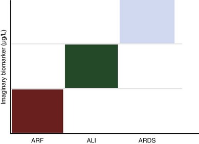
The use of simple thresholds for the diagnosis of disease processes of increasing prevalence in the general population is common. This is the case with the use of blood sugar for diabetes and hemoglobin for anemia. It appears improbable that in the case of ARDS, a biomarker alone will resolve this issue. Instead, a clinical prediction model or a combination of such a predictor model with a biomarker would provide a better definition of ALI and ARDS (Figure 36-6). Ideally, such a biomarker should be (1) 100% sensitive, (2) 100% specific, (3) easy to measure in blood, exhaled air, or any other biologic sample, (4) affected by treatment, and (5) cost-effective. There have been recent efforts to identify biologic markers in pulmonary edema fluid and in blood collected from patients with and without ARDS. It has been postulated that owing to increased permeability of the alveolar-capillary barrier, proteins leak into the circulation. Patients at risk for ARDS who have higher levels of interleukin-8 in bronchoalveolar lavage fluid subsequently progress to ARDS. Serial LBP measurements may offer a clinically useful biomarker for identification of patients likely to experience the worst outcomes and with the highest probability for development of sepsis-induced ARDS. A combination of biologic markers that reflect endothelial and epithelial pulmonary injury, inflammation, and coagulation will be superior to clinical predictors or biomarkers alone for predicting mortality or stratifying patients with ALI or ARDS. Finally, serial elevation of plasma levels of functionally relevant cell proteins (above a critical threshold) could be used for the differential diagnosis for ALI and ARDS. The measure of those biomarkers may inform clinicians of the development of ALI or ARDS.

An interesting direction for research would be identification of reliable biomarkers that are specific for the noninfectious, septic-like syndrome of VILI. It is hoped that the rapidly evolving sciences of genomics, proteomics, and computational biology can be used to model VILI and to tailor mechanical ventilation strategies to create an ideal inflammatory environment for minimal injury, tissue repair, cell regeneration, and organ function. Such a strategy would not only mitigate VILI but also decrease the incidence of other ventilator-induced complications.

Suggested Readings

Ashbaugh DG, Bigelow DB, Petty TL, Levine BE. Acute respiratory distress in adults. Lancet. 1967;2:319–323.

Bernard GR, Artigas A, Brigham KL, et al. Consensus Committee: The American-European Consensus Conference on ARDS. Am J Respir Crit Care Med. 1994;149:818–824.

Briel M, Meade M, Mercat A, et al. Higher vs lower positive end-expiratory pressure in patients with acute lung injury and acute respiratory distress syndrome: systematic review and meta-analysis. JAMA. 2010;303:865–873.

Murray JE, Matthay MA, Luce JM, Flick MR. An expanded definition of the adult respiratory distress syndrome. Am Rev Respir Dis. 1988;138:720–723.

Sud S, Friedrich JO, Taccone P, et al. Prone ventilation reduces mortality in patients with acute respiratory failure and severe hypoxemia: systematic review and meta-analysis. Intensive Care Med. 2010;36:585–599.

Tomashefski JFJr. Pulmonary pathology of acute respiratory distress syndrome. Clin Chest Med. 2000;21:435–466.

2000 Ventilation with lower tidal volumes as compared with traditional tidal volumes for acute lung injury and the acute respiratory distress syndrome. The Acute Respiratory Distress Syndrome Network. N Engl J Med. 2000;342:1301–1308.

Villar J, Kacmarek RM, Pérez-Méndez L, Aguirre-Jaime A. A high positive end-expiratory pressure, low tidal volume ventilatory strategy improves outcome in persistent acute respiratory distress syndrome: a randomized, controlled trial. Crit Care Med. 2006;34:1311–1318.

Villar J, Pérez-Méndez L, Basaldúa S, et al. HELP network: Age, plateau pressure and PaO2/FIO2 at ARDS onset predict outcome. Respir Care. 56, 2011.

Villar J, Pérez-Méndez L, Kacmarek RM. Current definitions of acute lung injury and the acute respiratory distress syndrome do not reflect their true severity and outcome. Intensive Care Med. 1999;25:930–935.

Villar J, Pérez-Méndez L, López J, et al. HELP Network: An early PEEP/FIO2 trial identifies different degrees of lung injury in patients with acute respiratory distress syndrome. Am J Respir Crit Care Med. 2007;176:795–804.

Ware LB, Matthay MA. The acute respiratory distress syndrome. N Engl J Med. 2000;342:1334–1349.