Chapter 64 Acute Neuromuscular Diseases and Disorders
This chapter is devoted to acute neuromuscular diseases that may be seen in a pediatric intensive care unit (PICU). Although clinicians may encounter a variety of neuromuscular illnesses, this chapter begins with the most common disorders seen in the PICU. Weakness due to spinal cord or other central nervous system abnormalities are discussed in a separate chapter.
Guillain-Barré Syndrome
The most common acute neuromuscular disease seen in the intensive care unit is Guillain-Barré syndrome (GBS). When given the history of an ascending paralysis, a clinician can easily place GBS in the differential diagnosis; however, this history may be difficult to obtain, particularly if the patient is a small child or an infant. GBS is the most common cause of acute flaccid paralysis in children. The incidence is estimated to be 0.38 to 1.1 per 100,000 in a population younger than 15 years.1,2 A prodromal respiratory or gastrointestinal illness is commonly found in the history. The prodromal illnesses may include Campylobacter jejuni and cytomegalovirus. In one study, 70% of patients reported an illness before the onset of symptoms, with 26% having documented cytomegalovirus.3
The neurologic symptoms typically present with progressive paralysis that is relatively symmetrical and may evolve to all extremities. Other symptoms include varying degrees of hyporeflexia or areflexia, or even respiratory embarrassment. Other presentations may include acute ataxia, pain, or cranial neuropathies.4,5 In one study, risk factors for patients requiring ventilation included cranial nerve involvement, elevated cerebrospinal fluid (CSF) protein during the first week of illness, and a short period between antecedent illness and onset of symptoms.6
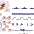
Autonomic symptoms, which may be overlooked, also are present in some cases. Autonomic instability, particularly cardiac arrhythmias, increase the morbidity of this disease. Cardiac monitoring of the R-R interval with reduction of beat-to-beat variability may possibly identify patients at risk for fatal arrhythmia.7 Cardiac arrhythmias induced by tracheal tube manipulation have been reported.8
Asbury and Cornblath9 have established criteria for the diagnosis of GBS. Per their criteria, the features that are required for the diagnosis include progressive motor weakness of more than one limb and areflexia. Symptoms that are strongly supportive of GBS include the relative symmetry of symptoms, mild sensory symptoms, cranial nerve involvement, autonomic symptoms, and recovery that usually begins 2 to 4 weeks after symptom progression discontinues. Sphincter disturbances rarely occur early in the course of GBS and are usually transient.10
Diagnostic studies include examination of the CSF and nerve conduction studies. The CSF reveals elevated protein amid a relative paucity of white blood cells, usually less than 10 cells/mL, with the protein increasing after the first week of symptoms.9 Electrodiagnostic testing reveals motor conduction velocities in the demyelinating range, conduction block, temporal dispersion, and prolonged F waves. Bradshaw and Jones4 reported that conduction block and temporal dispersion occurred in 74% of patients.
If the symptoms are severe, treatment options for GBS include plasmapheresis and administration of intravenous immunoglobulin (IVIg). In 2003 the American Academy of Neurology published a practice parameter after reviewing the adult literature and made several recommendations.11 First, plasmapheresis and IVIg both hasten recovery, and neither is more efficacious. Using these two treatments sequentially is not superior to using either treatment alone. Finally, steroids do not seem to help. The decision of which therapy to apply to children is controversial because a large randomized study has not been performed. Plasmapheresis may be technically difficult to perform in young or very small children; therefore immunoglobulin may be used with more ease. Results from one adult study in which the two methods were compared showed that 53% of the patients treated with immunoglobulin improved by one or more grades on the functional scale at 4 weeks compared with 34% of the patients treated with plasmapheresis.12 Favorable improvement in pediatric patients treated with immunoglobulin has been reported in several small series.13–16 More recently, a randomized trial in children showed that fewer relapses occurred if 2 g/kg of IVIg were divided over 5 days instead of 2 days.17 If additional courses of IVIg are necessary, a 2-day protocol is often well tolerated.
Several GBS variants exist. The best known are the Miller-Fisher variant and acute inflammatory axonal polyneuropathy, the axonal form of GBS. The neurologic triad found in the Miller-Fisher syndrome includes ataxia, areflexia, and ophthalmoparesis. Miller-Fisher syndrome has been linked to immunoglobulin G antibodies against ganglioside GQ1b.18 In some C. jejuni strains, molecular mimicry exists between the surface epitopes and ganglioside GQ1b.19 The GQ1b ganglioside is thought to cross-react in the brainstem area of the ophthalmic cranial nerves.20 The axonal form of GBS has been associated with a more prolonged recovery than the classic form of GBS, which is attributed to axonal involvement. Early research suggests that CSF levels of neurofilament correspond to levels of axonal damage on electromyographic (EMG) testing and may help complement EMG studies to help predict patients who will have more prolonged recoveries.21,22
Myasthenia Gravis
Myasthenia gravis (MG) has many forms that may present in the pediatric population. The juvenile form of MG is the most common and is clinically identical to the autoimmune adult form of MG. Overall, however, juvenile MG is rare and comprises 10% of all cases of MG in Western populations. Antibodies directed toward the acetylcholine receptor (AchR) at the postsynaptic neuromuscular junction cause this form of the disease. These antibodies result in blockade of the AchR, increase the degradation of the AchR, and also result in complement damage to the AchR.23 Fenichel24 reported that 75% of cases occur after age 10 years; however, this age of onset has been debated in recent years.25,26 AchR antibodies are found less frequently in juvenile MG compared with adult autoimmune MG and are more easily shown in the postpubertal patient population.27 Anticholinesterase antibody levels should be determined, however, in all patients with suspected MG. Newer assays are finding antibodies previously missed in older anticholinesterase antibody assays, including binding, blocking, and modulating, but these assays may need to be ordered separately.28–30
The most common heralding symptoms of weakness in MG include ptosis (with pupillary sparing) and diplopia (from restricted eye movements). These symptoms wax and wane, and the weakness may generalize to the extremities. The two clinical forms of juvenile MG are ocular and generalized. In ocular MG, symptoms include ptosis and diplopia, but the weakness does not progress to other areas of the body. Generalized MG may begin with ocular symptoms and progress to generalized weakness, usually within 1 year of onset; however, generalized weakness may be the initial presentation. The exact prevalence of generalized compared with ocular forms of juvenile MG is disputed. As in adults with MG, pediatric patients have the fewest symptoms in the morning or after rest, with increasing fatigability with exercise being a hallmark of this disease. The most troublesome symptoms seen in generalized MG are those involving bulbar and respiratory muscles, which may result in difficulty chewing or swallowing and exercise intolerance.
Antibodies have been found that can block, bind, or modulate AChR. Approximately 80% of patients will have antibodies to AChR found in standard assays. Antibodies directed against muscle specific kinase (MuSK) appear to account for some of the remaining 20%.28 Newer, more sensitive assays can also find antibodies with low affinity for AChR.30 Clinically, MuSK-positive patients tend to have more frequent bulbar involvement and respiratory crises than do AChR-positive patients and require larger doses of maintenance corticosteroids, although there is no clear difference in clinical outcomes.31 Seronegative (AChR-negative and MuSK-negative) patients have a disease severity between the other two groups but appear to have better clinical outcomes.31
Treatment of MG begins with anticholinesterase medications. The symptoms of MG usually respond to pyridostigmine bromide (Mestinon), the most common oral form of anticholinesterase medication. The dosage of pyridostigmine bromide is 7 mg/kg/day divided four to six times daily as needed for symptoms. Immunosuppressant agents, including prednisone, azathioprine, cyclophosphamide, and tacrolimus, may be added to the regimen for pyridostigmine nonresponders.32 Mycophenolate also may be prescribed, although it has not proved more efficacious than placebo in two randomized trials of patients already taking prednisone.33,34 Prednisone is usually initiated at 1 to 2 mg/kg/day. Clinicians must be careful when using prednisone because it may exacerbate weakness on initiation.
Many studies have suggested that the beneficial effects of thymectomy are best when it is performed early in the course of MG.27,35,36 Because of the spontaneous remission rate of 22.4 per 1000 person-years reported by Rodriguez et al.,37 as however, many clinicians are reluctant to proceed with early thymectomy, particularly with young children.
Myasthenic crisis is an exacerbation of myasthenic symptoms requiring ventilatory assistance. In adults with MG, myasthenic crisis has been reported to occur in 15% to 20% of patients, with 74% having their first crisis within 2 years of disease presentation.38,39 Anlar et al.40 reported that one third of patients with juvenile MG had at least one episode of crisis. Crisis duration in adults has been reported as having a median duration of 13 days by Thomas et al.39 Initial therapy during crisis includes mechanical ventilation, which provides rest for the weakened patient. Two retrospective studies suggest that the use of biphasic positive airway pressure) may prevent intubation and shorten hospital stay.41,42 In each study, hypercapnia was a predictor of biphasic positive airway pressure failure.41,42
Anticholinesterase medications should be discontinued because they increase secretions that could lead to mucous plugging. Myasthenic crisis is most commonly heralded by infection in 38% of patients; however, 30% of patients have no obvious trigger for their crisis other than respiratory or bulbar weakness.39 A thorough investigation for the cause of the crisis should be undertaken. The mortality rate has fallen with improvements in health care; however, Thomas et al.39 recently reported a 10% mortality rate in patients with myasthenic crisis.
Plasmapheresis and IVIg (2 g/kg over 2 to 5 days) also play a role in the treatment of myasthenic crisis and acute exacerbations of myasthenic symptoms. In cases of adult crisis, plasmapheresis has been shown to be more efficacious than IVIg; however, plasmapheresis has more deleterious adverse effects, including cardiovascular and infectious complications.43 In the first randomized adult trial between plasmapheresis and IVIg, no significant difference was found between the two treatments; however, this study also included myasthenic exacerbations, as well as crisis.44 Only small numbers of juvenile MG exacerbations or crisis treated with IVIg have been reported.45,46 These reports have been favorable for IVIg in acute exacerbations of MG.45,46 IVIg has been shown to be superior to placebo in a randomized controlled trial, with significant improvements seen as early as 14 days after infusion and lasting through 28 days.47 Several dosing regimens have been used. The previous study used 2 g/kg divided over 2 days; another study failed to find a difference between a 1 g/kg dosage of IVIg given over 1 day and a 2 g/kg dosage given over 2 days.48 Evidence supports the use of IVIg to treat patients experiencing myasthenic crisis or to treat an exacerbation in patients in whom plasmapheresis is not feasible.
Cholinergic crisis must also be a consideration in a patient with an MG exacerbation. Cholinergic crisis occurs with an overdose of anticholinesterase drugs in patients with MG. The overdose causes depolarization of skeletal muscles and muscarinic adverse effects, including increased secretions, diarrhea, lacrimation, sweating, and bradycardia. These symptoms will improve upon withdrawal of the anticholinesterase medications. Some authors argue that cholinergic crisis is rarely the cause for worsening myasthenic symptoms.38,49
The clinician must always be cautious when initiating new medications in patients with MG. Many drugs interfere with the neuromuscular junction; the drugs best known for doing so are the aminoglycoside medications. Steroids can exacerbate weakness in a patient with MG, although this potential is not well recognized. For this reason one must be cautious when initiating the use of prednisone in a patient with refractory MG; it is necessary to observe the patient closely for any initial increased weakness. Antibiotics that have been implicated in the worsening of myasthenic symptoms include ampicillin, ciprofloxacin, clindamycin, erythromycin, sulfonamide, tetracycline, and the peptide antibiotics (polymyxin A and B and colistin). Cardiovascular medications including antiarrhythmic agents (e.g., quinidine, procainamide, and lidocaine) and β-blockers (e.g., propranolol, timolol, and others) also have been reported to worsen symptoms. Thyroid replacement medications and phenytoin also may cause problems. The neuromuscular junction blockers, including vecuronium, rocuronium, and pancuronium, as well as succinylcholine, should be used with caution because the effects of these medications are prolonged in patients with MG.49,50 The Myasthenia Gravis Foundation of America maintains a list of medications to avoid and to use with caution on their Web site at www.myasthenia.org.
Additional immune diseases have been associated in approximately 16% of patients with juvenile MG.37 The autoimmune diseases may include asthma, rheumatoid arthritis, juvenile diabetes mellitus, hyperthyroidism, chronic inflammatory demyelinating polyneuropathy, and central nervous system demyelination.25,37,51,52 Seizures have occurred in 4% to 12% of patients with juvenile MG, although the exact cause is not known.25,37
Congenital and Transient Neonatal Myasthenia Gravis
The other forms of MG are congenital MG and neonatal transient MG. Neonatal transient MG is unique in neonates who are born to mothers with autoimmune MG. Neonates can manifest symptoms of neonatal transient MG even if the mothers were symptom free during pregnancy and delivery. Neonatal transient MG occurs in approximately 12% of infants born to mothers with MG.53 If a mother with MG gives birth to an infant with neonatal MG, her subsequent neonates are also at increased risk of having this transient disorder. Neonatal MG usually resolves in the first few weeks after birth, when the maternally derived antibody level diminishes in the neonate. Results from several studies have shown that even symptom-free infants born to mothers with MG have elevated titers of AchR antibodies.54 Additionally, the same phenomenon has been reported in infants born to mothers with anti-MuSK.40 It is not known why some infants appear to be more susceptible than others for having transient neonatal MG. The antibody concentration of the symptom-free neonate rapidly decreases when compared with the antibody concentration of a neonate with symptoms.55 The symptoms of neonatal transient MG usually include hypotonia, feeding problems (particularly fatigue), weak cry, and respiratory difficulty. These symptoms are treated with supportive care, and anticholinesterase medications are used for severe symptoms.
Congenital MG usually presents in childhood, with symptoms similar to those of juvenile MG. Many defects are responsible for causing symptoms in congenital MG, including congenital abnormalities resulting in presynaptic, synaptic, or postsynaptic defects of the neuromuscular junction.56 Congenital MG is always negative for Ach antibody, and a family history of congenital MG may or may not be present. The inheritance of congenital MG may be autosomal recessive or dominant, or sporadic.56 Treatment of congenital MG is different from the treatment of juvenile MG because immunosuppression obviously does not play a role. Symptoms of congenital MG may or may not respond to anticholinesterase medications.
Tick Paralysis
Affected patients are usually between the ages of 1 and 5 years. A review of 33 patients with tick paralysis reported that 82% were younger than 10 years, and 76% were female patients.57 Longer hairstyles have been speculated to be the cause of this female preponderance. A thorough search of the patient should ensue because more than one tick may be attached. The ticks most commonly implicated in North America are Dermacentor andersoni (wood tick) and Dermacentor variabilis (dog tick)58; however, other types of ticks have been documented. In Australia, the most common tick variety to cause paralysis is Ixodes holocyclus.58 The cause of the weakness is a neurotoxin that is secreted in the saliva of the gravid female tick. The neurotoxin is produced during the engorgement phase of feeding after mating. The neurotoxin inhibits the release of acetylcholine at the presynaptic terminal.58
The symptoms in North American hosts begin with vague complaints of fatigue, irritability, and pain. Vague symptoms may not begin until approximately 5 days after tick attachment, but they progress rapidly.59 Symptoms may include cerebellar signs, such as ataxia.59 If the tick remains attached, a symmetrical ascending flaccid paralysis with areflexia develops. Subsequently, bulbar and facial weakness as well as respiratory involvement occur. No systemic features are seen in tick paralysis. Patients are afebrile with normal vital signs, erythrocyte sedimentation rate, CSF, and mental status. The removal of the tick results in the rapid reversal of symptoms, usually within 24 hours.
Upon discovery, the tick needs to be promptly removed. Removal of the tick is performed with blunt curved forceps or tweezers. The tick should be grasped at the point of attachment, as close to the skin as possible. The tick should be pulled upward with steady pressure. Twisting or jerking motions may cause parts of the tick to break off, particularly the mouth parts. The tick should not be handled with bare hands. Needham60 evaluated various methods of tick removal including fingernail polish, petroleum jelly, 70% isopropyl alcohol, and a hot kitchen match. None of these passive techniques induced tick detachment.
Tick paralysis is more severe in Australia than in North America. The presenting symptoms are similar to those in the North American cases; however, ocular involvement with nonreactive pupils has been described.61 Flaccid paralysis may take days to evolve, unlike in North American hosts. The major difference in Australian tick paralysis occurs after the tick is removed. Australian patients must be carefully observed because maximal weakness may not occur until 48 hours after tick removal.61 Another distinguishing feature of Australian tick paralysis is the possible use of an antitoxin for treatment. The antitoxin, a canine hyperimmune serum, is used cautiously in humans because of potential reactions, including serum sickness.61 Efficacy of the antitoxin remains uncertain because no controlled studies have been performed.61
Periodic Paralyses
Clinicians may encounter several forms of periodic paralysis (PP), including hypokalemic, hyperkalemic, and normokalemic. Persons with most forms of the periodic paralyses have a family history of the disease. The weakness, which eventually results in paralysis, is associated with potassium response as demonstrated in hyperkalemic PP (HyperPP) or potassium serum levels in hypokalemic PP (HypoPP). PP also may be accompanied by cardiac abnormalities, as in Andersen-Tawil syndrome, and thus checking an electrocardiogram may be prudent, regardless of the serum potassium level.62
Hypokalemic Periodic Paralysis
HypoPP is the most common form of the periodic paralyses. The presentation of HypoPP usually occurs within the second decade of life. The number of attacks, which may be frequent, usually decrease as patients get older. The rate of occurrence of HypoPP is 1 in 100,000 people. The inheritance pattern of HypoPP is autosomal dominant, with boys/men more frequently affected, but one third of cases are sporadic.63,64 The most common mutation in familial HypoPP is the dihydropyridine receptor in the voltage sensitive Ca channel, located on chromosome 1q.65 Another common mutation is a voltage-sensitive sodium channel, SCN4A.64,66 In a minority of cases no mutation is found.67
The onset of symptoms in persons with HypoPP usually occurs after the consumption of a high-carbohydrate meal or after vigorous exercise followed by rest. Other provoking factors include cold temperature, emotional stress, menses, and pregnancy.64,66,68 In one study of a large affected family, Chinese food was specifically cited as a specific provocative factor.68 Weakness usually begins during sleep with the patient noticing weakness upon awakening. The weakness usually begins proximally in the legs and then progresses distally before the upper extremities become involved; it may progress to flaccid paralysis of all limbs with areflexia and normal sensation. Cranial nerve function remains normal, and swallowing and respiratory function are rarely affected. The patient remains alert with a normal mental status during the attack, and sensation remains intact. Weakness usually lasts a few hours but may last several days. Upon noticing the initial symptoms of mild muscle cramping or “heaviness,” however, some patients are able to abort an attack with light exercise.69 Sudden death from cardiac arrhythmias or respiratory failure has been reported.70,71 During paralytic attacks, patients have minimal urine output, with decreased potassium excretion and absent defecation.68,72 In persons with HypoPP, myotonia confined to the eyelids has been described.73 Before this report, myotonia was described as occurring only with hyperkalemic periodic paralysis.
Diagnosis of HypoPP can be confirmed with the identification of hypokalemia during an attack. Laboratory testing during HypoPP reveals a markedly diminished potassium level. Although serum potassium levels are decreased, the total body amount of potassium remains normal. The decreased potassium level is due to a shift of the potassium into the muscle cells, resulting in inexcitable muscle cells.74 During an attack, potassium levels usually fall below 3, but levels below 2 have been reported.75 Secondary causes of hypokalemia such as Bartter’s syndrome, use of corticosteroids and diuretics, hyperaldosteronism, ingestion of laxatives and licorice, renal tubular acidosis, amphotericin B, p-aminosalicylic acid, alcoholism, and villous adenoma must be ruled out.76
The paralytic attack may be reversed with normalization of the potassium level. The clinician must be careful when correcting the potassium level, remembering that the total body amount of potassium remains normal. Correction with orally administered potassium (0.2 to 0.4 mmol/kg every 15 to 30 minutes) should be considered. Patients with cardiac symptoms or an inability to swallow, however, require parentally administered potassium.63 While the potassium level is corrected, vigilant cardiac monitoring, monitoring of serial potassium levels, and muscle strength examinations should be used. Administration of intravenous fluids with dextrose or physiological saline solution should be avoided because they may prolong an attack or even induce cardiac arrhythmias.76,77 Griggs, Resnick, and Engel76 reported that a 5% mannitol solution should be considered as a diluent for intravenous potassium replacement.
Links et al.68 studied a large kindred with HypoPP and showed that all family members older than 50 years had permanent muscle weakness. Muscle biopsy specimens from patients with HypoPP reveal vacuoles in the muscle fibers.71 Vacuolar changes in the muscle also have been shown in family members of persons with HypoPP who have not had any paralytic attacks.68 Links et al.68 concluded from their study that all patients eventually exhibit permanent muscle weakness but that only 60% may have paralytic attacks.
Once a patient is known to have HypoPP, prophylactic medications should be initiated. Acetazolamide has been shown to prevent future attacks in patients with and without a family history of the disease when they take daily doses of 250 to 750 mg.78 Some patients, however, have been reported to have an exacerbation of attacks when taking acetazolamide.79 Another report revealed that acetazolamide prophylaxis improved strength between attacks in 80% of patients who displayed persistent weakness between paralytic attacks.78 Daily oral ingestion of potassium chloride shortens the duration of the attacks but does not appear to prevent attacks.78 Other medications used for prophylaxis of attacks include triamterene and spironolactone in patients who are not responsive to acetazolamide.78,79 Other considerations for the prevention of attacks include avoidance of high-sodium, high-carbohydrate meals as well as arduous exercise followed by prolonged rest.
Thyrotoxic periodic paralysis is another entity of weakness with concomitant hypokalemia. As the name implies, a thyrotoxic state is the impetus of this disease. It is mostly found in adult Asian boys/men, although it has been reported in the Asian-American pediatric population.80 The purpose of treatment of this disease is to alleviate the hyperthyroid state (also see Chapter 77).
Hyperkalemic Periodic Paralysis
The term hyperkalemic periodic paralysis (HyperPP) may be misleading because high, normal, and low levels of potassium have been reported in these attacks.81 The name “HyperPP” actually correlates to the response these patients have to potassium. HyperPP is also referred to as potassium-sensitive PP, a term that may be more appropriate and less confusing. HyperPP is autosomal dominant with a common gene located on chromosome 17q, affecting the alpha subunit of the sodium channel, but other sodium channels also may be affected.64,82,83 Sporadic cases have been reported.64,84 HyperPP usually presents in the first decade of life.
Light exercise can prevent an attack. Attacks may be provoked by potassium intake and relieved by glucose intake. Most attacks do not require treatment. In the rare severe attack, however, glucose can be administered intravenously.85 Cardiac monitoring is important if medical intervention is needed because cardiac arrhythmias may occur.86 Prophylactic therapy should be considered in these patients because permanent muscle weakness develops over time.87 Muscle biopsy specimens reveal vacuoles in the muscle cells.81 Acetazolamide and thiazide agents have been used for prophylaxis of this disease.88
Normokalemic Periodic Paralysis
Normokalemic periodic paralysis (NormPP) is a disputed phenomenon. Patients with NormPP have normal potassium levels during an attack and have been shown to be sensitive to potassium.89 Provoking factors include cold temperatures, alcohol intake, and stress.89 The kindred described by Poskanzer and Kerr89 also displayed autosomal dominant inheritance. Therefore it has been argued that this is actually HyperPP, which also is known to display normal values of potassium during an attack. Molecular testing has revealed NormPP mutations in genes associated with HyperPP and HypoPP.90,91 Treatment of this disease is similar to the treatment of HyperPP.
Botulism
Infantile botulism is a syndrome predominately found in infants ages 6 days to 12 months.92 In infantile botulism, Clostridium botulinum enters the body as a spore through ingestion, germination occurs, and the organism begins to produce the neurotoxin that is the cause of the symptoms.93 Infant botulism differs from foodborne botulism, in which the preformed toxin is actually ingested. In mouse models, the relationship of the gut and the spores are important, with the pH of the gut and the transient lack of competitive intestinal flora being essential in allowing the spores to germinate.92 Infants appear to be susceptible, as are adults who have abnormal intestinal flora from abdominal surgery, gut abnormalities, or antibiotic use.94–97
Most cases of infantile botulism occur in California, Pennsylvania, and Utah. In one report, more than 75% of the patients with botulism had C. botulinum in their home environment.98 Other sources include soil disruption from cultivation or construction and parental occupations that involve soil exposure.99 The consumption of honey and corn syrup is a risk factor; these products should not be fed to children who are younger than 12 months. The role of breast-feeding in botulism remains controversial.100 In one study of 44 patients with botulism, 100% of the patients were breast-fed.99 Nevertheless, breast milk has been cited as a protective influence against botulism. Arnon et al.101 reported that of the 10 patients who died of sudden infant death syndrome that was linked to C. botulinum infection, eight were fed formula exclusively, and the remaining two patients had not received breast milk 10 weeks before their death. They concluded that breast-fed infants are not completely protected from botulism but appear to have diminished severity of the disease at onset.101
The botulinum toxin irreversibly binds at the presynaptic segment of the neuromuscular junction, inhibiting acetylcholine release and causing neuromuscular weakness. The autonomic system is also affected because the toxin binds the acetylcholine-mediated preganglionic parasympathetic and sympathetic synapses, as well as the postganglionic parasympathetic synapse.102
The most common symptoms include weak cry, poor suck and feeding, decreased tone with decreased reflexes, weakness in a descending pattern, and constipation.102 Autonomic symptoms, which often are the first to appear, include constipation, tachycardia, fluctuating blood pressure, urinary retention, decreased tears and saliva, and flushed skin or pallor.92,103 L’Hommedieu and Polin103 proposed an algorithm of symptoms beginning with tachycardia and constipation progressing to loss of head control, difficulty feeding, and weak cry. A depressed gag reflex is followed by peripheral muscle weakness and, finally, diaphragmatic weakness. Because of the combination of autonomic and neuromuscular symptoms, the infant with botulism may mistakenly be thought to be septic or dehydrated. Enlarged, sluggishly reactive pupils also may be present but are less common than the other autonomic symptoms.104
The consequence of botulism that is of greatest cause for concern is respiratory embarrassment. Schreiner, Field, and Ruddy102 reported that only 24% of the patients reviewed did not require ventilation or an artificial airway. Patients with botulism also have become apneic during certain procedures, including lumbar puncture and placement of an intravenous catheter.102 Hypoxic ischemic encephalopathy resulting from respiratory arrest also has been described.104 Other complications that have been described include syndrome of inappropriate secretion of antidiuretic hormone, urinary tract infections, pneumonia, and autonomic instability.102 Aminoglycosides exacerbate the neuromuscular blockade and should be avoided in persons with botulism.105 L’Hommedieu et al.105 reported that in five patients with botulism who received aminoglycosides, clinical deterioration was apparent in all of them.
The diagnosis of botulism is clinical but confirmed by isolation of the organism or toxin in stool. EMG in patients with botulism reveals decreased compound motor action potential amplitude and facilitation of the compound motor action potential amplitude with high-frequency repetitive motor nerve stimulation.106
The management of patients with botulism has advanced in recent years. Previously, clinicians could offer only supportive care until axonal sprouting could reestablish the neuromuscular junction. The median length of hospital stay was 27 to 37 days, and patients typically required mechanical ventilation for a median of 13 to 16 days.98,104,107 In patients with respiratory compromise, mechanical ventilation should be instituted until the patient regains protective reflexes and respiratory strength. If patients are unable to tolerate eating orally, nasogastric or nasojejunal feeding should be initiated. Human botulism immune globulin (BIG) has provided the first direct pharmacologic treatment. A randomized, controlled trial has shown that BIG decreased mean hospital stay (5.7 weeks to 2.6 weeks), mean duration of mechanical ventilation, mean duration of feeding through a tube or intravenously, and mean hospital charges with no adverse effects related to BIG.108 A retrospective article spanning 30 years complemented the randomized trial.109 Resolution of symptoms occurs in the reverse pattern of presentation, with return of head control appearing to be a reliable measure of improving muscle function.103
Diphtheria
Although diphtheria was the leading killer of children in the early 20th century, the United States currently reports fewer than 10 cases of diphtheria annually. Epidemics have occurred in developing countries. Recently the former Soviet Union had a dramatic resurgence of diphtheria, with approximately 48,000 cases reported in 1994.110
Although several forms of diphtheria exist, the most common in children is the upper respiratory tract infection. Initial mild infection of the pharyngeal area is followed by tonsillar pseudomembrane formation. The pseudomembrane consists of necrotic epithelium and fibrin, as well as numerous colonies of the bacterium, and may cover the airways and pharynx to the main bronchi down into the smaller bronchi. It can lead to aspiration and ultimately complete obstruction of the airway. Extensive soft tissue edema and lymph node enlargement occur.111
Toxic myocardiopathy is estimated to occur in 10% to 25% of patients and is responsible for 50% to 60% of the deaths.112 Toxic myocardiopathy often arises in the second to third week of illness when the affected individual is improving. Abnormalities of the myocardium, the conductive system, and the pericardium occur. The conductive disturbances are in response to the toxin.113 Cardiac ectopy is 100% sensitive and specific to predict fatal outcome in children with severe diphtheria.112
When severe disease is present, neuropathy is seen in approximately three fourths of the patients.111 In a classic case of diphtheria, local paralysis of the soft palate occurs 2 to 3 weeks after the beginning of the oropharyngeal infection. Weaknesses of the pharyngeal, facial, and ocular nerves follow. The symmetrical polyneuropathy has a varied onset from 10 days to 3 months following the oropharyngeal infection. The axonal and demyelinating neuropathies are based on the circulating toxins and range from motor weakness to sensory abnormalities in a stocking-glove distribution.111,112
The distal ascending weakness and spinal fluid findings are described as being “indistinguishable” from GBS.112 Additionally, dysfunction of the autonomic function may occur with associated hypertension and cardiac failure, although this phenomenon is rare. Typically the patient recovers completely.
Diagnostically, cultures should be obtained from the nose, throat, and the infected mucocutaneous area. Giving the antitoxin is critical even when there is only a presumptive diagnosis. If the antitoxin is administered on the first day, the mortality rate is 1% compared with a rate of 20% if administration is delayed until the fourth day.112 Immunoglobulin preparations also have been hypothesized as helpful. The only antimicrobial agents that have been the subject of prospective studies proving efficacy are penicillin and erythromycin.112
Airway complications should be anticipated, as should the probability of congestive heart failure and, ultimately, malnutrition. Studies have revealed no difference in the occurrence of carditis, neuritis, or death in patients receiving steroids. Additionally, use of digitalis is associated with an increased occurrence of arrhythmias. Overall prognosis depends on multiple variables, including the delay in the administration of the antitoxin along with the immunization status and age of the individual. The fatality rate of 10% for respiratory tract diphtheria has not changed in 50 years.112
Acute Intermittent Porphyria
The most common of the four types of porphyria is acute intermittent porphyria (AIP). Clinical symptoms in acute attacks span multiple medical subspecialties and may be precipitated by numerous medications, hormonal variations, calorie restrictions, and alcohol. AIP most commonly occurs in girls/women with the age of onset between 15 and 40 years, and it rarely occurs before puberty.114
An acute neuropathy is found in approximately 40% of acute AIP attacks.115–117 The neuropathy typically follows the onset of the attack by 1 to 4 weeks but may do so as late as 11 weeks afterward. Although paresthesias and distal sensory changes may be a prodromal finding, the motor signs are much more prominent. Typically the patient has proximally symmetrical upper extremity weakness, but it may advance to involve the lower extremities. Generalized weakness is documented in approximately 42% of patients.115 In AIP’s most dramatic setting, the patient can have a rapid progression of weakness that leads to a flaccid involvement of all four extremities and respiratory compromise. When cranial nerves are involved, nerves VII and X are the most frequently affected. Vascular compromise has been documented in persons with vision loss, which may be monocular or total. Although this vision loss is usually transient, it can be permanent.115
AIP is often difficult to diagnose. The patient’s chief complaints are typically nonspecific abdominal and back pain. This colicky pain often leads to the consideration of surgical intervention. Notably, AIP is not associated with temperature elevation, leukocytosis, or rebound tenderness.115 Neurologic and psychiatric symptoms often accompany the onset of attack.
Another important issue is significant hyponatremia and associated seizures, which may be further precipitated by the use of intravenous fluids containing dextrose and water. Cardiovascular complications include hypertension and tachycardia. In its most extreme case, significant hypertension may be present with associated hypertensive encephalopathy and ischemic changes. Intravenous infusion of magnesium sulfate may be helpful.114 Nutritional support is important to avoid a catabolic state, which will further complicate the clinical picture. In the event that nutrition is required intravenously, high-glucose solutions with dextrose are recommended. Enteral feeding is preferred, with carbohydrates providing 50% to 60% of the energy needs.114
Diagnostically in persons with AIP, urine and stool can be tested for alpha aminolevulinic acid. In addition, a marked elevation of urinary porphobilinogen (PBG) is seen. Checking PBG deaminase levels in the blood is helpful because they are abnormal even between the acute attacks.114AIP should be a consideration in the differential diagnosis of progressive weakness. It is most often confused with GBS. The ascending qualities, which are classic in GBS, are rare in acute porphyria. Additionally, persons with acute porphyria do not have elevation of the CSF protein or abnormalities of the cellular contents. The associated abdominal discomfort and tachycardia that are seen in porphyria would not be anticipated in GBS. Differential considerations should include lead intoxication and hereditary tyrosinemia as well.115
Elder and Hift114 provided a review of AIP therapy. The two recommended approaches are carbohydrate loading and administration of heme. If the patient has severe symptoms such as seizures, hyponatremia, and initial signs of neuropathy, aggressive therapy is begun as early in the crisis as possible. In mild attacks it may be possible to wait 24 hours to determine if the attack will spontaneously reverse. Carbohydrate loading is delivered as a 20% glucose solution provided via a central venous catheter. Studies that support the use of heme are primarily noncontrolled and have difficulty reaching statistical significance, but the overall consensus is that it does provide benefit. Daily measurements of urinary alpha aminolevulinic acid or PBG may be a helpful monitor.
Spinal Muscular Atrophy
Spinal muscular atrophy (SMA), a disease of the anterior horn cell, is most commonly inherited in an autosomal recessive manner. The responsible gene is the survivor motor neuron gene on chromosome 5q13.118,119 SMA has three subtypes that present in childhood, and both autosomal dominant and X-linked inheritance have been reported. The combined incidence of all forms of SMA has been estimated as 1 case in 6000 to 25,700 live births.120,121 After cystic fibrosis, SMA is the next most common fatal disease with an autosomal recessive pattern of inheritance.120 The most severe form is previously known as Werdnig-Hoffmann disease but is now more commonly referred to as SMA type I. It usually presents shortly after birth but should be apparent before age 6 months and is defined by the patient never being able to achieve independent sitting. SMA type II usually presents between ages 6 and 18 months and is characterized by the patient sitting but never standing or walking. In SMA type III, patients do stand independently and walk.
In SMA type I, the examination reveals a floppy baby with proximal weakness greater than distal weakness. The lower extremities are more affected than the upper extremities, and the only spontaneous movement in these infants may be in the hands and feet. When supine, the infant will assume a frog-legged position. Polyminimyoclonus, a fine tremor most easily visualized in the hands, also may be present in these patients. Areflexia, tongue fasciculations, facial weakness, and normal sensation are also found.122,123 Retrospectively, some mothers will report decreased fetal movement during the pregnancy with the affected infant. Death usually occurs before age 2 years as a result of respiratory problems.120 In patients with clinical symptoms within the first day of life, life expectancy was between 2 and 6 months, with a mean age at death slightly before 4 months.124
Patients with SMA type II usually have delayed motor milestones after having normal motor development in infancy. Polyminimyoclonus is also present in these patients. Life expectancy is variable, with many patients not surviving past adolescence.120 Life expectancy can be enhanced, however, with fastidious respiratory care.125 Not surprising was the correlation that patients with an earlier onset of the disease had an earlier death.126
In SMA type III, weakness is again more proximal than distal, with the lower extremities being more severely affected. The gait exhibited in these patients has a waddling quality, and lumbar lordosis is also prominent. Once the patient loses the ability to ambulate, the inability to raise the hands above the head also occurs.127 If symptoms begin after age 2 years, ambulation may continue to a median age of 44 years.127 If symptoms begin before age 2 years, ambulation continues to a median age of 12 years.127 Life expectancy for patients with SMA type III may be the same as in the normal population because muscle weakness appears to stabilize in these patients.
Respiratory complications are the most aspect of this disease that cause the most concern and include aspiration, pneumonia, and respiratory failure. Respiratory failure may even be the presenting symptom in SMA type I.128 Respiratory muscle weakness results in restrictive lung disease with a weak cough and hypoventilation.123 Hypercapnia is also a consequence of restrictive lung disease, so supplemental oxygen may have devastating consequences, including apnea and death.123 If supplemental oxygen is needed, conventional ventilation or noninvasive ventilation should be instituted.
Other complications may occur over time. Scoliosis complicates pulmonary function over time because of chest wall alterations. Contractures, particularly in the lower extremities, are also common. In addition, feeding difficulties play a prominent role, particularly in the developing infant with SMA type I. If concerns arise, a feeding evaluation should be performed to rule out aspiration. Supplemental feeding through a nasogastric tube or gastrostomy may be necessary.
Aggressive symptomatic treatment, including more frequent use of ventilation and gastrostomy, has been associated with longer life spans. Specific pharmacologic treatments have not been successful.129 Phenylbutyrate was ineffective in a randomized, controlled trial.130 Although riluzole (which blocks glutamatergic neurotransmission in the central nervous system) is effective in persons with amyotrophic lateral sclerosis, it has not been adequately tested in children.131
Poliomyelitis
The paralytic form of polio represents only 1% to 2% of the actual infections. Aseptic meningitis represents less than 10% of infections and is often thought to be a nonspecific illness. The remaining 90% to 95% of those affected have no apparent infection. Patients who will have paralytic disorder show very high fevers and significant muscle pain with the lack of reflexes. Paralysis rapidly progresses to complete loss of motor use asymmetrically in one or more extremities over a few hours. The distribution of weakness is classically proximal and in the lower extremities. Cranial nerve abnormalities have been reported in 5% to 35% of the patients. Loss of function peaks at 5 days. The disorder can be associated with bowel and bladder problems over the initial 3 days. Sensory abnormalities are rare. Physical examination reveals meningeal findings with changes in the reflexes both superficially and deep. One of the classic signs described in the early reports is the “head drop.” As the examiner lifts the patient’s shoulders and raises the trunk, the head often falls backward in a limp fashion. It is thought that this phenomenon is not due to paralysis of the neck muscles because it can occur in the nonparalytic form. The clinical course may include significant respiratory muscle weakness. Involvement of the bulbar muscles, brainstem, the respiratory center, and cranial nerve pose difficulties in breathing and paralysis of the pharynx and vocal cords. Respiratory compromise leads to most deaths in the paralytic form.132 Typically, 50% of patients with any paralysis exhibit some degree of residual deficits, although most do improve. A 10% mortality rate is now reported in the patients with the paralytic from. Before the use of mechanical ventilation, 60% died.132
A throat and stool culture may reveal the poliovirus, which is shed early in the course of pharyngeal infection and later from the stool. It is difficult to isolate from the spinal fluid in affected patients. Usually the results of routine laboratory tests are unremarkable. CSF findings are characteristic of aseptic meningitis. A white blood cell count between 20 and 300 cells is expected with a predominance of lymphocytes and a normal glucose level. Normal or slightly elevated levels of protein may be found. In the first few hours after the onset of symptoms, polymorphonuclear leukocytes may predominate, but within 12 hours the predominance of lymphocytes is seen.132
This disorder has numerous clinical manifestations. In the viral myocarditis, the heart is extremely sensitive, and thus very small doses of digoxin have been suggested. Hypertension is well recognized and can be severe enough to cause encephalopathy. In the child with poliomyelitis, analgesics, including opiates, may be required for pain relief. Hot packs have been noted to be effective when applied every 2 to 4 hours. Constipation and bladder paralysis are major issues early in the course and should be monitored closely. Because of the risk of aspiration and airway compromise, a high level of vigilance must be maintained. If the patient demonstrates respiratory compromise, then a tracheostomy is indicated with accompanying mechanical ventilation.132 Use of antiviral agents is debated. Additionally, some authors argue that steroids are not indicated in enteroviral infections.132
Children who experience mild weakness generally have a full recovery. If paralysis is present, the recovery is ongoing for 2 years, with 80% realized by 6 months.132 Adults may have new symptoms later in life after paralytic poliomyelitis, including weakness and muscle atrophy that is related to continued normal attrition of anterior horn cells.133
Polio-like Syndromes
Polio-like syndromes have been reported, and antiviral intervention has been advocated.134 Interferon-α therapy within 24 hours of admission has been recommended on the basis of information from small case studies. The authors thought that this intervention altered the course and that improvement was evident within 1 to 2 days. West Nile Virus is now recognized as a possible etiologic agent for a polio-like syndrome.135 Magnetic resonance imaging and proton magnetic resolution spectroscopy have been recommended to monitor the functional activity of the neurons. It is difficult to determine without further study whether the natural course would have been almost complete recovery or if it was in the fact the interferon-α intervention that was responsible.136 Overall prognosis in patients with nonpolio enteroviral infections is very good.132
Organophosphate and Carbamate Poisoning
The clinician must always maintain a high index of suspicion and consider poisoning in the differential diagnosis in patients with altered mental status, respiratory symptoms, or weakness (also see Chapter 105). In their study of 37 children with organophosphate or carbamate poisonings, Zweiner and Ginsburg137 reported that 43% of these patients were evaluated by their primary care doctor, and pesticide toxicity was not suspected. Patients commonly do not have a known history of exposure. Exposure to these substances may occur as inhalation, ingestion, or dermal contact. In one study of 37 infants and children with organophosphate and carbamate poisonings, 76% of these patients ingested these substances (which were improperly stored), 16% had transcutaneous exposure (through contact with treated carpets, linens, and lawns), and 8% were poisoned by an unknown etiology.137 Cholinesterase, which is present in the neuromuscular junction, is irreversibly inhibited by organophosphates and reversibly inhibited by carbamate compounds. Therefore, a constellation of muscarinic, nicotinic, and central nervous system symptoms may occur.
Symptoms may originate from various systems. Muscarinic symptoms include miosis, excessive salivation, sweating, lacrimation, diarrhea, urination, and bradycardia. In severe poisonings, flaccid paralysis with areflexia is common. In moderate poisonings, muscle fasciculations may be present. Central nervous system symptoms include coma and seizures; however, seizures are less common in persons with carbamate toxicity.138 Pulmonary symptoms including bronchoconstriction, increased pulmonary secretions, and wheezing have been reported.139 In one study of 52 children with organophosphate or carbamate poisoning, 100% of these patients exhibited hypotonia, stupor, or coma.140 With further analysis of the 16 patients with organophosphate poisoning, the other common symptoms included miosis (56%), salivation (37%), pulmonary edema (37%), diarrhea (30%), and bradycardia (25%).140 Various cardiac rhythms may occur with pathologic signs of cardiotoxicity.141 Overall, carbamate poisonings are usually less severe and shorter in duration, although the symptoms are essentially the same as those found in organophosphate poisonings.142
If organophosphate and carbamate compounds are ingested, gastric lavage and activated charcoal should be initiated. If contaminated, the patient’s skin and hair should be rinsed and cleansed thoroughly with soap, and the clothes should be changed to reduce further exposure.138
In both forms of poisonings, atropine is used as an antidote for the muscarinic symptoms. Treatment with atropine, however, does not reverse the nicotinic symptoms, which include muscle weakness and respiratory failure. Atropine should be administered as quickly as possible and in adequate doses. In children older than 12 years, the dosing is 1 to 2 mg intravenously every 10 to 30 minutes.139 In children younger than 12 years, the initial dose is 0.05 mg/kg with maintenance doses of 0.02 to 0.05 mg/kg over 10 to 30 minutes.139 In organophosphate and carbamate poisonings, the atropine dose is 5 to 10 times greater than conventional atropine dosing.139 Atropine should be continued until the muscarinic symptoms begin to abate. The signs of atropinization include mydriasis, tachycardia, and xerostomia, and they help provide parameters for adequate dosing.143 Atropine should be continued for at least 24 hours after severe exposures and then tapered if symptoms are improving.139
Pralidoxime chloride, the only cholinesterase reactivator in the United States, is an antidote for only the nicotinic symptoms of organophosphate poisonings; therefore atropine must be used concomitantly. Pralidoxime chloride does not help in carbamate exposures. Various doses have been reported for pralidoxime in patients older than 12 years. A conservative dose is 0.5 to 1 g administered intravenously over 15 to 30 minutes, repeated every 10 to 12 hours, beginning 1 to 2 hours after the initial dose. More recently, another study suggests a 2 g loading dose with a continuous infusion of 1g/h for 48 hours.144 These doses have not been directly compared with each other. In patients younger than 12 years, the dose is 25 to 50 mg/kg administered intravenously over 15 to 30 minutes, repeated every 10 to 12 hours, beginning 1 to 2 hours after the initial dose.139 Further studies appear warranted because a meta-analysis showed either no benefit or possible harm of pralidoxime compared with best medical therapy.145
After the antidotes are given, the mainstay of treatment is supportive. If necessary, ventilation should be provided until the patient regains respiratory strength. Suctioning of secretions in both the oropharynx and in the respiratory tree is essential. Seizures should be treated with diazepam or lorazepam. Cardiac monitoring should be implemented because complex ventricular arrhythmias may occur.146,141 Early feeding may prolong the hospital stay.147 Death usually occurs as a result of respiratory arrest and pulmonary complications, including excessive secretions, edema, and bronchoconstriction.139
Rhabdomyolysis
Rhabdomyolysis refers to a process in which myoglobin is liberated from injured or damaged skeletal muscle into the blood and urine, resulting in myoblobinemia and myoglobinuria. Evidence of chronic rhabdomyolysis, the most common form in children, may be discovered incidentally during routine blood laboratory workups that include CPK levels.148 Acute episodes of rhabdomyolysis also may result from a myriad of causes. The history of the patient is extremely important in determining the exact cause. Acute episodes of rhabdomyolysis may be fatal because of electrolyte abnormalities, cardiac arrhythmias, and renal damage, which occur during this process.
Potential causes of rhabdomyolysis include environmental factors (extreme cold or heat); viral and bacterial infections; and metabolic abnormalities, including hypokalemia, hypernatremia, nonketotic hyperosmolar coma, and diabetic ketoacidosis.149,150 Less common causes include excessive muscle activity as seen in convulsive seizures, extreme exertion, drugs, toxins, venoms, physical trauma, malignant hyperthermia, and metabolic myopathies.
Muscle-related symptoms found in rhabdomyolysis include severe weakness, hypoactive reflexes, tenderness, edema, cramps, and localized pain.150,151 Muscle symptoms make the diagnosis more obvious; however, if the patient has decreased level of consciousness from metabolic abnormalities, trauma, drugs, or seizures, these muscle symptoms may be obscured.
Abnormal laboratory results include myoglobinuria and profound elevation of serum CPK. Muscle cell destruction results in the release of potassium and phosphorous into the blood, with consequential hyperkalemia, hyperphosphatemia, and eventually hypocalcemia.151,152 Elevations of aldolase, uric acid, lactic dehydrogenase, and transaminase (serum glutamate oxaloacetate transaminase and serum glutamate pyruvate transaminase) levels also occur.151,152
Complications from rhabdomyolysis may affect the heart, kidneys, and ventilation. The severe myoglobinuria in rhabdomyolysis may result in acute tubular necrosis, which can be fatal. Alkalinization of the urine, hydration, and osmotic diuresis have been used to prevent renal damage.152 Hypocalcemia, which results from the elevated potassium and phosphorous levels, may lead to cardiac arrhythmias. In the review by Robotham and Haddow,151 several types of cardiac disturbances were reported, including ventricular arrhythmias, intraventricular conduction delays, abnormal axis deviation, sinus bradycardias and tachycardias, ischemic changes, nonspecific ST segment and T wave changes, and T wave changes associated with hyperkalemia. Compartment syndrome also may occur because of severe muscle edema, and fasciotomy may be required to prevent neurovascular compression.152 If the diaphragm and intercostal muscles are affected, mechanical ventilation may be required. Bulbar weakness necessitating mechanical ventilation in rhabdomyolysis is rare.152 Overall, it is unusual for muscle weakness to be permanent, with full muscle strength usually returning in 1 to 6 weeks.152
Several diseases may result in recurrent rhabdomyolysis. McArdle’s disease, a metabolic myopathy that also goes by the names type V glycogenosis and myophosphorylase deficiency, is predominantly autosomal recessive. Patients with this disease exhibit exercise intolerance, muscle stiffness, and myalgia.153 Vigorous activity, including squatting, sprinting, and carrying heavy objects, may precipitate an episode of muscle rigidity with cramping and myoglobinuria resulting in rhabdomyolysis. Recognition of these symptoms will help achieve a timely diagnosis, prevent morbidity, and avoid recurrent rhabdomyolysis. Tarui’s disease, also known as glycogenosis type VII and phosphofructokinase deficiency, is also autosomal recessive. The clinical picture is similar to that found in McArdle’s disease and may also result in rhabdomyolysis.153 Carnitine palmitoyltransferase deficiency is an autosomal recessive disease that also may lead to rhabdomyolysis. Carnitine palmitoyltransferase catalyzes carnitine and fatty acid for transfer into the mitochondria. Rhabdomyolysis may be precipitated by prolonged exercise and fasting and may be prevented by eating frequent meals of low-fat, carbohydrate-rich foods with avoidance of both fasting and prolonged exercise.153
Malignant Hyperthermia
Malignant hyperthermia (MH) is a disease that is associated with certain anesthetic agents, including inhalation anesthesia such as halothane and depolarizing muscle relaxant agents such as succinylcholine (also see Chapter 12). MH occurs in approximately 1 in 12,000 children with anesthesia.154 MH has a variable clinical presentation as described by Kaus and Rockoff.155 In classic MH, the symptoms include tachypnea, tachycardia, blood pressure abnormalities, cyanosis, mottling, and diaphoresis. Hypoxia, hypercapnia, metabolic acidosis, and muscle rigidity resulting in severe rhabdomyolysis also occur. The severe hyperthermia, which denotes the disease, may exceed more than 42° C. A second presentation of MH occurs after the administration of succinylcholine and results in an abrupt onset of generalized muscle rigidity, cardiac arrest, and rhabdomyolysis. An additional presentation is masseter muscle spasm, which occurs after the administration of halothane and succinylcholine, and results in severe contracture of the jaw that lasts 5 to 20 minutes.155
Recent advances have been made in understanding the genetics of MH. The ryanodine receptor appears to be involved, and first- and second-degree family members of patients who have or are suspected of having MH should be considered at risk for MH until a gene test or muscle biopsy is performed.154,156 As more causative mutations are found, fewer patients will have unexplained causes of their MH. In addition to cases caused by mutations of the ryanodine receptor, some muscle diseases have been associated with MH, including Duchenne muscular dystrophy, Becker muscular dystrophy, central core disease, myotonia congenita, King-Denborough syndrome, Schwartz-Jampel syndrome, and other muscular dystrophies.155
Neuroleptic Malignant Syndrome
Neuroleptic malignant syndrome occurs in 0.5% to 1.4% of patients exposed to antipsychotic medications.157,158 Neuroleptic malignant syndrome has a constellation of signs and symptoms including hyperthermia, muscle rigidity, autonomic instability, tachycardia, tachypnea, diaphoresis, hypertension, and altered mental status (also see Chapter 124).159 Rhabdomyolysis may be a component of this syndrome, which includes elevated CPK levels and myoglobinuria; therefore, MH may be in the differential diagnosis of severe acute-onset weakness.
Inflammatory Myopathies
Dermatomyositis and Polymyositis
Dermatomyositis and polymyositis are inflammatory myopathies with symmetrical proximal muscle weakness that progresses over weeks to months (see also Chapter 98, Autoimmune Diseases and Their Treatment). The incidence ranges from 2.5 to 4.1 per million children in the United States and comprised 5% of all rheumatologic referrals in one review.160,161 Five major criteria exist for the diagnosis of polymyositis/dermatomyositis as described by Bohan and Peter.162 Of the five criteria, one clinical criterion is symmetrical proximal weakness that may include respiratory muscles. Another clinical criterion is dermatological, which includes the heliotrope rash and Gottron’s sign. In addition, a scaly erythematous rash of the face, neck, upper torso, knees, elbows, and median malleoli is also present. The two laboratory criteria include elevation of skeletal muscle enzymes, including CPK and adolase, and a muscle biopsy specimen with evidence of necrosis of type I and II fibers, phagocyotosis, and inflammatory exudates. The electrodiagnostic criterion includes EMG findings of spontaneous activity, myopathic motor units, and bizarre high-frequency repetitive discharges. Definitive diagnosis must include three or four criteria and the rash for dermatomyositis, and four criteria without the rash for polymyositis.
Systemic symptoms such as fatigue, lethargy, irritability, arthralgias, myalgias, weight loss, gastrointestinal discomfort, and low-grade fever may herald the onset of weakness.163 Complications of dermatomyositis and polymyositis include respiratory problems such as chronic interstitial pulmonary fibrosis and pneumothorax; gastrointestinal involvement with decreased esophageal motility, gastric ulceration, and bleeding; and cardiac problems such as arrhythmias, abnormal electrocardiograms, and pericarditis.163,164 The initial treatment of polymyositis and dermatomyositis is corticosteroids. The mortality of juvenile dermatomyositis is 3% in the United States.165
Benign Acute Childhood Myositis
Benign acute childhood myositis (BACM) is a self-limited process that usually affects boys more often than girls.166 The presentation of muscular symptoms occurs after a prodrome of viral upper respiratory illness. An acute onset of severe muscle pain usually involving the calf muscles, difficulty walking, and increased CPK levels usually follow the prodrome. Mackay et al.168 describe the prodromal symptoms of fever, cough, headache, rhinorrhea, sore throat, and vomiting as being the most common symptoms. In the 41 episodes of BACM that Mackay et al.166 reported, 42% of those tested were confirmed to be caused by a virus, with 50% of those cases confirmed as being caused by influenza B. The mean CPK level was 14 times normal, but resolution of symptoms occurred within 1 week. BACM resolves rapidly. Bed rest may be necessary until the pain resolves; otherwise, no treatment is needed. This disease process rarely progresses into a severe life-threatening form.
References are available online at http://www.expertconsult.com.
1. Rantala H., Uhari M., Niemela M. Occurrence, clinical manifestations, and prognosis of Guillain-Barre syndrome. Arch Dis Child. 1991;66:706-709.
2. Hart D., Rojas L., Rosario J., et al. Childhood Guillain-Barre syndrome in Paraguay, 1990 to 1991. Ann Neurol. 1994;36:859-863.
3. Ammache Z., Afifi A., Brown C., et al. Childhood Guillain-Barre syndrome: clinical and electrophysiologic features predictive of outcome. J Child Neurol. 2001;6:477-483.
4. Bradshaw D., Jones H. Guillain-Barre syndrome in children: clinical course, electrodiagnosis, and prognosis. Muscle Nerve. 1992;15:500-506.
5. Hood Guillain-Barre syndrome, clinical presentation, diagnosis, and therapy. J Child Neurol. 1996;11:4-12.
6. Rantala H., Uhari M., Cherry J., et al. Risk factors of respiratory failure in children with Guillain-Barre syndrome. Pediatric Neurol. 1995;13:289-292.
7. Oakley C. The heart in the Guillain-Barre syndrome,. BMJ. 1984;288:94.
8. Emmons P., Blume W., DuShane J. Cardiac monitoring and demand pacemaker in Guillain-Barre syndrome. Arch Neurol. 1975;32:59-61.
9. Asbury A., Cornblath D. Assessment of current diagnostic criteria for Guillain-Barre syndrome. Ann Neurol. 1990;27(Suppl):S21-S24.
10. Jones H. Guillain-Barre syndrome in children. Curr Opin Pediatr. 1995;7:663-668.
11. Hughes R.A.C., Wijdicks E.F.M., Barohn R., et al. Practice parameter: Immunotherapy for Guillain-Barré syndrome: report of the Quality Standards Subcommittee of the American Academy of Neurology. Neurology. 2003;61:736-740.
12. Van der Meche F., Schmitz P., Group tDG- B.S. A randomized trial comparing intravenous immune globulin and plasma exchange in Guillain-Barre syndrome,. N Engl J Med. 1992;326:1123-1129.
13. Al-Quadah A. Immunoglobulins in the treatment of Guillain-Barre syndrome in early childhood. J Child Neurol. 1994;9:178-180.
14. Shahar E., Roifman C., Shorer Z., et al. High-dose intravenous serum gamma globulins are effective in severe pediatric Guillain-Barre syndrome: a prospective follow-up study of 23 cases. Ann Neurol. 1994;36:503.
15. Vajsar J., Sloane A., Wood E., et al. Plasmapheresis vs intravenous immunoglobulin in childhood Guillain-Barre syndrome. Arch Pediatr Adolesc Med. 1994;148:1210-1212.
16. Korinthenberg R., Monting J. Natural history and treatment effects in Guillain-Barre syndrome: a multicentre study. Arch Dis Child. 1996;74:281-287.
17. Korinthenberg R., Schessl J., Kirschner J., et al. Intravenously administered immunoglobulin in the treatment of childhood Guillain-Barré syndrome: a randomized trial. Pediatrics. 2005;116:8-14.
18. Chiba A., Kusunoki S., Shimizu T., et al. Serum IgG antibody to ganglioside GQ1b is a possible marker of Miller Fisher syndrome. Ann Neurol. 1992;31:677-679.
19. Jacobs B., Endtz H., van der Meche F., et al. Serum anti-GQ1b IgG antibodies recognize surface epitopes on Campylobacter jejuni from patients with Miller Fisher syndrome. Ann Neurol. 1995;37:260-264.
20. Chiba A., Kusunoki S., Obata H., et al. Serum anti-GQ1b IgG antibody is associated with ophthalmoplegia in Miller Fisher syndrome and Guillain-Barre syndrome: clinical and immunohistochemical studies. Neurology. 1993;43:1911-1917.
21. Petzold A., Brettschneider J., Jin K., et al. CSF protein biomarkers for proximal axonal damage improve prognostic accuracy in the acute phase of Guillain-Barré Syndrome. Muscle Nerve. 2009;40:42-49.
22. Petzold A., Hinds N., Murray N.M.F., et al. CSF neurofilament levels: a potential prognostic marker in Guillain-Barre syndrome. Neurology. 2006;67:1071-1073.
23. Drachman D. Myasthenia gravis. N Engl J Med. 1994;330:1797-1810.
24. Fenichel G. Myasthenia gravis. Pediatr Ann. 1989;18:432-438.
25. Snead O., Benton J., Dwyer D., et al. Juvenile myasthenia gravis. Neurology. 1980;30:732-739.
26. Afifi A., Bell W. Tests for juvenile myasthenia gravis: comparative diagnostic yield and prediction of outcome. J Child Neurol. 1993;8:403-411.
27. Andrews P., Massey J., Howard J., et al. Race, sex, and puberty influence onset, severity, and outcome in juvenile myasthenia gravis. Neurology. 1994;44:1208-1214.
28. Bartoccioni E., Scuderi F., Minicuci G.M., et al. Anti-MuSK antibodies: correlation with myasthenia gravis severity. Neurology. 2006;67:504-507.
29. Niks E.H., Verrips A., Semmekrot B.A., et al. A transient neonatal myasthenic syndrome with anti-musk antibodies,. Neurology. 2008;70:1215-1216.
30. Leite M.I., Jacob S., Viegas S., et al. IgGI antibodies to acetylcholine receptors in ‘seronegative’ myasthenia gravis. Brain. 2008;131:1940-1952.
31. Deymeer F., Gungor-Tuncer O., Yilmaz V., et al. Clinical comparison of anti-MuSK- vs anti-AChR-positive and seronegative myasthenia gravis. Neurology. 2007;68:609-611.
32. Ponseti J.M., Azem J., Fort J.M., et al. Long-term results of tacrolimus in cyclosporine- and prednisone-dependent myasthenia gravis. Neurology. 2005;64:1641-1643.
33. The Muscle Study Group. A trial of mycophenolate mofetil with prednisone as initial immunotherapy in myasthenia gravis,. Neurology. 2008;71:394-399.
34. Sanders D.B., Hart I.K., Mantegazza R., et al. An international, phase III, randomized trial of mycophenolate mofetil in myasthenia gravis. Neurology. 2008;71:390-391.
35. Youssef S. Thymectomy for myasthenia gravis in children. J Pediatr Surg. 1983;18:537-541.
36. Adams C., Theodorescu D., Murphy E., et al. Thymectomy in juvenile myasthenia gravis. J Child Neurol. 1990;5:215-218.
37. Rodriguez M., Gomez M., Howard F., et al. Myasthenia gravis in children: long-term follow-up. Ann Neurol. 1983;13:504-510.
38. Fink M. Treatment of the critical ill patient with myasthenia gravis. In: Ropper A., editor. Neurological and neurosurgical intensive care. New York: Raven Press, 1993.
39. Thomas C., Mayer S., Gungor Y., et al. Myasthenic crisis: clinical features, mortality, complications, and risk factors for prolonged intubation. Neurology. 1997;48:1253-1260.
40. Anlar B., Ozdirim E., Renda Y., et al. Myasthenia gravis in childhood. Acta Paedriatr. 1995;85:838-842.
41. Rabinstein A., Wijdicks E.F.M. BiPAP in acute respiratory failure due to myasthenic crisis may prevent intubation. Neurology. 2002;59:1647-1649.
42. Seneviratne J., Mandrekar J., Wwijdicks E.F.M., et al. Noninvasive ventilation in myasthenic crisis. Arch Neurol. 2008;65:54-58.
43. Qureshi A., Choudhry M., Akbar M., et al. Plasma exchange versus intravenous immunoglobulin treatment in myasthenic crisis. Neurology. 1999;52:629-632.
44. Gajdos P., Chevret S., Clair B., et al. Clinical trial of plasma exchange and high-dose intravenous immunoglobulin in myasthenia gravis. Ann Neurol. 1997;41:789-796.
45. Herrmann D., Carney P., Wald J. Juvenile myasthenia gravis: treatment with immune globulin and thymectomy. Pediatr Neurol. 1998;18:63-66.
46. Selcen D., Dabrowski E., Michon A., et al. High-dose intravenous immunoglobulin therapy in juvenile myasthenia gravis. Pediatr Neurol. 2000;22:40-43.
47. Zinman L., Ng E., Bril V. IV immunoglobulin in patients with myasthenia gravis: a randomized controlled trial. Neurology. 2007;68:837-841.
48. Gajdos P., Tranchant C., Clair B., et al. Treatment of myasthenia gravis exacerbation with intravenous immunoglobulin: a randomized double-blind clinical trial. Arch Neurol. 2005;62:1689-1693.
49. Mayer S. Intensive care of the myasthenic patient. Neurology. 1997;48(Suppl 5):S70-S75.
50. Adams S., Mathews J., Grammer L. Drugs that may exacerbate myasthenia gravis. Ann Emerg Med. 1984;13:532-538.
51. Kimura K., Nezu A., Kimura S., et al. A case of myasthenia gravis in childhood associated with chronic inflammatory demyelinating polyradiculoneuropathy,. Neuropediatrics. 1998;29:108-112.
52. Gotkine M., Fellig, Abramsky O. Occurrence of CNS demyelinating disease in patients with myasthenia gravis. Neurology. 2006;67:881-883.
53. Namba T., Brown S., Grob D. Neonatal myasthenia gravis: report of two cases and review of the literature. Pediatrics. 1970;45:488-504.
54. Ohta M., Matsubara F., Hayashi K., et al. Acetylcholine receptor antibodies in infants of mothers with myasthenia gravis. Neurology. 1981;31:1019-1022.
55. Lefvert A., Osterman P. Newborn infants to myasthenic mothers: a clinical study and an investigation of acetylcholine receptor antibodies in 17 children. Neurology. 1983;33:133-138.
56. Engel A., Ohno K., Milone M. Congenital myasthenic syndromes caused by mutations in acetylcholine receptor genes. Neurology. 1997;48(Suppl 5):S28-S35.
57. Dworkin M., Shoemaker P., Anderson D. Tick paralysis: 33 human cases in Washington State, 1946-1996. Clin Infect Dis. 1999;29:1435-1439.
58. Greenstein P. Tick paralysis. Med Clin North Am. 2002;86:441-446.
59. Gorman R., Snead O. Tick paralysis in three children. Clin Pediatr. 1978;17:249-251.
60. Needham G. Evaluation of five popular methods for tick removal. Pediatrics. 1985;75:997-1002.
61. Grattan-Smith P., Morris J., Johnston H., et al. Clinical and neurophysiological features of tick paralysis. Brain. 1997;120:1975-1987.
62. Donaldson M.R., Yoon G., Fu Y.H., et al. Andersen-Tawil syndrome: a model of clinical variability, pleiotropy, and genetic heterogenity. Ann Med. 2004;36(Suppl 1):92-97.
63. Ahlawat S., Sachdev A. Hypokalaemic paralysis. Postgrad Med J. 1997;75:193-197.
64. Miller T.M., Dias da Silva M.R., Miller H.A., et al. Correlating phenotype and genotype in the periodic paralyses. Neurology. 2004;63:1647-1655.
65. Ptacek L., Tawil R., Griggs R., et al. Dihydropyridine receptor mutations cause hypokalemic periodic paralysis. Cell. 1994;77:863-868.
66. Sugiura Y., Makita N., Li L., et al. Cold induces shifts of voltage dependence in mutant SCN4A, causing hypokalemic periodic paralysis. Neurology. 2003;61:914-918.
67. Matthews E., Labrum R., Sweeney M.G., et al. Voltage sensor charge loss accounts for most cases of hypokalemic periodic paralysis. Neurology. 2009;72:1544-1547.
68. Links T., Smit A., Molenaar W., et al. Familial hypokalemic periodic paralysis clinical, diagnostic and therapeutic aspects. J Neurol Sci. 1994;122:33-43.
69. Schiller T., Auerbach P. Hypokalemic periodic paralysis: two case reports. Pediatr Emerg Care. 1988;4:183-186.
70. Smith W. Periodic paralysis: report of two fatal cases. J Nerv Men Dis. 1937;90:210-215.
71. Talbott J. Periodic paralysis: a clinical syndrome. Medicine. 1941;20:85-143.
72. Pudenz R., McIntosh J., McEachern D. Role of potassium in familial periodic paralysis. JAMA. 1938;111:2253-2258.
73. Resnick J., Engel W. Myotonic lid lag in hypokalaemic periodic paralysis. J Neurol Neurosurg Psychiatry. 1967;30:47-51.
74. Zierler K., Andres R. Movement of potassium into skeletal muscle during spontaneous attack in family periodic paralysis. J Clin Invest. 1957;36:730-737.
75. Charness M. Clinical conferences at The Johns Hopkins Hospital. Hypokalemic periodic paralysis. Johns Hopkins Med J. 1978;143:148-153.
76. Griggs R., Resnick J., Engel W. Intravenous treatment of hypokalemic periodic paralysis. Arch Neurol. 1983;40:539-540.
77. Kunin A., Surawicz B., Sims E. Decrease in serum potassium concentrations and appearance of cardiac arrhythmias during infusion of potassium with glucose in potassium-depleted patients. N Engl J Med. 1962;266:228-233.
78. Griggs R., Engel W., Resnick J. Acetazolamide treatment of hypokalemic periodic paralysis: prevention of attacks and improvement of persistent weakness. Ann Internal Med. 1970;73:39-48.
79. Torres C., Griggs R., Moxley R., et al. Hypokalemic periodic paralysis exacerbated by acetazolamide. Neurology. 1981;31:1423-1428.
80. Miller J., Quillian W., Cleveland W. Nonfamilial hypokalemic periodic paralysis and thyrotoxicosis in a 16-year-old male. Pediatrics. 1997;100:412-414.
81. Gamstorp I. Adynamia episodica hereditaria. Acta Paedriatr. 1956;45(Suppl 108):1-126.
82. George A.L.Jr., Ledbetter D.H., Kallen R.G., et al. Assignment of a human skeletal muscle sodium channel alpha-subunit gene (SCN4A) to 17q23.1-25.3. Genomics. 1991;9:555-556.
83. Fontaine B., Khurana T., Hoffman E., et al. Hyperkalemic periodic paralysis and the adult muscle sodium channel alpha-subunit gene. Science. 1990;250:1000-1002.
84. Dyken M., Timmons G. Hyperkalemic periodic paralysis with hypocalcemic episode. Arch Neurol. 1963;9:508-517.
85. Herman R.H., McDowell M.K. Hyperkalemic paralysis (adynamia episodica hereditaria). Report of four cases and clinical studies. Am J Med. 1963;35:749-767.
86. Lisak R., Lebeau J., Tucker S., et al. Hyperkalemic periodic paralysis and cardiac arrhythmia. Neurology. 1972;22:810-815.
87. McArdle B. Adynamia episodica hereditaria and its treatment. Brain. 1962;85:121-148.
88. Layzer R., Lovelace R., Rowland L. Hyperkalemic periodic paralysis. Arch Neurol. 1967;16:455-472.
89. Poskanzer D., Kerr D. A third type of periodic paralysis, with normokalemia and favourable response to sodium chloride,. Am J Med. 1961;31:328-342.
90. Chinnery P., Walls T., Hanna M., et al. Normokalemic periodic paralysis revisited: does it exist? Ann Neurol. 2002;52:251-252.
91. Vicart S., Sternberg D., Fournier E., et al. New mutations of SCN4A cause a potassium-sensitive normokalemic periodic paralysis. Neurology. 2004;63:2120-2127.
92. Long S. Infant botulism. Pediatr Infect Dis J. 2001;20:707-709.
93. Midura T., Arnon S. Infant botulism: identification of Clostridium botulinum and its toxins in feces. Lancet. 1976;2:934-935.
94. Shapiro R., Hatheway C., Swerdlow D. Botulism in the United States: a clinical and epidemiologic review. Ann Intern Med. 1998;129:221-228.
95. Chia J., Clark J., Ryan C., et al. Botulism in an adult associated with food-borne intestinal infection with Clostridium botulinum. N Engl J Med. 1986;315:239-241.
96. Griffin P., Hatheway C., Rosenbaum R., et al. Endogenous antibody production to botulinum toxin in an adult with intestinal colonization botulism and underlying Crohn’s disease. J Infect Dis. 1997;175:633-637.
97. McCroskey L., Hatheway C. Laboratory findings in four cases of adult botulism suggest colonization of the intestinal tract. J Clin Microbiol. 1988;26:1052-1054.
98. Long S., Gajewski J., Brown L., et al. Clinical, laboratory, and environmental features of infant botulism in Southeastern Pennsylvania. Pediatrics. 1985;75:935-941.
99. Long S. Epidemiologic study of infant botulism in Pennsylvania: report of the Infant Botulism Study Group. Pediatrics. 1985;75:928-934.
100. Spika J., Shaffer N., Hargrett-Bean N., et al. Risk factors for infant botulism in the United States. Am J Dis Child. 1989;143:828-832.
101. Arnon S., Damus K., Thompson B., et al. Protective role of human milk against sudden death from infant botulism. J Pediatr. 1982;100:568-573.
102. Schreiner M., Field E., Ruddy R. Infant botulism: a review of 12 years’ experience at the Children’s Hospital of Philadelphia. Pediatrics. 1991;87:159-165.
103. L’Hommedieu C., Polin R. Progression of clinical signs in severe infant botulism. Therapeutic implications. Clin Pediatr (Phila). 1981;20:90-95.
104. Thompson J., Glasgow L., Warpinski J., et al. Infant botulism: clinical spectrum and epidemiology. Pediatrics. 1980;66:936-942.
105. L’Hommedieu C., Stough R., Brown L., et al. Potentiation of neuromuscular weakness in infant botulism by aminoglycosides. J Pediatr. 1979;95:1065-1070.
106. Cornblath D., Sladky J., Sumner A. Clinical electrophysiology of infantile botulism. Muscle Nerve. 1983;6:448-452.
107. Wilson R., Morris J., Snyder J., et al. Clinical characteristics of infant botulism in the United States: a study of the non-California cases. Pediatr Infect Dis. 1982;1:148-150.
108. Arnon S.S., Schechter R., Maslanka S.E., et al. Human botulism immune globulin for the treatment of infant botulism. N Engl J Med. 2006;354:462-471.
109. Underwood K., Rubin S., Deakers T., et al. Infant botulism: a 30-year experience spanning the introduction of botulism immune globulin intravenous in the intensive care unit at Children’s Hospital Los Angeles. Pediatrics. 2007;120:e1380-e1385.
110. Perles Z., Nir A., Cohen E., et al. Atrioventricular block in a toxic child: do not forget diphtheria. Pediatr Cardiol. 2001;21:282-283.
111. Hadfield T.L., McEvoy P., Polotsky Y., et al. The pathology of diphtheria,. J Infect Dis. 2000;181(Suppl 1):S10-S22.
112. Overturf G.D. Corynebacterium diphtheria. In: Long S.S., Pickering L.K., Prober C.G., editors. Principles and practice of pediatric infectious diseases. New York: Churchill Livingstone, 2003.
113. Rosenberg R.N., Prusiner S.B., Dimauro S., et al. The molecular and genetic basis of neurological disease, ed 2. Boston: Butterworth-Heinemann; 1997.
114. Elder G.H., Hift R.J. Treatment of acute porphyria. Hosp Med. 2001;62:422-425.
115. Bissel D.M. The porphyrias. In: Rosenberg R.N., Prusiner S.B., DiMauro S., editors. editors: The molecular and genetic basis of neurological disease. ed 2. Boston: Butterworth-Heinemann; 1997: 1255 1269.
116. Cavanagh J.B., Ridley A.R. The nature of the neuropathy complicating acute intermittent porphyria,. Lancet. 1967;2:1023-1024.
117. Ridley A. The neuropathy of acute intermittent porphyria. Q J Med. 1969;8:307-309.
118. Brzustowicz L., Lehner T., Castilla L., et al. Genetic mapping of chronic childhood-onset spinal muscular atrophy to chromosome 5q11.2-13.3. Nature. 1990;344:540-541.
119. Oskoui M., Levy G., Garland C.J., et al. The changing natural history of spinal muscular atrophy type 1,. Neurology. 2007;69:1931-1936.
120. Talbot K. Spinal muscular atrophy. J Inherit Metab Dis. 1999;22:545-554.
121. Pearn J.H. The gene frequency of acute Werdnig-Hoffmann disease (SMA type 1). A total population survey in North-East England,. J Med Genet. 1973;10:260-265.
122. Iannaccone S., Browne R., Samaha F., et al. Prospective study of spinal muscular atrophy before age 6 years. Pediatr Neurol. 1993;9:187-193.
123. Iannaccone S., Burghes A. Spinal muscular atrophies. Adv Neurol. 2002;88:83-98.
124. Thomas N., Dubowitz V. The natural history of type I (severe) spinal muscular atrophy,. Neuromuscul Disord. 1994;4:497-502.
125. Gozal D. Pulmonary manifestations of neuromuscular disease with special reference to Duchenne muscular dystrophy and spinal muscular atrophy. Pediatr Pulmon. 2000;29:141-150.
126. Zerres K., Rudnik-Schoneborn S., Forrest E., et al. A collaborative study on the natural history of childhood and juvenile onset proximal spinal muscular atrophy (type II and III SMA): 569 patients,. J Neurol Sci. 1997;146:67-72.
127. Russman B., Buncher C., White M., et al. Function changes in spinal muscular atrophy II and III. Neurology. 1996;47:973-976.
128. McWilliam R., Gardner-Medwin D., Doyle D., et al. Diaphragmatic paralysis due to spinal muscular atrophy. Arch Dis Child. 1985;60:145-149.
129. Bosboom W.M., Vrancken A.F., van den Berg L.H., et al. Drug treatment for spinal muscular atrophy type I. Cochrate Database Syst Rev. 2009;1:CD006281.
130. Mercuri E., Bertini E., Messina S., et al. Randomized, double-blind, placebo-controlled trial of phenylbutyrate in spinal muscular atrophy. Neurology. 2007;68:51-55.
131. Russman B.S., Iannaccone S.T., Samaha F.J. A phase 1 trial of riluzole in spinal muscular atrophy,. Arch Neurol. 2003;60:1601-1603.
132. Feigin R.D., Cherry J.D. ed 4 Textbook of pediatric infectious diseases 1998:vol 2 WB Saunders Philadelphia
133. Thorsteinsson G. Management of postpolio syndrome. Mayo Clin Proc. 1997;72:627-638.
134. Yoshimura K., Kurashige T. A case of poliomyelitis-like syndrome,. Brain Dev. 1997;20:540-542.
135. Gordon S.M., Isada C.M. West Nile fever: lessons from the 2002 season. Cleve Clin J Med. 2003;70:449-454.
136. Arya S. Antiviral therapeutic intervention in poliomyelitis-like syndrome. Brain Dev. 1999;21:567.
137. Zwiener R., Ginsberg C. Organophosphate and carbate poisoning in infants and children. Pediatrics. 1988;81:121-126.
138. Morgan D. Recognition and management of pesticide poisonings. Washington, D.C: U.S. Environmental Protection Agency Office of Pesticide Program; 1976.
139. Mortensen M. Management of acute childhood poisonings caused by selected insecticides and herbicides. Pediatr Clin North Am. 1986;33:421-445.
140. Lifshitz M., Shahak E., Sofer S. Carbamate and organophosphate poisoning in young children. Pediatr Emerg Care. 1999;15:102-103.
141. Anand S., Singh S., Saikia U.N., et al. Cardiac abnormalities in acute organophosphate poisoning. Clin Toxicol. 2009;47:230-235.
142. Lifshitz M., Rotenberg M., Sofer S., et al. Carbamate poisoning and oxime treatment in children: a clinical and laboratory study. Pediatrics. 1994;93:652-655.
143. Goswamy R., Chaudhuri A., Mahashur A. Study of respiratory failure in organophosphate and carbamate poisoning. Heart Lung. 1994;23:466-472.
144. Pawar K.S., Bhoite R.R., Pillay C.P., et al. Continuous pralidoxime infusion versus repeated bolus injection to treat organophosphorus pesticide poisoning: a randomised controlled trial. Lancet. 2006;368:2136-2141.
145. Peter J.V., Moran J.L., Graham P. Oxime therapy and outcomes in human organophosphate poisoning: an evaluation using meta-analytic techniques. Crit Care Med. 2006;34:502-510.
146. Tafuri J., Roberts J. Organophosphate poisoning. Ann Emerg Med. 1987;16:193-202.
147. Moses V., Mahendri N.V., John G., et al. Early hypocaloric enteral nutritional supplementation in acute organophosphate poisoning—a prospective randomized trial. Clin Toxicol. 2009;47:419-424.
148. Brumback R., Feeback D., Leech R. Rhabdomyolysis in childhood. A primer on normal muscle function and selected metabolic myopathies characterized by disordered energy production,. Pediatr Clin North Am. 1992;39:821-858.
149. Watemberg N., Leshner R., Armstrong B., et al. Acute pediatric rhabdomyolysis. J Child Neurol. 2000;15:222-227.
150. Mannix R., Tan M.L., Wright R., et al. Acute pediatric rhabdomyolysis: causes and rates of renal failure. Pediatrics. 2006;118:2119-2125.
151. Robotham J., Haddow J. Rhabdomyolysis and myoglobinuria in childhood. Pediatr Clin North Am. 1976;23:279-301.
152. Chamberlain M. Rhabdomyolysis in children: a 3-year retrospective study. Pediatr Neurol. 1991;7:226-228.
153. Griggs R., Mendell J., Miller J. Evaluation and treatment of myopathies. Philadelphia: FA Davis; 1995.
154. Sessler D. Malignant hyperthermia. J Pediatr. 1986;109:9-14.
155. Kaus S., Rockoff M. Malignant hyperthermia. Pediatr Clin North Am. 1994;41:221-237.
156. Litman R.S., Rosenberg H. Malignant hyperthermia update on susceptibility testing. JAMA. 2005;293:2918-2924.
157. Pope H., Keck P., McElroy S. Frequency and presentation of neuroleptic malignant syndrome in a large psychiatric hospital. Am J Psychiatry. 1986;143:1227-1232.
158. Knight M., Roberts R. Phenothiazine and butyrophenone intoxication in children. Pediatr Clin North Am. 1986;33:299-309.
159. Silva R., Munoz D., Alpert M., et al. Neuroleptic malignant syndrome in children and adolescents. J Am Acad Child Adolesc Psychiatry. 1999;38:187-194.
160. Denardo B., Tucker L., Miller L., et al. Demography of a regional pediatric rheumatology patient population. J Rheumatol. 1994;21:1553-1561.
161. Mendez E.P., Lipton R., Ramsey-Goldman R., et al. US incidence of juvenile dermatomyositis, 1995-1998: results from the National Institute of Arthritis and Musculoskeletal and Skin Diseases Registry. Arthritis Rheum. 2003;49:300-305.
162. Bohan A., Peter J. Polymyositis and dermatomyositis. N Engl J Med. 1975;292:344-347.
163. Spiro A. Childhood dermatomyositis and polymyositis. Pediatr Rev. 1984;6:163-172.
164. Pachman L. Inflammatory myopathy in children. Rheum Dis Clin North Am. 1995;20:919-942.
165. Pachman L. Juvenile dermatomyositis. Pathophysiology and disease expression. Pediatr Clin North Am. 1995;42:1071-1098.
166. Mackay M., Kornberg A., Shield L., et al. Benign acute childhood myositis: laboratory and clinical features. Neurology. 1999;53:2127-2131.