Actinomyces

Published on 25/03/2015 by admin

Filed under Pediatrics

Last modified 22/04/2025

Print this page

rate 1 star rate 2 star rate 3 star rate 4 star rate 5 star
Your rating: none, Average: 2 (1 votes)

This article have been viewed 2036 times

Chapter 182 Actinomyces

Actinomyces organisms are slow-growing, gram-positive bacteria that are part of the endogenous oral flora in humans. Their filamentous structure gives them a fungus-like appearance. Infection caused by these bacteria is termed actinomycosis, which is a chronic, granulomatous, suppurative disease characterized by direct extension to contiguous tissue across natural anatomic barriers with the formation of numerous draining fistulas and sinus tracts. These infections usually involve the cervicofacial, thoracic, abdominal, or pelvic regions.

Etiology

Actinomyces is a member of the order Actinomycetales, which includes gram-positive filamentous bacteria such as Nocardia, Streptomyces, and mycobacteria. Actinomyces israelii is the predominant species causing human actinomycosis. Other implicated species, in order of importance, are Propionibacterium propionicum, Actinomyces turicensis, Actinomyces odontolyticus, Actinomyces meyeri, Actinomyces naeslundii, Actinomyces viscosus, Actinomyces europaeus, and Actinomyces radingae. Arcanobacterium pyogenes (previously in the Actinomyces genus) also causes human actinomycosis.

Actinomyces organisms are non–spore-forming, gram-positive, non–acid-fast, nonmotile, facultative or strictly anaerobic bacilli with variable morphology ranging from diphtheroid to mycelial with short branching forms. Actinomyces organisms are part of the endogenous flora of mucous membranes and are often found in clinical specimens such as sputum, bronchial washes, purulent exudates, and tissues obtained surgically or at necropsy. Staining of crushed tissue specimens rinsed with sterile saline or purulent exudate stained with Gram or acid-fast procedures may reveal organisms within the classic sulfur granules, which are characteristically associated with pulmonary disease caused by A. israelii or A. meyeri. Cultures on brain-heart infusion agar incubated at 37°C anaerobically (95% nitrogen and 5% carbon dioxide) and a separate set incubated aerobically reveal organisms within the lines of streak at 24-48 hr. A. israelii colonies appear as loose masses of delicate, branching filaments with a characteristic spider-like growth. Colonies of A. naeslundii, A. viscosus, and P. propionicum may have similar growth characteristics. Biochemical testing is frequently used for speciation but is limited by the complexity within this group. Newer speciation methods are based on 16S recombinant RNA sequence analysis.

Pathogenesis

The 3 significant sites of Actinomyces infection are, in order of frequency, cervicofacial, abdominal and pelvic, and pulmonary, although infection may involve any organ in the body. Infection typically follows introduction of the organism into tissues after trauma or surgery. The hallmark of actinomycosis is spread that fails to respect tissue or fascial planes. The use of intrauterine devices (IUDs) may predispose to development of pelvic actinomycosis. Pulmonary actinomycosis occurs after inhalation or aspiration of organisms, introduction of a colonized foreign body, or spread from an existing cervicofacial or abdominal actinomycotic infection.

Infection spreads contiguously and, rarely, hematogenously. Actinomycosis is a chronic, suppurative, scarring inflammatory process. Sites of infection show dense cellular infiltrates and suppuration that form many interconnecting abscesses and sinus tracts. This may be followed by cicatricial healing from which the organism spreads by burrowing along fascial planes, causing deep, communicating scarred sinus tracts. Sulfur granules are characteristic of actinomycosis. On hematoxylin-eosin staining, they appear as an adherent mass of polymorphonuclear neutrophils attached to the radially arranged eosinophilic clubs of the granule, which is the host immune response. They may be microscopic or macroscopic and are typically yellow, accounting for their name, but may be white, gray, or brown.

Actinomycosis, even in closed infections, is usually, if not always, polymicrobial in nature, involving mixed bacteria. In a large study of more than 650 cases, infection with Actinomyces was identified in pure culture in only 1 case and in others was usually identified with other oral flora, most notably members of the HACEK group, which includes Haemophilus aphrophilus, Actinobacillus actinomycetemcomitans, Cardiobacterium hominis, Eikenella corrodens, and Kingella kingae. A. actinomycetemcomitans is a fastidious, gram-negative bacillus that is part of the oral flora and has been implicated as a pathogen in periodontal disease. Other bacterial species frequently isolated concomitantly in human actinomycosis include Fusobacterium, Bacteroides, Capnocytophaga, Staphylococcus, Streptococcus, Enterococcus, and Enterobacteriaceae.

Clinical Manifestations

The 3 major forms of actinomycosis—cervicofacial, abdominal and pelvic, and pulmonary—arise by different routes but may progress to other forms of the disease. Actinomycosis in children suggests an underlying immunodeficiency, especially chronic granulomatous disease (Chapter 124).

Cervicofacial Actinomycosis

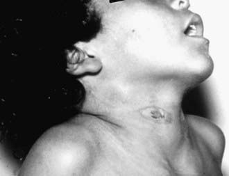
In the patient with cervicofacial actinomycosis, there is often a history of oral trauma, oral surgery, dental procedures, or caries facilitates entry of organisms into cervicofacial tissues. Cervicofacial actinomycosis usually manifests as a painless, slow-growing, hard mass and can produce cutaneous fistulas, a condition commonly known as lumpy jaw (Fig. 182-1). Less frequently, cervicofacial actinomycosis manifests clinically as an acute pyogenic infection with a tender, fluctuant mass with trismus, firm swelling, and fistulas with drainage containing the characteristic sulfur granules. Bone is not involved early in the disease, but periostitis, mandibular osteomyelitis, or perimandibular abscess may develop. Infection may spread by way of sinus tracts to the cranial bones, possibly giving rise to meningitis. The ability of Actinomyces to burrow through tissue planes and even bone is a key difference between actinomycosis and nocardiosis. The cervicofacial form of actinomycosis has the best prognosis and is usually cured with surgical debridement and excision combined with antibiotic therapy.

Abdominal and Pelvic Actinomycosis

Characteristically in abdominal and pelvic actinomycosis, there is some disruption of the mucosa of the gastrointestinal tract, usually as a result of an acute gastrointestinal perforation or abdominal trauma. Patients often present with a history of gastrointestinal surgery, diverticulitis, or appendicitis. Of all the forms of actinomycosis, delayed diagnosis is most typical for abdominal and pelvic infection. Gastrointestinal disease clinically develops as appendicitis in 25% of cases but can be manifested as various ulcerative diseases. Infection classically appears after appendectomy as a firm, irregular mass in the ileocecal area that softens and then drains externally through a fistula. Hepatic involvement occurs in approximately 15% of cases of abdominal actinomycosis, with solitary or multiple liver abscesses or in a miliary pattern. The clinical course is indolent, with chills, fever, night sweats, and weight loss, and the presentation is similar to that of tuberculous peritonitis. Infection usually spreads by direct extension or, rarely, hematogenously, possibly involving any tissue or organ, including muscle, spleen, kidneys, fallopian tubes, ovaries, uterus, testes, bladder, and rectum.

Women using IUDs are at risk for development of pelvic actinomycosis, which classically manifests as vaginal discharge, pelvic pain, abdominal pain, menorrhagia, fever, pelvic mass, and a history of pelvic inflammatory disease. The risk is higher if the IUD has been in place for >2-3 yr.

Pulmonary Actinomycosis

Neither the clinical nor the radiographic presentation of pulmonary actinomycosis is specific. Pulmonary actinomycosis may manifest as an endobronchial infection, a tumor-like lesion, diffuse pneumonia, or a pleural effusion. Principal symptoms include fever, productive cough, chest pain, and weight loss. Infection frequently dissects along tissue planes and may extend through the chest wall or diaphragm, characteristically producing numerous sinus tracts that contain small abscesses and purulent drainage. Other complications include bony destruction of adjacent ribs, sternum, and vertebral bodies. Pyogenic mediastinitis has been attributed to A. odontolyticus in lung transplant recipients. Multiple lobe involvement of the lungs is occasionally found. Predisposing conditions include dental caries, aspiration, thermal or chemical inhalation injury, introduction of a colonized foreign body, and pre-existing cervicofacial or abdominal disease. The classic radiographic triad of thoracic actinomycosis is chronic lower lobe pulmonary consolidation, empyema, and wavy periostitis of the ribs. Accurate diagnosis is difficult because of the propensity of Actinomyces to infect pre-existing pulmonary cavities. Diagnosis can be confirmed by examination of purulent sinus tract drainage for sulfur granules, and with appropriate cultures. The significance of the presence of Actinomyces in sputum or bronchoscopy specimens is limited because these organisms are normal oral flora.

Diagnosis and Differential Diagnosis

Microscopic examination with appropriate stains and culture of purulent drainage from fistulas, abscesses, draining sinus tracts, bronchoalveolar lavage, and sputum can reveal Actinomyces. Except for A. meyeri, which is nonbranching, Actinomyces organisms appear as branching, filamentous rods. Inoculation of anaerobic and aerobic cultures enhances the yield of cultures. Gram, Gomori methenamine silver, or Giemsa stains of purulent material or tissue reveal diagnostic filamentous, branching bacteria at the periphery of sulfur granules. Nocardia is indistinguishable from Actinomyces on Gram stain, but unlike Actinomyces, Nocardia stains with the modified acid-fast stain.

Cranial CT or MRI is important to evaluate the possibility of cerebral actinomycosis in patients with cervicofacial disease or neurologic findings. Infection that invades across tissue planes and ignores anatomic boundaries is highly suggestive of actinomycosis. Abdominal CT may be helpful in identifying the presence of a contrast-enhancing, multicystic lesion, which could be approached by CT-guided needle biopsy for culture.

The masslike lesion of actinomycosis may manifest as a tumor, necessitating invasive approaches for diagnosis. Actinomycosis must be differentiated from other chronic inflammatory infections, including tuberculosis, nocardiosis, polymicrobial bacterial infections, and fungal infections. Actinomycosis may mimic appendicitis, pseudoappendicitis caused by Yersinia enterocolitica, amebiasis, hepatic abscess, lung abscess, and osteomyelitis.

Treatment

The mainstay of treatment for actinomycosis is an appropriate surgical approach to sinus tracts and abscesses, prolonged antibiotic therapy, and management of complications such as hemoptysis. Large abscesses usually require complete surgical excision. Bone disease may require multiple debridements. Prompt initiation of antibiotics results in a high cure rate. Actinomycosis is treated with penicillin G (250,000 U/kg/day intravenously [IV] divided every 4 hr; maximum, 18-24 million U/day). Other appropriate antibiotics are tetracycline, clindamycin, and carbapenems. Although controversy still exists about the optimal dosage and duration of therapy, appropriate therapy usually includes parenteral antibiotics for 2-6 wk followed by oral antibiotics for 3-12 mo. The oral antibiotic of choice is penicillin V (100 mg/kg/day divided every 6 hr by mouth [PO]). Hepatic abscesses or other deep tissue infections should be treated for 6-12 mo. Although most A. israelii strains are sensitive to penicillin with minimum inhibitory concentrations of 0.03-0.5 µg/mL, some resistant strains have been identified. Antibiotic susceptibility testing should be performed on all isolates from patients who have significant disease or are immunocompromised.

A. actinomycetemcomitans is a co-pathogen in at least 30% of actinomycotic infections. It is important to consider also treating this organism empirically, especially in the critically ill patient. Failure to recognize this organism and treat it adequately has resulted in clinical relapse and deterioration in patients with actinomycosis. A. actinomycetemcomitans is susceptible to cephalosporins, amoxicillin-clavulanate, rifampin, trimethoprim-sulfamethoxazole, aminoglycosides, ciprofloxacin, tetracycline, and azithromycin. It is susceptible to penicillin and ampicillin in vitro, but test results do not correlate necessarily with clinical outcome. In some patients with periodontitis associated with A. actinomycetemcomitans, mechanical periodontal treatment combined with metronidazole plus amoxicillin is effective for subgingival suppression.

Bibliography

Bartlett AH, Rivera AL, Krishnamurthy R, et al. Thoracic actinomycosis in children: case report and review of the literature. Pediatr Infect Dis J. 2008;27:165-169.

Clarridge JEIII, Zhang Q. Genotypic diversity of clinical Actinomyces species: phenotype, source, and disease correlation among genospecies. J Clin Microbiol. 2002;40:3442-3448.

Feder HMJr. Actinomycosis manifesting as an acute painless lump of the jaw. Pediatrics. 1990;85:858-864.

Olah E, Berger C, Boltshauser E, et al. Cerebral actinomycosis before adolescence. Neuropediatrics. 2004;35:239-241.

Park JK, Lee HK, Ha HK, et al. Cervicofacial actinomycosis: CT and MR imaging findings in seven patients. Am J Neuroradiol. 2003;24:331-335.

Reddy I, Ferguson DAJr, Sarubbi FA. Endocarditis due to Actinomyces pyogenes. Clin Infect Dis. 1997;25:1476-1477.

Robison JL, Vaudry WL, Dobrovolsky W. Actinomycosis presenting as osteomyelitis in the pediatric population. Pediatr Infect Dis J. 2005;24:365-369.

Sabbe LJM, Van De Merwe D, Schouls L, et al. Clinical spectrum of infections due to the newly described Actinomyces species A. turicensis, A. radingae, and A. europaeus. J Clin Microbiol. 1999;37:8-13.

Sharma M, Briski LE, Khatib R. Hepatic actinomycosis: an overview of salient features and outcome of therapy. Scand J Infect Dis. 2002;34:386-391.

Smego RAJr, Foglia G. Actinomycosis. Clin Infect Dis. 1998;26:1255-1261.

Tyrrell J, Noone P, Prichard JS. Thoracic actinomycosis complicated by Actinobacillus actinomycetemcomitans: case report and review of literature. Respir Med. 1992;86:341-343.