Abnormalities of the Optic Nerve

Published on 25/03/2015 by admin

Filed under Pediatrics

Last modified 22/04/2025

Print this page

rate 1 star rate 2 star rate 3 star rate 4 star rate 5 star
Your rating: none, Average: 0 (0 votes)

This article have been viewed 1872 times

Chapter 623 Abnormalities of the Optic Nerve

Optic Nerve Hypoplasia

Hypoplasia of the optic nerve is a nonprogressive condition characterized by a subnormal number of optic nerve axons with normal mesodermal elements and glial supporting tissue. In typical cases, the nerve head is small and pale, with a pale or pigmented peripapillary halo or double-ring sign.

This anomaly is associated with defects of vision and of visual fields of varying severity, ranging from blindness to normal or near-normal vision. It may be associated with systemic anomalies that most commonly involve the central nervous system (CNS). Protean CNS defects such as hydranencephaly or anencephaly or more focal lesions compatible with continued development of a patient can accompany optic nerve hypoplasia, but unilateral or bilateral optic nerve hypoplasia may be found without any concomitant defects.

Optic nerve hypoplasia is a principal feature of septo-optic dysplasia of de Morsier, a developmental disorder characterized by the association of anomalies of the midline structures of the brain with hypoplasia of the optic nerves, optic chiasm, and optic tracts; typically noted are agenesis of the septum pellucidum, partial or complete agenesis of the corpus callosum, and malformation of the fornix, with a large chiasmatic cistern. Patients can have hypothalamic abnormalities and endocrine defects ranging from panhypopituitarism to isolated deficiency of growth hormone, hypothyroidism, or diabetes insipidus. Neonatal hypoglycemia and seizures are important presenting signs in affected infants (Chapter 585).

MRI is preferred for evaluating CNS abnormalities in patients with optic nerve hypoplasia. During MRI, special attention should be directed to the pituitary infundibulum, where ectopia of the posterior pituitary may be found. Posterior pituitary ectopia appears on MRI as an absence of the pituitary infundibulum with an abnormal bright spot at the upper infundibulum area. This abnormality is present in approximately 15% of patients and suggests posterior pituitary hormone deficiency, requiring further endocrinologic work-up.

Bilateral subtle hypoplasia may be difficult to diagnose from the appearance of the disc alone because no comparison with a contralateral uninvolved eye is possible. However, it is important to establish the diagnosis because this eliminates confusion with optic atrophy or glaucoma and can explain the cause of decreased vision in a patient unresponsive to amblyopia therapy. Endocrine function should be watched closely in patients with optic nerve hypoplasia.

The cause of optic nerve hypoplasia remains unclear. Early gestational injuries to midline CNS structures with secondary axonal injury or a disruption of normal neuronal guidance mechanisms that affect both the optic nerve and cerebral neurons might account for these commonly associated disorders. Optic nerve hypoplasia can occur with somewhat increased incidence in infants of diabetic mothers and has been associated with maternal use of phenytoin, quinine, LSD (lysergic acid diethylamide), and alcohol during pregnancy. Preterm labor, gestational vaginal bleeding, low maternal weight gain, and weight loss during pregnancy in young primaparas have also been identified as risk factors.

Children with periventricular leukomalacia display an unusual form of optic nerve hypoplasia. The optic nerves demonstrate a large cup within a normal-sized optic disc. This form of optic nerve hypoplasia occurs secondary to transsynaptic degeneration of optic axons caused by the primary bilateral lesion in the optic radiation (periventricular leukomalacia).

Optic Nerve Coloboma

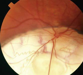
Optic nerve colobomas can be unilateral or bilateral. The visual acuity can range from normal to complete blindness. The coloboma develops secondary to incomplete closure of the embryonic fissure. The defect can produce a partial or total excavation of the optic disc (Fig. 623-1) Chorioretinal and iris colobomas can also occur. Optic nerve colobomas may be seen in a multitude of ocular and systemic abnormalities including the CHARGE association (coloboma, heart disease, atresia choanae, retarded growth and development and/or central nervous system anomalies, genetic anomalies and/or hypogonadism, ear anomalies and/or deafness).

Papilledema

The term papilledema is reserved to describe swelling of the nerve head secondary to increased intracranial pressure (ICP). Clinical manifestations of papilledema include edematous blurring of the disc margins, fullness or elevation of the nerve head, partial or complete obliteration of the disc cup, capillary congestion and hyperemia of the nerve head, generalized engorgement of the veins, loss of spontaneous venous pulsation, nerve fiber layer hemorrhages around the disc, and peripapillary exudates (see Fig. 584-2). In some cases, edema extending into the macula can produce a fan- or star-shaped figure. Concentric peripapillary retinal wrinkling (Paton lines) may be noted. Transient obscuration of vision can occur, lasting seconds and associated with postural changes. Vision, however, is usually normal in acute papilledema. Normally, when the ICP is relieved, the papilledema resolves and the disc returns to a normal or nearly normal appearance within 6-8 wk. Sustained chronic papilledema or long-standing unrelieved increased ICP can, however, lead to permanent nerve fiber damage, atrophic changes of the disc, macular scarring, and impairment of vision.

The pathophysiology of papilledema is probably as follows: elevation of intracranial subarachnoid cerebrospinal fluid (CSF) pressure, elevation of CSF pressure in the sheath of the optic nerve, elevation of tissue pressure in the optic nerve, stasis of axoplasmic flow and swelling of the nerve fibers in the optic nerve head, and secondary vascular changes and the characteristic ophthalmoscopic signs of venous stasis. Associated neuro-ophthalmic signs of increased ICP in infants and children include 6th cranial nerve palsy and attendant esotropia, lid retraction, paresis of upward gaze, tonic downward deviation of the eyes, and convergent nystagmus.

The common etiologies of papilledema in childhood are intracranial tumors and obstructive hydrocephalus, intracranial hemorrhage, the cerebral edema of trauma, meningoencephalitis, toxic encephalopathy, and certain metabolic diseases. Whatever the cause, the optic disc signs of increased ICP in early childhood occasionally can be modified by the distensibility of the young skull. In the absence of conditions associated with early closure of sutures and early obliteration of the fontanel (craniosynostosis, Crouzon disease, and Apert syndrome), infants with increased ICP might not develop papilledema.

The differential diagnosis of papilledema includes structural changes of the disc (pseudopapilledema, pseudoneuritis, drusen, and myelinated nerve fibers), with which it may be confused, and the disc swelling of papillitis associated with optic neuritis in addition to the disc changes of hypertension and diabetes mellitus. Unless retinal hemorrhage or edema involves the macular area, the preservation of good central vision and the absence of an afferent pupillary defect (Marcus Gunn pupil) help to differentiate acute papilledema from the edema of the optic nerve head found in acute optic neuritis.

Papilledema is a neurologic emergency. It can be accompanied by other signs of increased ICP, including headaches, nausea, and vomiting. Neuroimaging should be performed; if no intracranial masses are detected, a lumbar puncture and determination of CSF pressure should follow.

Optic Neuritis

Optic neuritis is any inflammation or demyelinization of the optic nerve with attendant impairment of function. The process is usually acute, with rapidly progressive loss of vision. It may be unilateral or bilateral. Pain on movement of the globe or pain on palpation of the globe can precede or accompany the onset of visual symptoms. There is decreased visual activity, decreased color vision and contrast sensitivity, a relative afferent pupillary defect, and a normal macula and peripheral retina.

When the retrobulbar portion of the nerve is affected without ophthalmoscopically visible signs of inflammation at the disc, the term retrobulbar optic neuritis is applied. When there is ophthalmoscopically visible evidence of inflammation of the nerve head, the term papillitis or intraocular optic neuritis is used. When there is involvement of both the retina and papilla, the term optic neuroretinitis is used.

In childhood, optic neuritis can occur as an isolated condition or as a manifestation of a neurologic or systemic disease. Optic neuritis may be secondary to inflammatory diseases, infections, toxin exposure or nutrient deficiency. Inflammatory diseases include systemic lupus erythematosus, sarcoidosis, Behçet disease, and autoimmune optic neuritis. Infections include tuberculosis, syphilis, Lyme disease, meningitis, viral encephalitis, and HIV; optic neuritis can also be a postinfectious reaction. Toxins include methanol, lead, and ethambutol, and vitamin B12 is the common deficiency.

Optic neuritis can signify one of the many demyelinating diseases of childhood (Chapters 593; 593.2). Although a significant percentage of adults who experience an episode of optic neuritis eventually develop other symptoms associated with multiple sclerosis (MS), young children with optic neuritis are seemingly at less risk (risk of MS is 19% within 20 yr). Bilateral optic neuritis in children may be associated with neuromyelitis optica (Devic disease; Chapter 593.2). This syndrome is characterized by rapid and severe bilateral vision loss accompanied by transverse myelitis and paraplegia. Optic neuritis may also be a complication of long-term high-dose treatment with chloramphenicol or vincristine therapy. Extensive pediatric neurologic and ophthalmic investigation, including MRI and lumbar puncture, is usually required.

In most cases of acute optic neuritis, some improvement in vision begins within 1-4 wk after onset, and vision can improve to normal or near normal within weeks or months. The course varies with cause. Although central vision can fully recover, it is common to find permanent defects in other areas of visual function (contrast sensitivity, color, brightness sense, and motion perception). Optic neuritis can recur, especially, but not universally, in patients who go on to develop MS.

A treatment trial demonstrated that high-dose intravenous methylprednisolone can help to speed the visual recovery in young adults, and it can prevent the development of MS in those at risk. Orally administered corticosteroids should not be used because they are associated with a significant increase in the recurrence rate of optic neuritis. It is unknown to what degree the results of the aforementioned trial may be extrapolated to optic neuritis in childhood. In adults with MRI white matter lesions, treatment with interferon-β (1a or 1b) reduces the incidence of MS.

Optic Atrophy

Optic atrophy is degeneration of optic nerve axons, with attendant loss of function. The ophthalmoscopic signs of optic atrophy are pallor of the disc and loss of substance of the nerve head, sometimes with enlargement of the disc cup. The associated vision defect varies with the nature and site of the primary disease or lesion.

Optic atrophy is the common expression of a wide variety of congenital or acquired pathologic processes. The cause may be traumatic, inflammatory, degenerative, neoplastic, or vascular; intracranial tumors and hydrocephalus are principal causes of optic atrophy in children. In some cases, progressive optic atrophy is hereditary. Dominantly inherited infantile optic atrophy is a relatively mild heredodegenerative type that tends to progress through childhood and adolescence. Autosomal recessively inherited congenital optic atrophy is a rare condition that is evident at birth or develops at a very early age; the visual defect is usually profound. Behr optic atrophy is a hereditary type associated with hypertonia of the extremities, increased deep tendon reflexes, mild cerebellar ataxia, some degree of mental deficiency, and possibly external ophthalmoplegia. This disorder afflicts principally boys aged 3-11 yr. Some forms of heredodegenerative optic atrophy are associated with sensorineural hearing loss, as can occur in some children with type 1 diabetes mellitus. In the absence of an obvious cause, optic atrophy in an infant or child warrants extensive etiologic investigation.

Optic Nerve Glioma

Optic nerve glioma (Chapter 491), more properly referred to as juvenile pilocytic astrocytoma, is the most common tumor of the optic nerve in childhood. This neuroglial tumor can develop in the intraorbital, intracanalicular, or intracranial portion of the nerve; the chiasm is often involved.

The tumor is a cytologic benign hamartoma that is generally stationary or only slowly progressive. The principal clinical manifestations when the tumor occurs in the intraorbital portion of the nerve are unilateral loss of vision, proptosis, and deviation of the eye; optic atrophy or congestion of the optic nerve head can occur. Chiasmal involvement may be attended by defects of vision and visual fields (often bitemporal hemianopia), increased ICP, papilledema or optic atrophy, hypothalamic dysfunction, pituitary dysfunction, and sometimes nystagmus or strabismus. Juvenile pilocytic astrocytomas occur more in patients with neurofibromatosis.

Treatment of optic pathway gliomas is controversial. The best management is usually periodic observation with serial radiography (preferably MRI). Only symptomatic and radiographically progressing optic nerve gliomas require strong consideration for treatment. Surgical removal may be appropriate when the tumor is confined to the intraorbital, intracanalicular, or prechiasmal portion of the nerve if a patient has unsightly proptosis with complete or nearly complete loss of vision of the affected eye. When the chiasm is involved, resection is not usually indicated and radiation and chemotherapy may be necessary.

Bibliography

Auw-Haedrich C, Staubach F, Witschel H. Optic disk drusen. Surv Ophthalmol. 2002;47:515-532.

Balcer LJ. Optic neuritis. N Engl J Med. 2006;354:1273-1280.

Birkebaek NH, Patel L, Wright NB, et al. Endocrine status in patients with optic nerve hypoplasia: relationship to midline central nervous system abnormalities and appearance of the hypothalamic-pituitary axis on magnetic resonance imaging. J Clin Endocrinol Metab. 2003;88:5281-5286.

Garcia-Filion P, Fink C, Geffner ME, Borchert M. Optic nerve hypoplasia in North America: a re-appraisal of perinatal risk factors. Acta Ophthalmol. 2010;88:527-534.

Hickman SJ, Dalton CM, Miller DH, et al. Management of acute optic neuritis. Lancet. 2002;360:1953-1962.

Hu K, Davis A, O’Sullivan E. Distinguishing optic disc drusen from papilledema. BMJ. 2009;338:1207-1209.

Lam BL, Feuer WJ, Abukhalil F, et al. Leber hereditary optic neuropathy gene therapy clinical trial recruitment. Arch Ophthalmol. 2010;128(9):1129-1135.

Massaro M, Thorarensen O, Liu GT, et al. Morning glory disc anomaly and moyamoya vessels. Arch Ophthalmol. 1998;116:253-254.

Nicolin G, Parkin P, Mabbott D, et al. Natural history and outcome of optic pathway gliomas in children. Pediatr Blood Cancer. 2009;53:1231-1237.

Optic Neuritis Study Group. Visual function 5 years after optic neuritis. Experience of the Optic Neuritis Treatment Trial. Arch Ophthalmol. 1997;115:1545-1552.

Repka MX, Miller NR. Optic atrophy in children. Am J Ophthalmol. 1988;106:191-193.

Skarf B, Hoyt CS. Optic nerve hypoplasia in children: association with anomalies of the endocrine and CNS. Arch Ophthalmol. 1984;102:62-67.

Weiss AH, Beck RW. Neuroretinitis in childhood. J Pediatr Ophthalmol Strabismus. 1989;26:198-203.