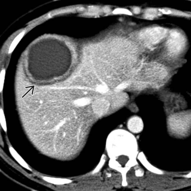
Simple fluid density (0-10 HU) or slightly hyperdense

 “Abscess” suggests a discrete, drainable fluid collection: Differentiate from ill-defined inflammation and fluid that is not drainable (i.e. phlegmon)

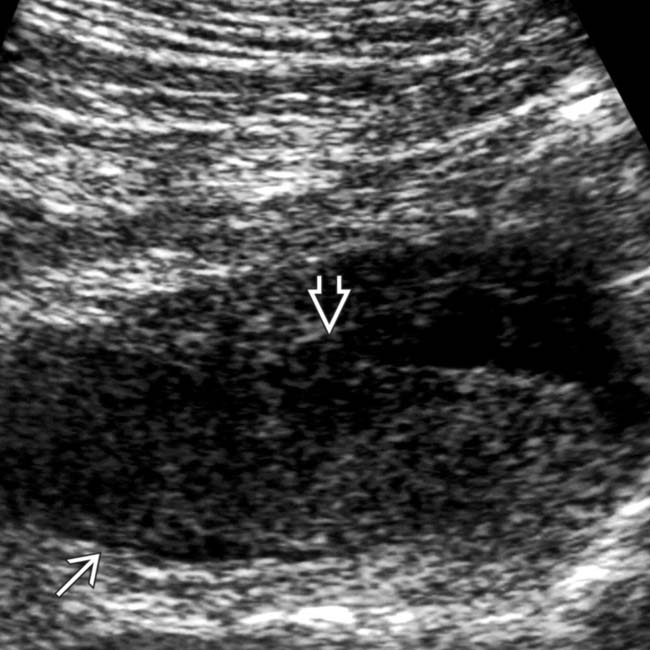
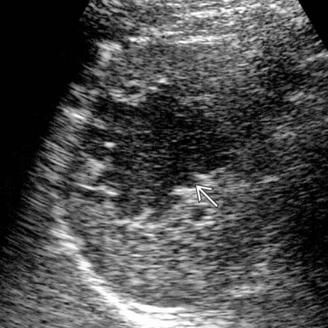
• US: Complex fluid collection with internal low-level echoes, membranes, or septations


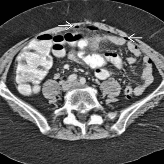
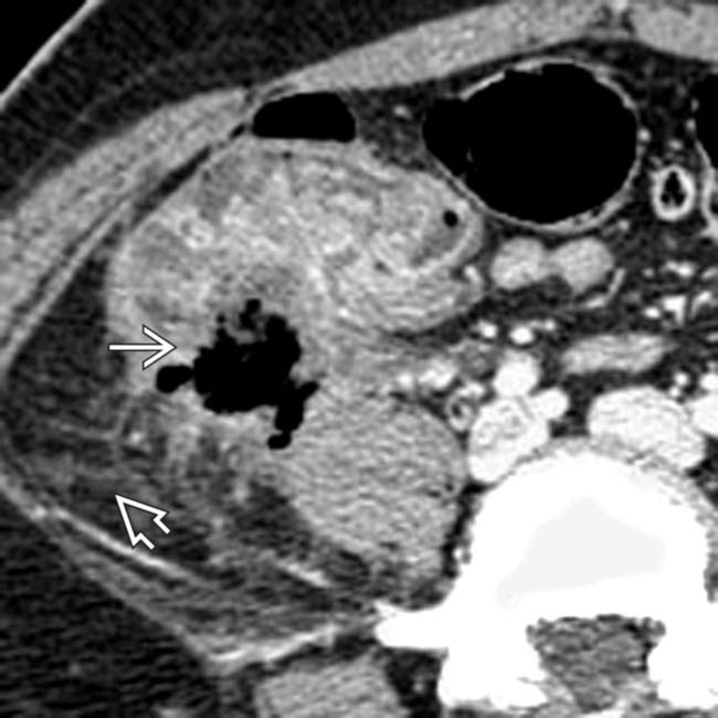
 with gas bubbles
  and an enhancing capsule
  , findings diagnostic for an abdominal abscess.
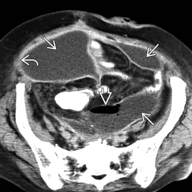
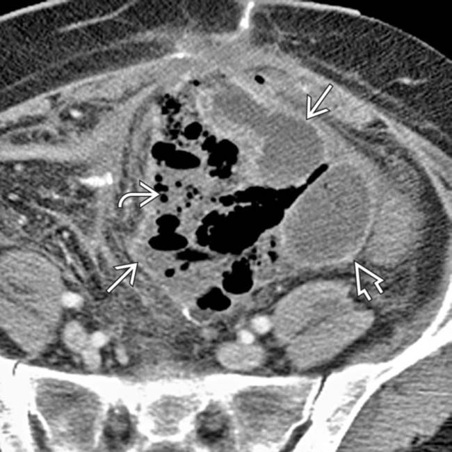
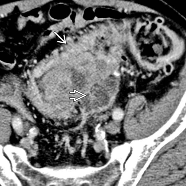
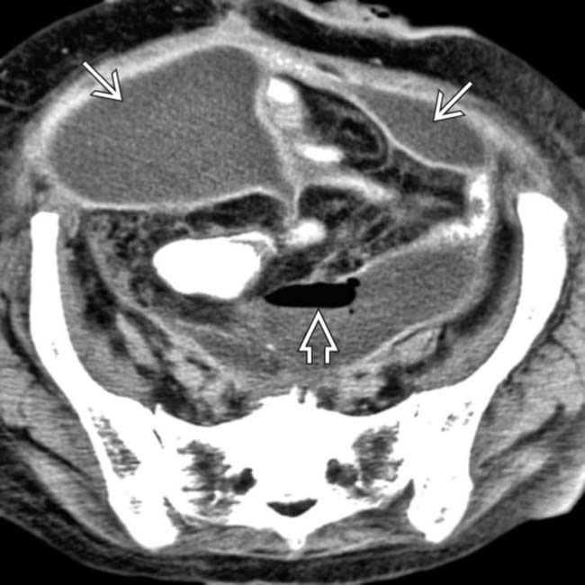
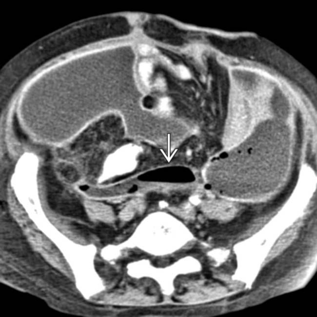
 with prominently enhancing capsules
  and mass effect on adjacent structures, representing abdominal abscesses. Note the air-fluid level
  within one of the abscesses.
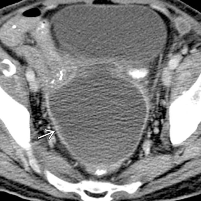
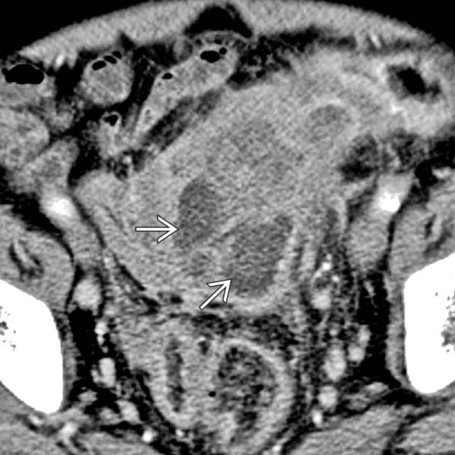
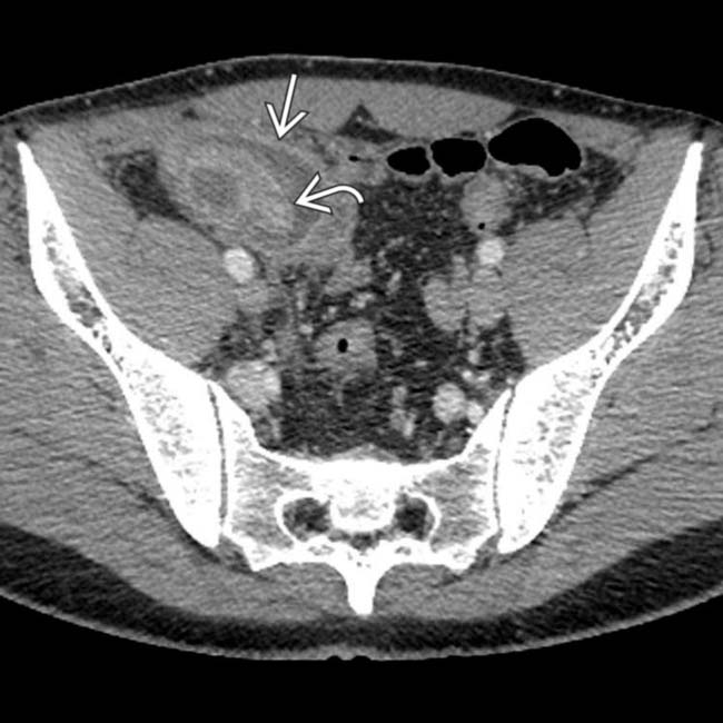
 following hysterectomy. Note the presence of a discrete enhancing rim and mass effect on adjacent loops of bowel and the bladder.
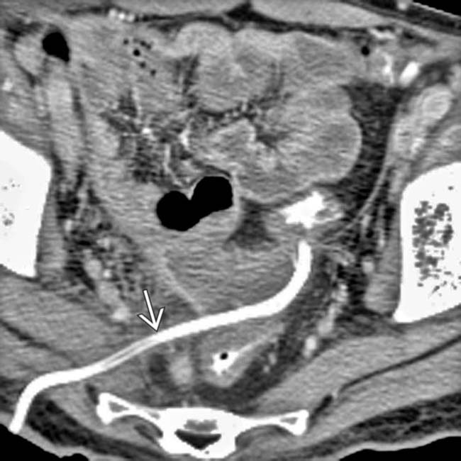
 using a transgluteal approach. The abscess has almost completely resolved following drainage.
IMAGING
General Features


CT Findings


• Presence of internal gas (∼ 50% of cases) in absence of intervention highly suspicious for infected collection


Ultrasonographic Findings
• Complex fluid collection with internal low-level echoes, membranes, or septations on US


CLINICAL ISSUES
Presentation


Treatment
• Options, risks, complications
 Percutaneous abscess drainage (PAD)
 Contraindications for PAD related to abscess
Percutaneous abscess drainage (PAD)
– Best candidates have well-defined, encapsulated, fluid-filled abscesses > 3 cm with safe catheter access route
– Drainage can be performed under CT or US guidance, with multiple approaches possible (transcutaneous, transgluteal, transrectal, transvaginal)
 Contraindications for PAD related to abscess


 .
.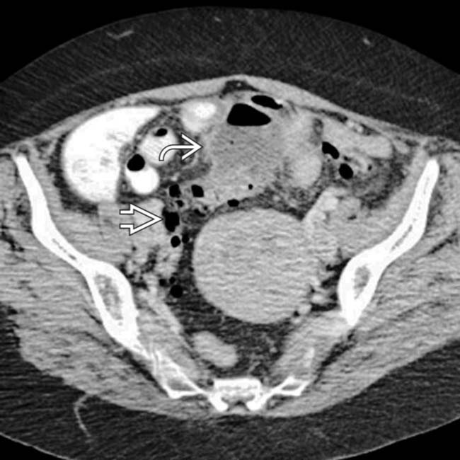
 immediately adjacent to a segment of sigmoid colon with extensive diverticulosis
  , which proved to be the source of the free air and abscesses.
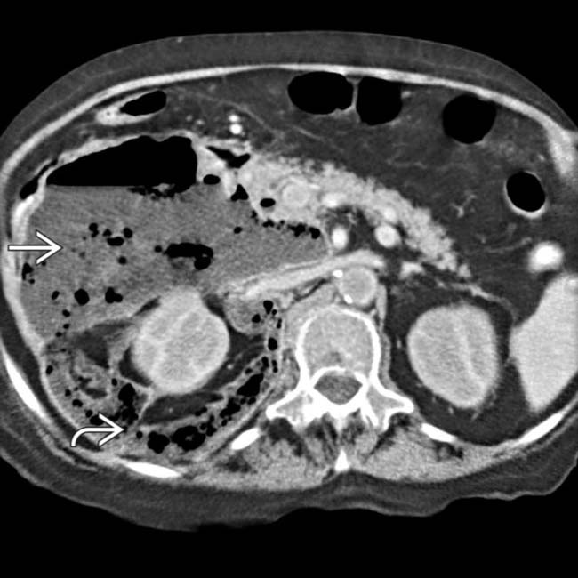
 dissecting through the retroperitoneal spaces, especially the anterior pararenal space and the interfascial plane
  .
.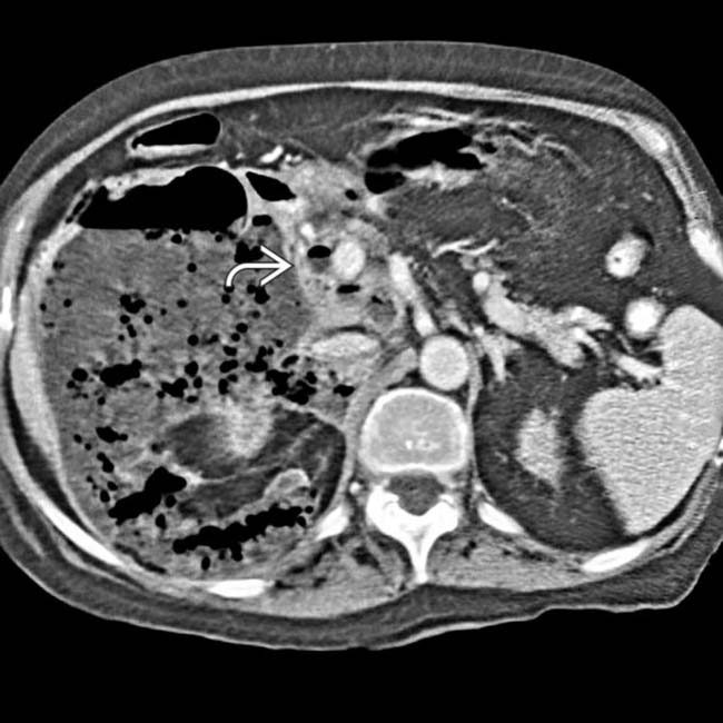
 at the papillotomy site.
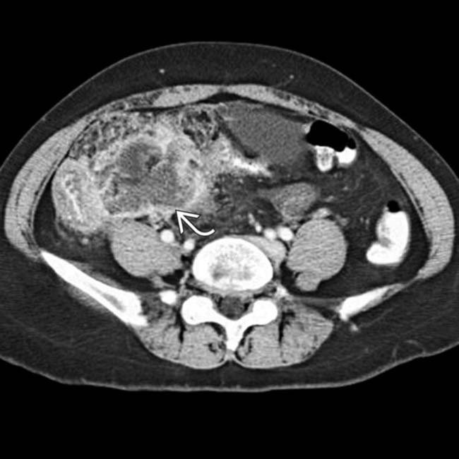
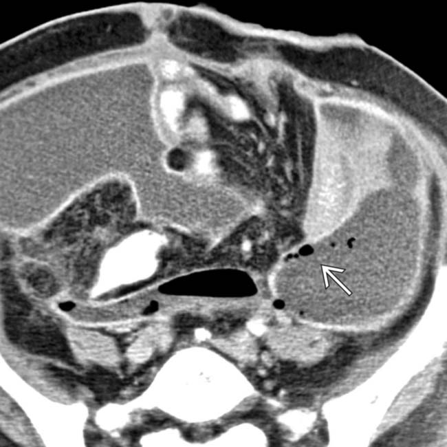
 with mass effect displacing the small bowel, bladder, and uterus.
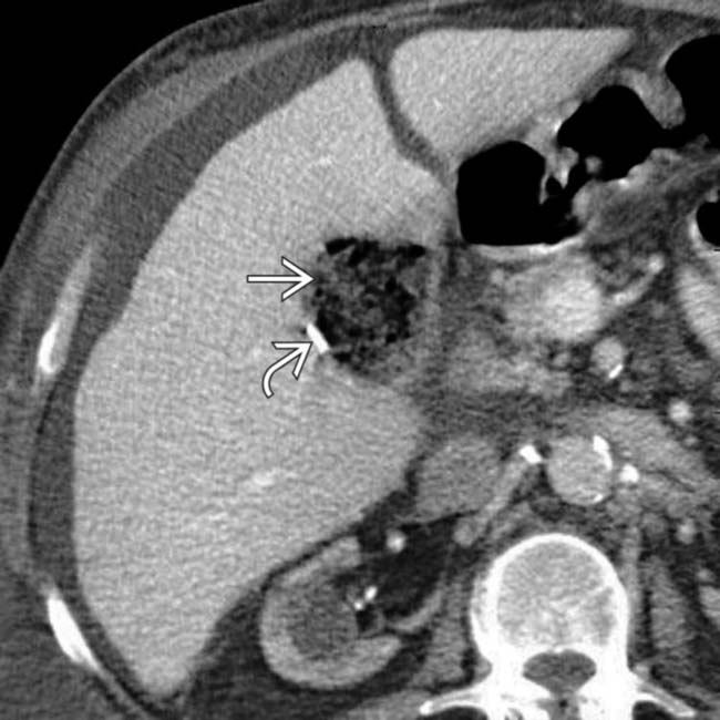
 , but very little fluid, in the cholecystectomy bed. Surgical clips
  are also seen. This represents oxidized cellulose (Surgicel), which was placed for hemostasis at surgery, and not an abscess.
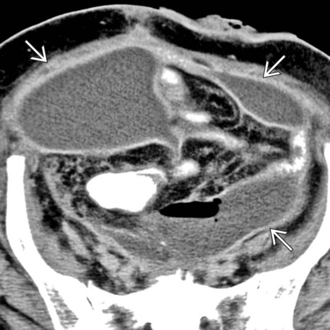
 .
.
 .
.
 , cecal thickening, and adjacent fat stranding
  .
.
 with fluid-fluid level
  .
.
 and ill-defined adjacent fluid collection
  .
.
 .
.
 after bowel resection. Note multiple fluid collections with enhancing rims; gas is seen only in pelvic abscess
  .
.
 , but not in other collections.
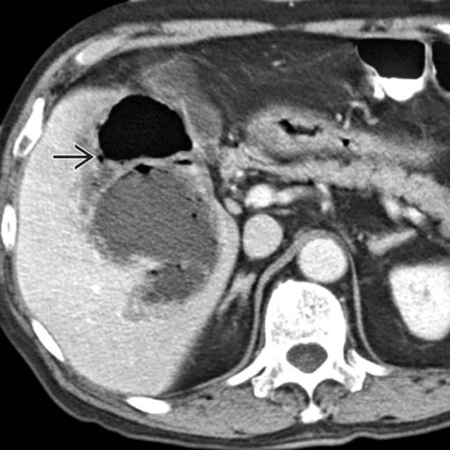
 .
.
 .
.
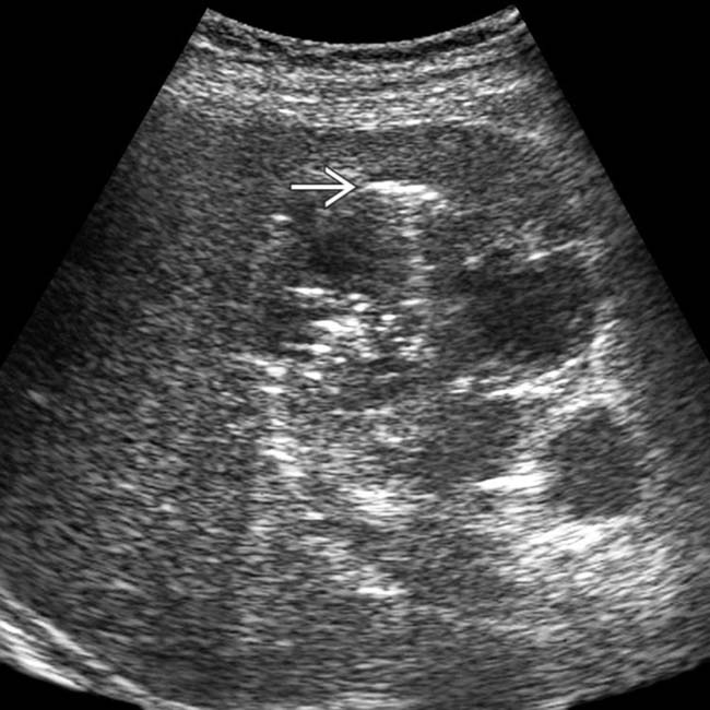
 surrounding the abscess.
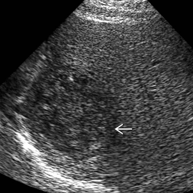
 within the hypoechoic mass, and the lack of distal acoustic enhancement.
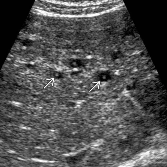
 .
.
 .
.
 with peripheral rim enhancement in the right lower quadrant. In the midst of the fluid collection is a tubular structure
  , representing a ruptured appendix.
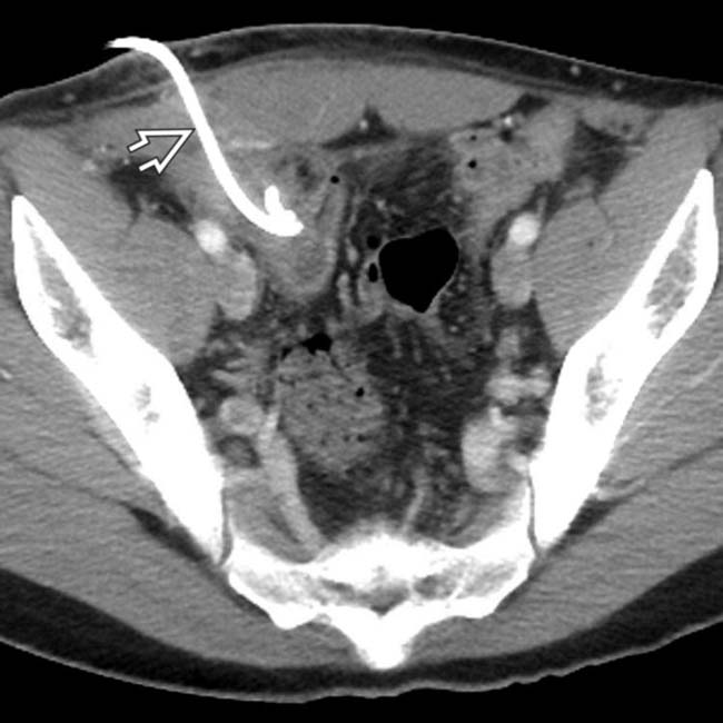
 placed percutaneously in the collection, with near resolution of the fluid within the abscess.


