 Central Venous Catheter Insertion (Perform)
PREREQUISITE NURSING KNOWLEDGE
• Knowledge of the normal anatomy and physiology of the cardiovascular system is needed.
• Clinical and technical competence in central line insertion and suturing is important.
• Knowledge of the principles of sterile technique is essential.
• Knowledge of the anatomy and physiology of the vasculature and adjacent structures of the neck, groin, and arm is needed.
• Competence in chest radiographic interpretation is necessary.
• Advanced cardiac life support knowledge and skills are needed.
• Indications for a central venous catheter (CVC) include the following4,7:

 Hypotension after major surgery
 Right ventricular ischemia or infarction

 Administration of total parenteral nutrition
 Lack of peripheral venous access
 Assessment of hypovolemia or hypervolemia

 Long-term infusions of medications


• Placement of a CVC can guide treatment after major surgery and during active bleeding.
• The central venous pressure (CVP) can be helpful in the differentiation of right ventricular failure from left ventricular failure.
• The CVP is commonly elevated during or after right ventricular failure, ischemia, or infarction because of decreased compliance of the right ventricle while the pulmonary artery occlusion pressure is normal.
• The CVP can be helpful in the determination of hypovolemia. The CVP value is low if the patient is hypovolemic. Venodilation also decreases CVP.
• Relative contraindications of CVC insertion include the following4,7:


• The CVP provides information regarding right heart filling pressures and right ventricular function and volume.
• The CVP historically was measured with a water manometer system but is now measured with a single-pressure transducer system (see Procedures 70 and 76).
• The CVP waveform is identical to the right atrial pressure (RAP) waveform.
• The normal CVP value is 2 to 6 mm Hg.
• Electrocardiographic (ECG) monitoring is essential in the accurate interpretation of the CVP value.
• Understanding is needed of a, c, and v waves. The a wave reflects right atrial contraction; the c wave reflects closure of the tricuspid valve; and the v wave reflects right atrial filling during ventricular systole (see Figs. 70-1 and 73-7).
• Dysrhythmias may alter CVP or RAP waveforms.
• The risk for a pneumothorax is minimized with use of an internal jugular vein. The preferred site for catheter insertion is the right internal jugular vein. The right internal jugular vein is a “straight shot” to the right atrium.
• The right or left subclavian veins are also sites for central catheter placement. Placement of a CVC through the right subclavian vein is a shorter and more direct route than the left subclavian vein because it does not cross the midline of the thorax.4,7
• Femoral veins may be accessed but have the disadvantages of limiting the patient to bed rest with immobilization of the leg and increasing the patient’s risk of infection.
• The presence of a pacemaker may alter the choice of placement of a CVC because of a risk for dislodging pacemaker leads with insertion of a CVC.
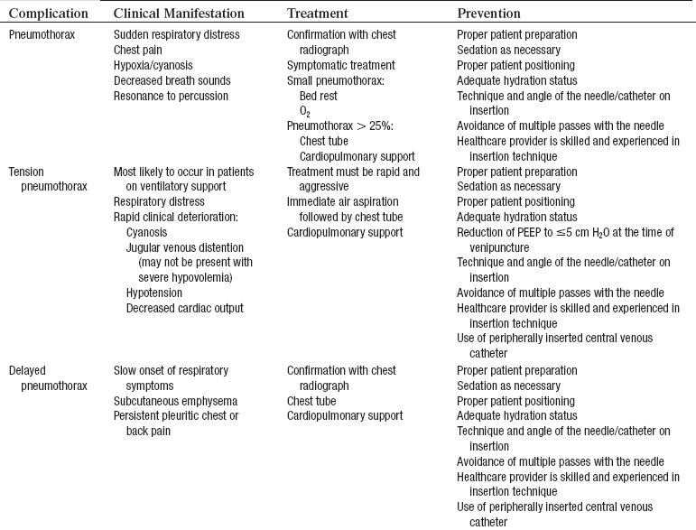
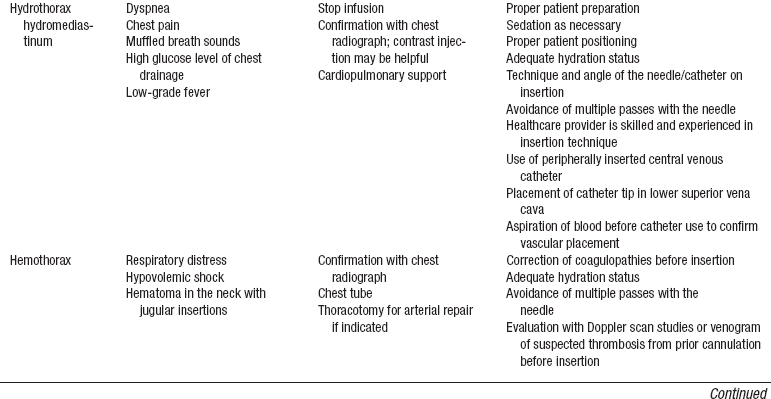
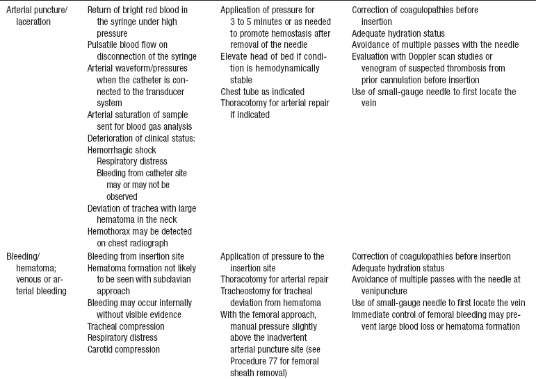
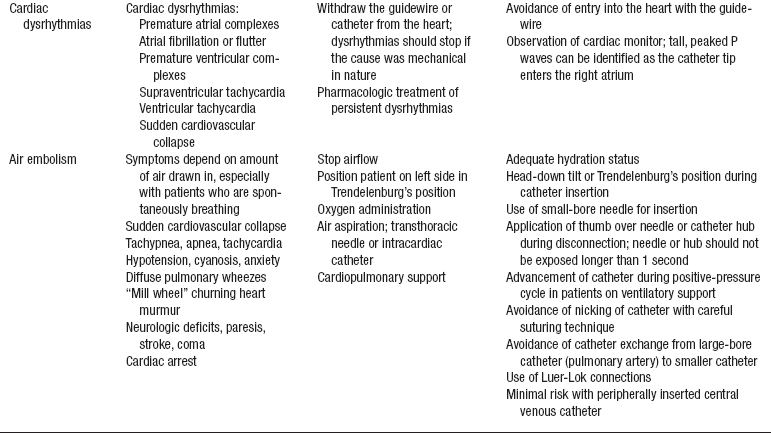
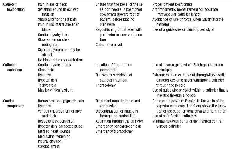
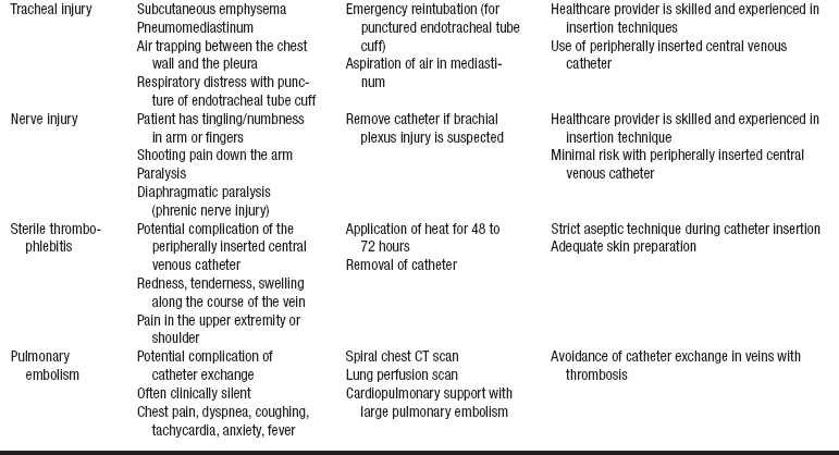
• Complications may occur during or after insertion of a central venous catheter (see Table 81-1).
EQUIPMENT
• CVC of choice (single, dual, or triple lumen) usually supplied with insertion needle, dilator, syringe, and guidewire.
• Large sterile drapes or towels
• 1% lidocaine without epinephrine
• Large package of 4 × 4 gauze sponges
• Suture kit (hemostat, scissors, needle holder)
• 3-0 or 4-0 nylon suture with curved needle
• Syringes: one 10- to 12-mL syringe; two 3- to 5-mL syringes; two 22-gauge, 1½-inch needles
• Masks, head coverings, goggles (shield and mask combination may be used), sterile gloves, and sterile gowns
• Skin protectant pads or swab sticks
• Chlorhexidine-impregnated sponge
• Antiseptic solution (e.g., 2% chlorhexidine-based preparation)
• Normal saline flush syringes or 0.9% sodium chloride vials, 10- to 30-mL
Additional equipment as needed includes the following:
• Hemodynamic monitoring system (see Procedure 76)
• Sutureless catheter securement device
• Intravenous (IV) solution with Luer-Lok administration set for IV infusion
• Bedside monitor and oscilloscope with pulse oximetry
• Supplemental oxygen supplies
• Package of alcohol pads or swab sticks
PATIENT AND FAMILY EDUCATION
• Explain the need for the CVC insertion and assess patient and family understanding.  Rationale: Clarification and understanding of information decrease patient and family anxiety levels.
• Explain the procedure and the time involved.  Rationale: Explanation increases patient cooperation and decreases patient and family anxiety levels.
• Explain the need for sterile technique and that the patient’s face may be covered.  Rationale: The explanation decreases patient anxiety and elicits cooperation.
• Explain the benefits and potential risks for the procedure.  Rationale: Information is offered so that the patient can make an informed decision.
PATIENT ASSESSMENT AND PREPARATION
Patient Assessment
• Determine the patient’s medical history of cervical disk disease or difficulty with vascular access.  Rationale: Baseline data are provided.
• Determine the patient’s medical history of pneumothorax or emphysema.  Rationale: Patients with emphysematous lungs may be at increased risk for puncture and pneumothorax, depending on the approach.
• Determine the patient’s medical history of anomalous veins.  Rationale: Patients may have a history of dextroacardia or transposition of the great vessels, which leads to greater difficulty in catheter placement.
• Assess the intended insertion site.  Rationale: Scar tissue may impede placement of the catheter. Permanent pacemakers or implantable cardioverter defibrillators may preclude placement. Previous surgery and previous placement of a CVC may cause a thrombus to be present.
• Assess the patient’s cardiac and pulmonary status.  Rationale: Some patients may not tolerate a supine or Trendelenburg’s position for extended periods of time.
• Assess vital signs and pulse oximetry.  Rationale: Baseline data are provided.
• Assess electrolyte levels (e.g., potassium, magnesium, calcium).  Rationale: Electrolyte abnormalities may increase cardiac irritability.
• Assess the patient for heparin sensitivity or allergy.  Rationale: CVCs are heparin-bonded, although non–heparin-bonded catheters are available. If the patient has a heparin allergy or has a history of heparin-induced thrombocytopenia, use a non–heparin-coated catheter.
• Assess for a coagulopathic state and determine whether the patient has recently received anticoagulant or thrombolytic therapy.  Rationale: These patients are more likely to have complications related to bleeding and may need interventions before insertion of the CVC.
Patient Preparation
• Verify correct patient with two identifiers.  Rationale: Prior to performing a procedure, the nurse should ensure the correct identification of the patient for the intended intervention.
• Ensure that the patient and family understand preprocedural teaching. Answer questions as they arise, and reinforce information as needed.  Rationale: Understanding of previously taught information is evaluated and reinforced.
• Obtain informed consent.  Rationale: Informed consent protects the rights of the patient and makes a competent decision possible for the patient; however, in emergency circumstances, time may not allow for this form to be signed.
• Perform a pre-procedure verification and time out, if non-emergent.  Rationale: Ensures patient safety.
• Prescribe sedation or analgesics as needed.  Rationale: The patient may need sedation or analgesics to promote comfort and to ensure adequate cooperation and appropriate placement.
• If the patient is obese or muscular and the preferred site is the internal jugular vein or subclavian vein, place a towel posteriorly between the shoulder blades.  Rationale: This placement helps extend the neck and provide better access to the subclavian and internal jugular veins.
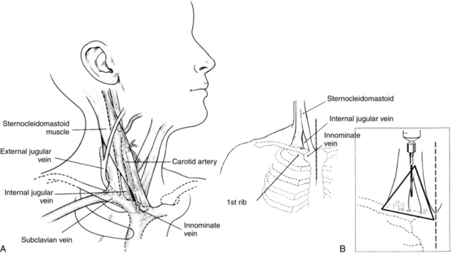
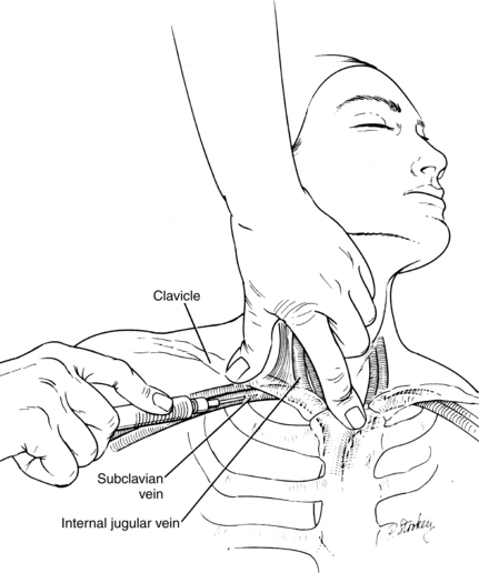
Figure 81-1 Anatomy of the jugular vein. A, Anatomy of the internal jugular vein showing its lower location within the triangle formed by the sternocleidomastoid muscle and the clavicle. B, Triangle drawn over the clavicle and sternal and clavicular portions of the sternocleidomastoid muscle is centered over the internal jugular vein (inset). (FromDailey EK, Schroeder JS: Techniques in bedside hemodynamic monitoring, St Louis, 1994, Mosby; and Daily PO, Griepp RB, Shumway NE: Percutaneous internal jugular vein cannulation, Arch Surg 101:534-536, 1970. Copyright 1970, American Medical Association.)
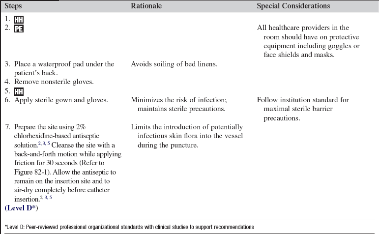
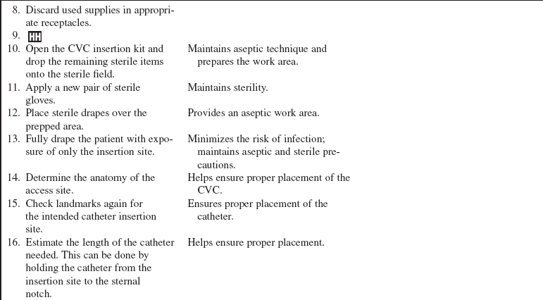
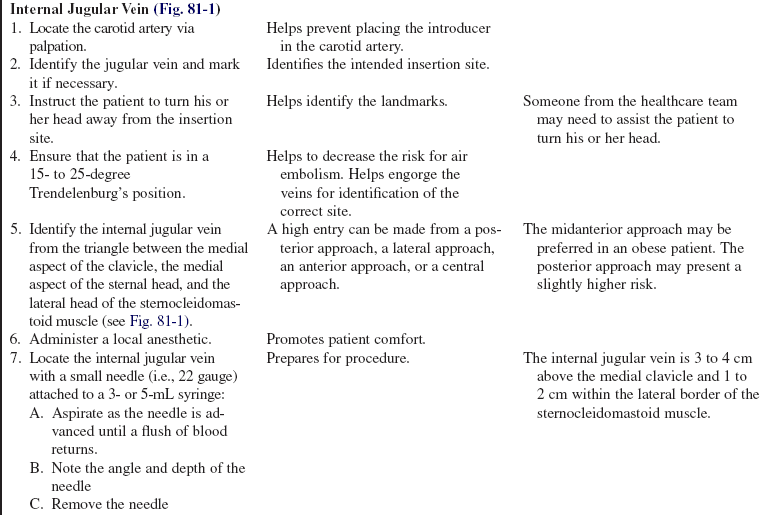
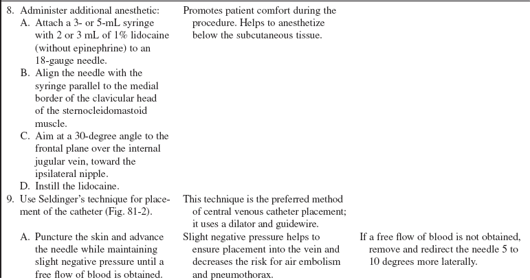

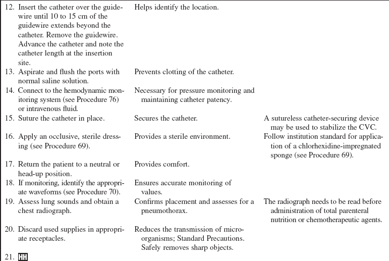

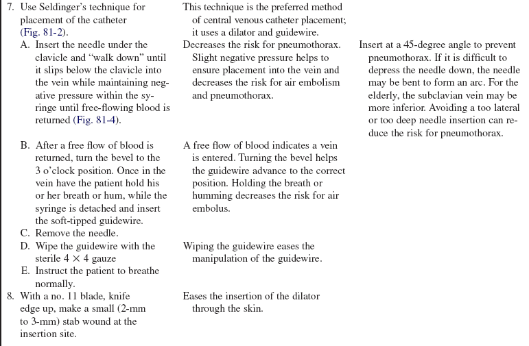
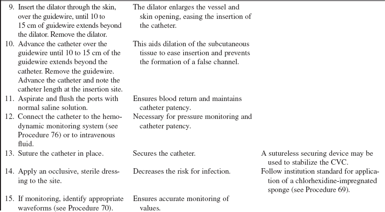


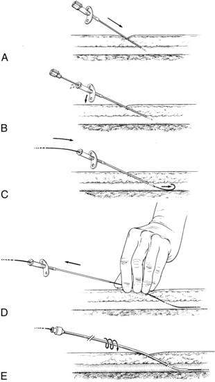
Figure 81-2 Basic procedure for Seldinger’s technique. A, The vessel is punctured with the needle at a 30- to 40-degree angle. B, The stylet is removed, and free blood flow is observed; the angle of the needle is then reduced. C, The flexible tip of the guidewire is passed through the needle into the vessel. D, The needle is removed over the wire while firm pressure is applied at the site. E, The tip of the catheter or sheath is passed over the wire and advanced into the vessel with a rotating motion. (From Dailey EK, Schroeder JS: Techniques in bedside hemodynamic monitoring, St Louis, 1994, Mosby.)
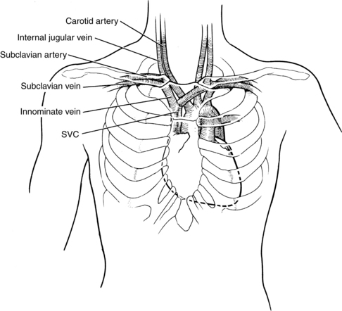
Figure 81-3 Anatomic location of the subclavian vein and surrounding structures. The subclavian vein joins the internal jugular vein to become the innominate vein at about the manubrioclavicular junction. The innominate vein becomes the superior vena cava (SVC) at about the level of the mid manubrium. (From Dailey EK, Schroeder JS: Techniques in bedside hemodynamic monitoring, St Louis, 1994, Mosby.)
References
![]() 1. Eyer, S, et al, Catheter-related sepsis. prospective, -randomized study of three methods of long-term catheter maintainence. Crit Care Med 1990; 18:1073–1079.
1. Eyer, S, et al, Catheter-related sepsis. prospective, -randomized study of three methods of long-term catheter maintainence. Crit Care Med 1990; 18:1073–1079.
2. Infusion Nurses Society. Policies and procedures for infusion nursing, ed 3. Norwood, MA: AuthorINS; 2006.
3. Intravenous Nurses Society. Infusion nursing standards of practice. J Infus Nurs. 2006; 29(1S):S1–S90.
![]() 4. Kumar, A, Darovic, GO. Establishment of cardiovascular access. In: Hemodynamic monitoring invasive and noninvasive clinical application. Philadelphia: Saunders; 2000.
4. Kumar, A, Darovic, GO. Establishment of cardiovascular access. In: Hemodynamic monitoring invasive and noninvasive clinical application. Philadelphia: Saunders; 2000.
![]() 5. O’Grady, NP, et al. Guidelines for the prevention of intravascular catheter-related infections. Am J Infect Control. 2002; 30:476–489.
5. O’Grady, NP, et al. Guidelines for the prevention of intravascular catheter-related infections. Am J Infect Control. 2002; 30:476–489.
![]() 6. Safdar, N, Klugar, DM, Maki, DG, A review of risk factors for catheter-related bloodstream infection caused by -percutaneously inserted, noncuffed central venous catheters. implications for preventive strategies. Medicine 2002; 81:466–472.
6. Safdar, N, Klugar, DM, Maki, DG, A review of risk factors for catheter-related bloodstream infection caused by -percutaneously inserted, noncuffed central venous catheters. implications for preventive strategies. Medicine 2002; 81:466–472.
7. Venus, G, Mallory, DL, Vascular cannulationCritical CareCivetta J, Taylor W, Kirby R. ed 5. Lippincott, Williams, Wilkins, Philadelphia, 2009.
Dailey, EK, Schroeder, JS. Techniques in bedside hemodynamic monitoring, ed 5. St Louis: Mosby; 1994.
Darovic, GO. Hemodynamic monitoring invasive and noninvasive clinical application. Philadelphia: Saunders; 2000.
Eggimann, P, et al. Impact of a prevention strategy targeted at vascular-access care on incidence of infections acquired in intensive care. Lancet. 2000; 355:1864–1868.
Friesinger, GC, Williams, SV, ACP/AHA Task Force on Clinical Privileges in Cardiology. clinical competence in hemodynamic monitoring. J Am Coll Cardiol. 1990; 15:1460.
Lorente, L, Henry, C, Martin, M, et al. Central venous catheter-related infection in a prospective and observational study of 2,595 catheters. Crit Care. 2005; 9:R631–R635.
