47. Role of Left Atrial Pressure Monitoring in the Management of Heart Failure

Published on 26/02/2015 by admin

Filed under Cardiovascular

Last modified 22/04/2025

Print this page

rate 1 star rate 2 star rate 3 star rate 4 star rate 5 star
Your rating: none, Average: 0 (0 votes)

This article have been viewed 1154 times

History

This 60-year-old man had coronary atherosclerosis, left bundle branch block (LBBB) with a QRS duration of 154 ms, peripheral vascular disease, and cardiomyopathy. He sought an opinion regarding management of his cardiomyopathy, which was diagnosed 10 years previously after he went to a local emergency department with chest tightness and shortness of breath. A coronary angiogram was performed that showed total occlusion of his right coronary artery, with no other significant coronary disease. His left ventricular ejection fraction (LVEF) at that time was 40%. He was managed medically, his symptoms improved markedly, and he returned to his baseline functional capacity. He was riding his bicycle regularly, working full time as a high school teacher, and doing heavy housework without difficulty. Approximately 2 years earlier, he noticed a decline in his exercise tolerance and developed intermittent dyspnea on exertion. His medical regimen was optimized, but he had no improvement in his symptoms. His LVEF was noted to have declined to 32%. A workup for ischemia was negative, and given his underlying LBBB and New York Heart Association (NYHA) class III symptoms, he was then referred for a biventricular pacemaker and treated with cardiac resynchronization therapy (CRT). Despite an optimal medical regimen and CRT, he had progressive dyspnea on exertion and worsening functional capacity. He reported development of lower extremity edema over the past 6 months and had frequent episodes of congestion. He required hospitalization five times in the previous 6 months for treatment of acute decompensated heart failure. His most recent two-dimensional echocardiogram revealed an LVEF of 29%, mild right ventricular dysfunction, and a moderately dilated left ventricle with mild mitral regurgitation. He has orthopnea that requires four pillows and frequently sleeps in a recliner. After his most recent hospitalization, he was enrolled in a home telemonitoring program, but did not experience improvement in his symptoms. He is obese and was recently diagnosed with sleep apnea, for which he uses continuous positive airway pressure at night.

Comments

The patient had chronic heart failure with progressive worsening in symptoms despite an optimized medical regimen. His volume status was difficult to manage over the past year, even with the addition of home telemonitoring. He should be counseled about the importance of sodium restriction, medication, and dietary compliance. Given his obesity, his volume status might be difficult to assess, and evaluation of his hemodynamics by right catheterization would be helpful in establishing his true volume status. He might require advanced therapies for heart failure in the near future, including heart transplantation or a left ventricular assist device, and consideration should be given for other novel therapies that might assist in symptom management. Controlling volume status will help delay disease progression.

Current Medications

The patient was taking carvedilol 25 mg twice daily, lisinopril 40 mg daily, torsemide 80 mg twice daily, metolazone 2.5 mg as needed for weight gain of more than 5 lb in 48 hours, spironolactone 25 mg daily, digoxin 0.125 mg daily, atorvastatin 20 mg daily, and aspirin 81 mg daily.

Comments

He was on a standard medication regimen to promote neurohormonal blockade and manage heart failure.

Current Symptoms

The patient reported orthopnea for the previous 2 weeks and noted chronic lower extremity edema over the past 6 to 7 months. He was unable to walk more than 20 feet without significant dyspnea.

Physical Examination

Laboratory Data

Comments

The patient has hyponatremia and renal insufficiency, both of which can be attributable to high filling pressures and volume overload.

Electrocardiogram

Findings

The electrocardiogram shows an atrial paced, biventricular paced rhythm.

Focused Clinical Questions and Discussion Points

Question

What implantable monitors are available for this patient that may assist in management of his condition?

Discussion

Many available implantable cardioverter-defibrillators have the ability to measure impedance, which may be a marker of pulmonary edema,8 as well as other indicators of impending heart failure such as heart rate variability2 and activity level. Several investigational implantable sensors have been tested, including a pulmonary artery sensor and a left atrial pressure sensor. These devices remain investigational as of this publication and are available only in clinical trials. Other sensors have been tested as part of pacemaker or defibrillator leads and can measure hemodynamics. The Chronicle Offers Management to Patients with Advanced Signs and Symptoms of Heart Failure (COMPASS HF) trial assessed the efficacy of using a right ventricular pressure sensor to aid physician-guided management in patients with NYHA class III to IV heart failure symptoms.5 This study reached its safety end points, but did not reach its primary end point of reducing the rate of heart failure–related events. A separate analysis demonstrated a reduction in time to first heart failure rehospitalization. The Reducing Decompensation Events Utilizing Intracardiac Pressures in Patients with Chronic Heart Failure (REDUCE hf) trial assessed the use of continuous hemodynamic monitoring using right ventricular pressures in patients with NYHA class II to III heart failure symptoms; however, the study was terminated early because of lead failures and thus clinical efficacy was unable to be assessed.3 A pulmonary artery sensor was tested in the CardioMEMS Heart Sensor Allows Monitoring of Pressure to Improve Outcomes in NYHA Class III Patients (CHAMPION) trial, in which qualifying patients received a pulmonary artery sensor that was embolized into a branch of the right or left pulmonary artery using catheters. A total of 550 patients were enrolled; patients were randomized in a 1:1 fashion with the control group receiving standard medical therapy for heart failure and the treatment group receiving the pulmonary sensor. Those in the treatment group received medical therapy tailored to pulmonary artery pressure goals. Results of the study revealed a significant reduction in hospitalization for heart failure at 6 months (30% reduction, p = 0.022), a reduction in mean pulmonary artery pressure at 6 months, and improvement in quality of life.1 This device is not currently available and has not received U.S. Food and Drug Administration approval. The second device now available in clinical trials is a left atrial pressure sensor. This device was tested in a feasibility study, the Home Self-Therapy in Severe Heart Failure Patients (HOMEOSTASIS) trial,7 which suggested that direct left atrial sensing, in combination with physician-directed patient self-management, had the potential to improve outcomes in patients with advanced heart failure. It demonstrated a reduction in mean left atrial pressure of 3.6 mm Hg at 12 months. The left atrial pressure sensor is currently available as part of a large, randomized controlled trial called the Left Atrial Pressure Monitoring to Optimize Heart Failure Therapy Study (LAPTOP-HF) and is limited to investigational use only.

Question

Is this patient a good candidate for the use of implantable sensors?

Discussion

This patient was an ideal candidate for implantable sensor technology. His symptoms were difficult to manage, and his physical examination was unreliable in assessing his volume status. Most hospitalizations for heart failure are related to worsening volume status and congestion. Filling pressures have been shown to increase 5 to 7 days before the development of symptoms severe enough to warrant hospitalization4; therefore knowledge of his true filling pressures will allow tight control of volume status and potentially reduce heart failure symptoms and need for hospitalization.

Question

What is the appropriate medical regimen for patients with systolic heart failure?

Discussion

Major society guidelines, based on evidence-based practice, recommend the use of an angiotensin-converting enzyme (ACE) inhibitor and beta blockers in patients with reduced LVEF (≤0.40) with or without symptoms of heart failure. Three beta blockers have proved beneficial in patients with heart failure with reduced LVEF: carvedilol, metoprolol succinate, and bisoprolol. Angiotensin-receptor blockers (ARBs) should be used in patients who are intolerant of ACE inhibitors. ARBs are recommended as additive therapy in patients who have moderately severe to severe heart failure symptoms and can be monitored for hyperkalemia and or worsening renal function.6

Final Diagnosis

This patient had advanced heart failure with a reduced LVEF. He was placed on an optimal medical regimen and treated with CRT. Despite this, he remained unresponsive to these therapies and had persistent heart failure symptoms.

Plan of Action

After discussion, the patient was given a left atrial pressure sensor.

Intervention

The patient was taken to the electrophysiology laboratory, where he was placed under general anesthesia and prepared and draped using standard sterile precautions. The left atrium was inspected using intracardiac echocardiography. After the right atrium was accessed by an inferior approach from the right femoral vein, the left atrium was inspected and found to be free of thrombus and then accessed by a transseptal approach. The left atrial pressure sensor was anchored into the intraatrial septum and pressure measurements obtained. The left atrial pressure was 20 mm Hg. Placement was confirmed using intracardiac echocardiography (Figure 47-1), followed by obtaining pulmonary and lateral chest radiographs (Figure 47-2).

Outcome

The patient was given instructions on how to measure left atrial pressure twice daily using a handheld device. This device—the patient advisory module—had a physician-directed algorithm built in that directed his medication regimen based on direct left atrial pressure measurement beginning at 3 months after implantation. After treatment, he experienced significant improvement in heart failure symptoms, with a reduction in NYHA class to NYHA II. Additionally, he remained free of hospitalization for heart failure for the subsequent year. His mean left atrial pressure fell from 32 mm Hg at 3 months to 9.8 mm Hg at 1 year after implantation (Figure 47-3).
image

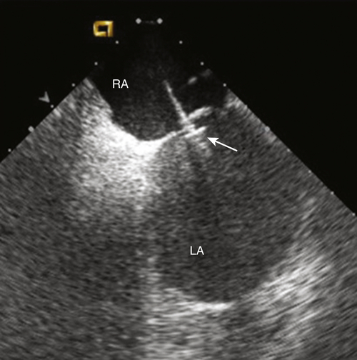
FIGURE 47-1 Intracardiac echocardiogram showing the left atrial pressure sensor in situ, anchored across the intraatrial septum from the right atrium. CAUTION: This is an investigational device, restricted by U.S. law to investigational use. LA, Left atrium; RA, right atrium.

image

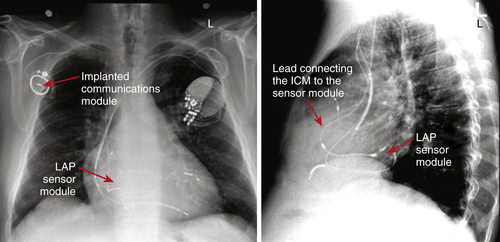
FIGURE 47-2 Pulmonary artery and lateral chest radiograph demonstrating proper placement of the left atrial pressure (LAP) monitoring system. The implanted communications module is used to transmit the pressure readings from the sensor module. The patient also has a cardiac resynchronization therapy device with an implantable cardioverter-defibrillator. CAUTION: This is an investigational device, restricted by U.S. law to investigational use. ICM, Implanted Communications Module.

image

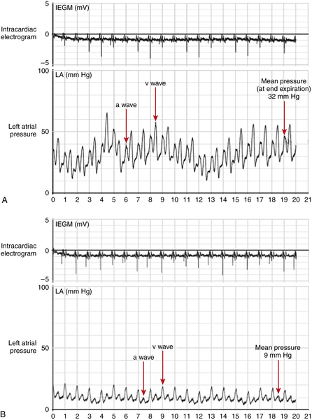
FIGURE 47-3 A, Left atrial pressure tracing obtained 3 months after implantation of the left atrial pressure (LAP) sensor, before initiation of a tailored regimen based on pressure readings. The waveform can be correlated with the atrial electrocardiogram and demonstrates significantly elevated pressure. B, LAP tracing obtained 12 months after implantation of the LAP sensor, after treatment was tailored using pressure readings. The waveform demonstrates a significant reduction in LAP in contrast to 9 months previously CAUTION: This is an investigational device, restricted by U.S. law to investigational use. IEGM, Intracardiac electrogram.

Selected References

1. Abraham W., Adamson P., Bourge R. et al. Wireless pulmonary artery haemodynamic monitoring in chronic heart failure: a randomised controlled trial. Lancet. 2011;377:658–666.

2. Adamson P.B. Continuous heart rate variability from an implanted device: a practical guide for clinical use. Congest Heart Fail. 2005;11:327–330.

3. Adamson P.B., Gold M.R., Bennett T. et al. Continuous hemodynamic monitoring in patients with mild to moderate heart failure: results of The Reducing Decompensation Events Utilizing Intracardiac Pressures in Patients. Congest Heart Fail. 2011;17:248–254.

4. Adamson P.B., Magalski A., Braunschweig F. et al. Ongoing right ventricular hemodynamics in heart failure: clinical value of measurements derived from an implantable monitoring system. J Am Coll Cardiol. 2003;41:565–571.

5. Bourge R.C., Abraham W.T., Adamson P.B. et al. Randomized controlled trial of an implantable continuous hemodynamic monitor in patients with advanced heart failure: the COMPASS-HF study. J Am Coll Cardiol. 2008;51:1073–1079.

6. Hunt S.A., Abraham W.T., Chin M.H. et al. 2009 focused update incorporated into the ACC/AHA 2005 Guidelines for the Diagnosis and Management of Heart Failure in Adults: a report of the American College of Cardiology Foundation/American Heart Association Task Force on Practice Guidelines: developed in collaboration with the International Society for Heart and Lung Transplantation. Circulation. 2009;119  e391.

7. Ritzema J., Troughton R., Melton I. et al. Physician-directed patient self-management of left atrial pressure in advanced chronic heart failure. Circulation. 2010;121:1086–1095.

8. Yu C.M., Wang L., Chau E. et al. Intrathoracic impedance monitoring in patients with heart failure: correlation with fluid status and feasibility of early warning preceding hospitalization. Circulation. 2005;112:841–848.

Share this: