44. Significant Residual or Worsening Mitral Regurgitation (MitraClip)

Published on 26/02/2015 by admin

Filed under Cardiovascular

Last modified 22/04/2025

Print this page

rate 1 star rate 2 star rate 3 star rate 4 star rate 5 star
Your rating: none, Average: 0 (0 votes)

This article have been viewed 1171 times

History

This 76-year-old female patient was referred to us for persistent heart failure symptoms in New York Heart Association [NYHA] class III to IV and severe functional mitral regurgitation (FMR) despite optimal therapeutic management combining medication, previous biventricular implantable cardioverter-defibrillator (ICD) (cardiac resynchronization therapy defibrillator [CRT-D]) implant, and atrioventricular node ablation for competing atrial fibrillation rhythm. Four months previously a CRT-D device was implanted based on conventional class I indication—drug-refractory symptomatic heart failure (NYHA class III to IV) because of postactinic cardiomyopathy (the patient had previously received radiotherapy for non-Hodgkin lymphoma) with severe left ventricular systolic dysfunction (left ventricular ejection fraction [LVEF], 29%); the electrocardiogram (ECG) showed significant ventricular conduction delay with complete left bundle branch block and 130-msec QRS complex duration. Atrial fibrillation was the underlying atrial rhythm. Besides severe left ventricular dysfunction, the transthoracic echocardiogram performed before CRT-D device implant showed a mildly dilated left ventricle (end-diastolic voume 186 mL and end-systolic volume 132 mL) and severe functional mitral regurgitation determined by failed coaptation of mitral valve leaflets resulting from symmetric dilation of the mitral anulus. Shortly after CRT-D implant, the patient underwent catheter ablation of the atrioventricular node as a result of recurring high ventricular rate atrial fibrillation.
After 4 months of CRT, both symptoms and mitral regurgitation remained unchanged. The patient was therefore evaluated for percutaneous edge-to-edge mitral valvuloplasty with a MitraClip (Abbott Laboratories, Abbott Park, Ill).1

Current Medications

The patient was taking acenocoumerol 1 mg adjusted to international normalized ratio value, captopril 10 mg daily, carvedilol 12.5 mg daily, spironolactone 50 mg daily, torsemide 20 mg adjusted according to weight, and metolazone 2.5 mg daily if body weight greater than 50 kg.

Comments

The medication profile of the patient is typical for advanced-phase heart failure, with low dosages of an angiotensin-converting enzyme inhibitor and beta blockers and weight-adjusted dosages of diuretics.

Current Symptoms

The patient was unable to walk up a single flight of 12 steps without stopping because of breathlessness (NYHA class III to IV) and unable to perform the 6-Minute Walk Test.

Physical Examination

Laboratory Data

Comments

Life expectancy considering the patient’s demographic, clinical, and laboratory data was calculated using the Seattle Heart Failure Model.2 Estimated life expectancy for this patient was considered to be very poor, with a median life expectancy of 2.6 years and estimated survival of only 69%, 48%, and 16% at 1, 2, and 5 years, respectively.

Electrocardiogram

Findings

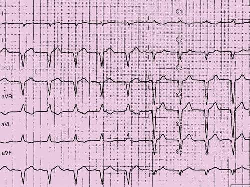
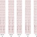
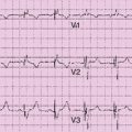
Constant biventricular paced rhythm was seen in VVI modality at 70 bpm (Figure 44-1). The vertical axis in the peripheral leads demonstrated a QRS duration of 120 msec. The underlying atrial rhythm was atrial fibrillation.

Echocardiogram

Findings

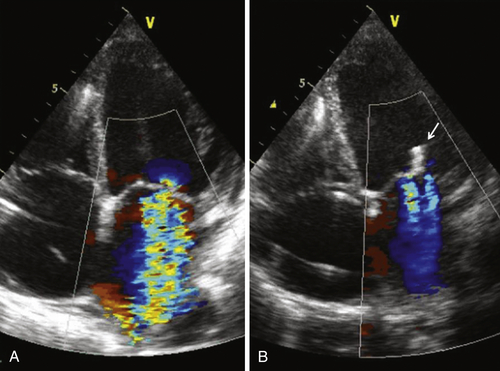
Figure 44-2 shows the transthoracic echocardiographic apical four-chamber view with color Doppler before (see Figure 44-2, A) MitraClip positioning.

Comments

A preprocedural transthoracic echocardiogram showed a mildly dilated left ventricle (end-diastolic volume 186 mL and end-systolic volume 132 mL) with severe systolic dysfunction (LVEF 26%). Severe functional mitral regurgitation was present, with a marked central regurgitant jet resulting from symmetric dilation of the annulus and consequent lack of leaflet coaptation. Lack of important alterations of leaflet morphology and movement implicate anatomomorphologic integrity of valve leaflets, chordae, and papillary muscles. Increased hemodynamic stress of mitral regurgitation is suggested by biatrial dilation, increased artery pulmonary pressure (45 mm Hg), hypokinesia of the right ventricle, and grade II tricuspid insufficiency.
image

FIGURE 44-1 

Focused Clinical Questions and Discussion Points

Question

Why is the patient considered a suitable candidate for percutaneous mitral valve repair rather than surgical valve repair?

Discussion

The patient is an extremely compromised CRT nonresponder who continued to be symptomatic because of persistent severe functional mitral regurgitation. Based on EUROSCORE II,3 the patient’s surgical mortality risk was estimated at 17.8%. Furthermore, surgical approach in patients with CRT with indication for valve repair is not supported by prospective, randomized evidence.
As far as percutaneous edge-to-edge mitral valvuloplasty by MitraClip is concerned, one multicenter, prospective, longitudinal study has demonstrated the clinical benefit in this patient subgroup.4 This study has shown that MitraClip repair of clinically and hemodynamically significant FMR after CRT improves symptoms and produces reversal of maladaptive remodeling in approximately 70% of these patients.

Question

What kind of mitral regurgitation disorder is most amenable to correction by MitraClip?

Discussion

Functional mitral regurgitation is most amenable to repair by MitraClip. Different from primary mitral regurgitation, functional mitral regurgitation (also termed secondary mitral regurgitation) is determined by geometric distortion of the subvalvular apparatus, secondary to left ventricular enlargement and remodeling as a result of idiopathic cardiomyopathy or coronary artery disease. The underlying condition in a patient with CRT produces tethering (apical and lateral papillary muscle displacement, and annular dilation) and reduced closing forces because of left ventricular dysfunction (reduced contractility or left ventricular dyssynchrony). It is important to emphasize that in functional mitral regurgitation, valve leaflets and chordae are structurally normal.5 Besides precise evaluation of the mitral regurgitation grade, echocardiographic examinations (both transthoracic and transesophageal) should assess the following aspects to prepare the patient for mitral valve repair by MitraClip:
1. Presence of annulus dilation resulting from underlying heart disease
2. Anatomomorphologic integrity of the other anatomic constituents of the mitral valve, namely the leaflets and chordae
3. Structural and hemodynamic “repercussions” of mitral regurgitation on other cardiac chambers and on pulmonary arterial pressure

image

FIGURE 44-2 

image

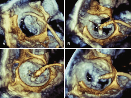
FIGURE 44-3 

Question

What is the recommended time from CRT implantation to indicate MitraClip in case of persistent moderate-to-severe mitral regurgitation? Why is such a time frame recommended?

Discussion

The patient with CRT should be considered eligible for MitraClip in the case of persisting symptoms associated with unchanged moderate or severe mitral regurgitation after 3 to 6 months of CRT. This preset time frame is recommended based on the established knowledge that CRT-induced reversal of maladaptive left ventricular remodeling, which usually occurs within the first 6 months after device implantation, may reduce mitral regurgitation.6,7 In the present case, it is important to emphasize that MitraClip intervention was not delayed to 6 months, because the clinical condition of the patient was extremely compromised with little or no therapeutic margin.

Final Diagnosis

This patient had persistent severe functional mitral regurgitation and was a symptomatic CRT nonresponder.

Plan of Action

Mitral regurgitation was addressed by a percutaneous approach based on positioning of the MitraClip.

Intervention

In general anesthesia and under continuous hemodynamic and transesophageal monitoring, the MitraClip delivery system is positioned by the transseptal catheterization approach. This system includes the clip at the tip, a steerable guiding catheter, and a clip delivery system to open, close, and release three-dimensional images.
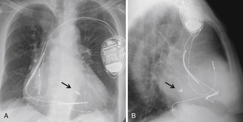
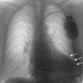
Figure 44-3 shows intraprocedural transesophageal echocardiographic images during MitraClip positioning (arrows). After transseptal catheterization, the clip, located at the tip of the MitraClip delivery system, is advanced across the mitral valve and into the left ventricle (see Figure 44-3, A) with the clip closed. The clip is then opened (see Figure 44-3, B), and slight retraction of the system toward the atrium in a central position is performed, thus capturing the valve leaflets. The clip is then closed (see Figure 44-3, C) at the desired position. If the position is suboptimal or inadequate, the maneuver may be repeated several times. After a satisfactory position has been achieved with resulting effective mitral regurgitation reduction, the clip is released (see Figure 44-3, D) and the delivery system removed. Postprocedural chest radiograph shows the correct position of the MitraClip device (arrows) in anteroposterior (Figure 44-4, A) and lateral (see Figure 44-4, B) projections.

Outcome

The outcome in this patient was satisfactory.
image

FIGURE 44-4 

Findings

Transthoracic echocardiogram showed that MitraClip allowed a remarkable reduction of mitral regurgitation from grade IV to grade II. As a result, artery pulmonary pressure reduced to 35 mm Hg, right ventricular hypokinesia was no longer present, and tricuspid insufficiency reduced to grade I (see Figure 43-2).
After 2 years and 9 months of follow-up, the patient was clinically stable in NYHA class III. The echocardiogram revealed persistence of severe left ventricular (LVEF, 25%) systolic dysfunction, but residual grade II mitral regurgitation and left ventricular diameters (end-diastolic diameter to end-systolic diameter ratio, 66/58 mm) remained stable.
Calculation of Seattle Heart Failure Score based on the patient’s demographic, clinical, laboratory, and therapeutic characteristics at last follow-up yielded an estimated mean life expectancy of 5.6 years, suggesting an increase in life expectancy of 2 years, compared to the preprocedural estimate.

Selected References

1. Feldman T., Kar S., Rinaldi M. et al. EVEREST Investigators Percutaneous mitral repair with the MitraClip system: safety and midterm durability in the initial EVEREST (Endovascular Valve Edge-to-Edge REpair Study) cohort. J Am Coll Cardiol. 2009;54:686–694.

2. Levy W.C., Mozaffarian D., Linker D.T. et al. The Seattle Heart Failure Model: prediction of survival in heart failure. Circulation. 2006;113:1424–1433.

3. Roques F., Michel P., Goldstone A.R. et al. The logistic EuroSCORE. Eur Heart J. 2003;24:882–883.

4. Auricchio A., Schillinger W., Meyer S. et al. PERMIT-CARE Investigators Correction of mitral regurgitation in nonresponders to cardiac resynchronization therapy by MitraClip improves symptoms and promotes reverse remodeling. J Am Coll Cardiol. 2011;58:2183–2189.

5. Vahanian A., Alfieri O., Andreotti F. et al. Guidelines on the management of valvular heart disease (version 2012). Eur Heart J. 2012;33:2451–2496.

6. Boriani G., Gasparini M., Landolina M. et al. InSync/InSync ICD Italian Registry Investigators. Impact of mitral regurgitation on the outcome of patients treated with CRT-D: data from the InSync ICD Italian Registry. Pacing Clin Electrophysiol. 2012;35:146–154.

7. Di Biase L., Auricchio A., Mohanty P. et al. Impact of cardiac resynchronization therapy on the severity of mitral regurgitation. Europace. 2011;13:829–838.