42. Nonresponders to Cardiac Resynchronization Therapy

Published on 02/03/2015 by admin

Filed under Cardiovascular

Last modified 22/04/2025

Print this page

rate 1 star rate 2 star rate 3 star rate 4 star rate 5 star
Your rating: none, Average: 0 (0 votes)

This article have been viewed 1221 times

History

This 79-year-old man with type II diabetes, hypertension, 2.5 pack per day smoking history for 46 years, and a strong family history of premature coronary disease (father died at 61 years of age of myocardial infarction) suffered a myocardial infarction in 1974 and had cardiac arrest as a result of ventricular fibrillation on the third day of that hospitalization. Coronary revascularization was undertaken with a four-vessel bypass in February, 1999. He developed symptoms of heart failure in 2006, with an echocardiogram showing akinesis of the inferior and posterobasal walls and an ejection fraction of 20% with moderate mitral regurgitation. He was treated with furosemide, carvedilol, candesartan, and statins and continued with New York Health Association (NYHA) class II to III symptoms, with an electrocardiogram demonstrating left bundle branch block (LBBB) with variable QRS durations of 120 to 130 msec. He was thought perhaps to be a candidate for cardiac resynchronization therapy. Outpatient tissue Doppler study confirmed an ejection fraction of 20% but fell short of the usual criteria for left ventricular dyssynchrony. Electrophysiologic study demonstrated easily inducible sustained monomorphic ventricular tachycardia, both at baseline and after procainamide challenge, prompting implantation of a single-chamber Medtronic defibrillator using the left cephalic vein. His ejection fraction was 25% in 2009. With progressive symptoms of heart failure in 2010 despite maximal medical therapy, and with a QRS duration of 140 msec, the decision was made to upgrade his device to a cardiac resynchronization therapy (CRT) system. The left subclavian system was still patent, allowing for passage of an active fixation atrial lead and a coronary sinus lead. Venography demonstrated that his coronary venous anatomy was limited, with few branches available and most atretic, consistent with his diabetes. One branch served as a reasonable target, with acceptable pacing thresholds without phrenic nerve stimulation. His paced QRS duration narrowed, but symptoms of heart failure have persisted. His level of depression worsened with the death of his wife, and he had occasional dietary indiscretion. Atrioventricular optimization was performed. With ongoing symptoms despite maximal medical therapy and dietary modification, and in an effort to maximize longevity of the implantable cardioverter-defibrillator (ICD), the left ventricular lead was inactivated 1 year later.

Current Medications

The patient was taking carvedilol 6.25 mg twice daily, furosemide 100 mg daily, potassium chloride 20 mEq daily, glyburide 2.5 mg daily, simvastatin 20 mg daily, amlodipine 5 mg daily, fish oil 1000 mg daily, and aspirin 81 mg daily.

Current Symptoms

The patient experienced dyspnea at 100 yards and one flight of stairs and had occasional orthopnea.

Physical Examination

Laboratory Data

Electrocardiogram

Findings

Figure 42-1 shows a biventricular paced sinus rhythm, heart rate of 66 bpm, and paced QRS of 136 msec.

Chest Radiograph

Findings

The coronary sinus venogram obtained at the time of implant shows the posteroapical branch selected and final position of the leads (Figures 42-2 and 42-3). Figure 42-4 demonstrates lead position in a different patient in whom the apical position of the coronary sinus lead is even more exemplary of an apical and nonbasal position.

Focused Clinical Questions and Discussion Points

Question

What defines a nonresponder in the CRT population, and at what point would this patient be considered a nonresponder?

Discussion

It is widely recognized that the definition of response and nonresponse to CRT varies considerably, whether by clinical parameters (i.e., changes in NYHA classification or hospitalizations for heart failure), imaging criteria (i.e., changes in ejection fraction, dyssynchrony, or stroke volume), or survival.18 Until universal agreement is achieved on this subject, it is difficult to define a predictor of success or, conversely, when to define failure. The patient described is on maximal medical therapy, has ongoing symptoms of heart failure despite CRT with persistent depression of ejection fraction, and would be considered a nonresponder by most clinicians. The length of time to be waited in any individual case before considering CRT a failure is unclear. This is an important issue because prematurely deactivating the coronary sinus lead, although minimizing current drain on the device, may preclude demonstration of a delayed response to CRT.
image

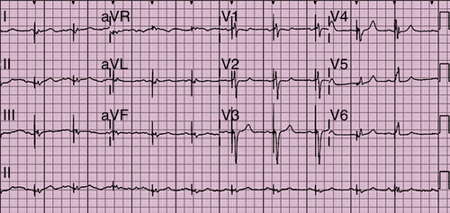
FIGURE 42-1 Electrocardiogram.

image

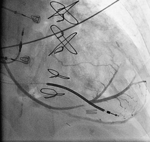
FIGURE 42-2 Coronary sinus venogram.

image

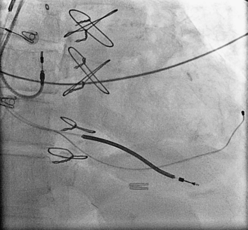
FIGURE 42-3 Chest radiograph of final coronary sinus lead position corresponding to venogram in Figure 42-2.

image

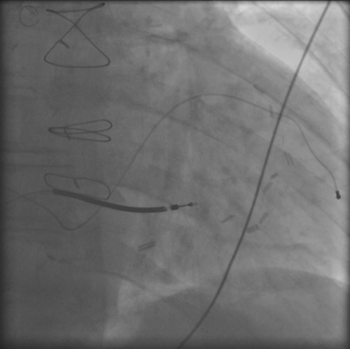
FIGURE 42-4 Chest radiograph in a different patient in whom the apical position of the coronary sinus lead is a better example of an apical coronary sinus lead position.

Question

What may have contributed to this patient’s nonresponse to CRT?

Discussion

As noted, predictors of response have been the subject of much investigation and a comprehensive understanding of this issue has yet to be achieved. A longer baseline QRS duration, as suggested by various trials (LBBB >150 msec), more basal position of the left ventricular lead, and lack of demonstration of significant intraventricular dyssynchrony by tissue Doppler imaging may have contributed to a lack of response in this case.

Question

What alternative strategies were available to this patient?

Discussion

Given the observation that the patient had atretic diabetic changes in the coronary venous tree, little else was available in the way of otherwise optimizing lead position because only one branch was sizable enough to engage. Interventional procedures such as venodilation have been employed in highly experienced centers provided the anatomy is reasonable and the approach feasible. Employment of newer quadripolar coronary sinus leads allowing for alternative pacing polarities and locations would not likely have helped in this individual.9 Epicardial lead placement is an alternative approach, but it is more invasive and there is no guarantee that it would resolve the issue of nonresponse in this case. Atrioventricular optimization was undertaken in this patient without success.

Question

Under what other circumstances might the left ventricular lead be deactivated or left inactive from the time of implant—that is, in addition to being a CRT nonresponder?

Discussion

If the lead dislodges and no other stable position is an option, attempting invasive repositioning of the coronary sinus lead may be deemed a greater risk. If multiple positions have been assessed and the safety margin between effective left ventricular pacing and phrenic nerve stimulation is too narrow, the decision may be made to inactivate the left ventricular lead. The same applies if the left ventricular pacing threshold is unacceptably high. In some situations, a prophylactic coronary sinus lead may have been implanted, such as in a patient with a rapid ventricular response to atrial fibrillation who may require eventual atriventricular node ablation, resulting in cardiac desynchronization. In such cases in which a high incidence of right ventricular pacing may be anticipated in the future, the coronary sinus lead may be left inactive until coronary sinus pacing is required.

Final Diagnosis

This patient was nonresponsive to CRT, possibly reflecting more apical or less basal position of coronary sinus lead versus shorter baseline QRS duration or less evidence of baseline mechanical dyssynchrony.

Plan of Action

The plan for this patient was deactivation of the coronary sinus lead after continued observation of nonresponse and continuation of medical therapy.

Intervention

Deactivation of coronary sinus lead was performed by programming left ventricular lead function to the “off” position.

Outcome

At the time of publication, the patient was still alive, albeit with ongoing symptoms of heart failure and profound depression. ICD deactivation also could be considered in the future if comorbid conditions prevail, as suggested by Heart Rhythm Society guidelines on management of patients with cardiac implantable electronic devices.10

Selected References

1. Chung E.S., Leon A.R., Tavzzi L. et al. Results of the predictors of response to CRT (PROSPECT) Trial. Circulation. 2008;117:2608–2616.

2. Delgado V., Van Bommel R.J., Bertini M. et al. Relative merits of left ventricular dyssynchrony, left ventricular lead position, and myocardial scar to predict long-term survival of ischemic heart failure patients undergoing cardiac resynchronization therapy. Circulation. 2011;123:70–78.

3. Fornwalt B.K., Sprague W.W., BeDell et al., et al. Agreement is poor among current criteria used to define response to cardiac resynchronization therapy. Circulation. 2010;121:1985–1991.

4. Goldenberg I., Moss A.J., Hall W.J. et al. Predictors of response to cardiac resynchronization therapy in the multicenter automatic defibrillator implantation trial with cardiac resynchronization therapy (MADIT-CRT). Circulation. 2011;124:1527–1536.

5. Hsing J.M., Selzman K.A., Leclercq C. et al. Paced left ventricular QRS width and ECG parameters predict outcomes after cardiac resynchronization therapy: PROSPECT-ECG substudy. Circ Arrhythm Electrophysiol. 2011;4:851–857.

6. Khan F.Z., Virdee M.S., Palmer C.R. et al. Targeted left ventricular lead placement to guide cardiac resynchronization therapy: the TARGET study: a randomized, controlled trial. J Am Coll Cardiol. 2012;59:1509–1518.

7. Knappe D., Pouleur A.C., Shah A.M. et al. Dyssynchrony, contractile function, and response to cardiac resynchronization therapy. Circ Heart Fail. 2011;4:433–440.

8. Singh J.P., Klein H.U., Huang D.T. et al. Left ventricular lead position and clinical outcome in the Multicenter Automatic Defibrillator Implantation Trial-Cardiac Resynchronization Therapy (MADIT-CRT) Trial. Circulation. 2011;123:1159–1166.

9. Burger H., Schwarz T., Ehrlich W. et al. New generation of transvenous left ventricular leads: first experience with implantation of multipolar left ventricular leads. Exp Clin Cardiol. 2011;16:23–26.

10. Lampert R., Hayes D.L., Annas G.J. et al. HRS expert consensus statement on the management of cardiovascular implantable electronic devices (CIEDs) in patients nearing end of life or requesting withdrawal of therapy. Heart Rhythm. 2010;7:1008–1026.