41. Cardiac Contractility Modulation in a Nonresponder to Cardiac Resynchronization Therapy

Published on 02/03/2015 by admin

Filed under Cardiovascular

Last modified 22/04/2025

Print this page

rate 1 star rate 2 star rate 3 star rate 4 star rate 5 star
Your rating: none, Average: 0 (0 votes)

This article have been viewed 1436 times

History

A 53-year-old man of Greek origin was diagnosed with dilated cardiomyopathy in 1996, at which time his left ventricular ejection fraction (LVEF) was described as moderatelely to severely reduced and coronary artery disease was initially excluded by angiography. The patient was in New York Heart Association (NYHA) class II and received beta blockers and angiotensin-converting enzyme inhibitors. Cardiac risk factors included arterial hypertension, medically treated type 2 diabetes mellitus, and ongoing smoking. His family history showed no cases of cardiomypathy or sudden cardiac death. His medical history included thalassemia minor (with normal hemoglobin values), hypothyroidism, and chronic gastritis.
In September 2001 the patient was diagnosed with stage III Hodgkin’s lymphoma with involvement of mediastinal and cervical lymph nodes and the spleen. In the ensuing months he received chemotherapy according to a modified escalated protocol of bleomycin, etoposide, doxorubicin (Adriamycin), cyclophosphamide, oncovin-vincristine, procarbazine, and prednisone (BEACOPP), followed by radiation therapy. Cardiotoxic anthracyclines were avoided because of the underlying heart condition. Insulin therapy was started for his worsening diabetes.
Over the next few years the patient was followed closely at regular intervals in the oncology outpatient clinic, with imaging studies that excluded relapse of the lymphoma. Echocardiographic and clinical evaluation findings of the heart were stable during this time.
In February 2009 the patient was admitted to the hospital with cardiac decompensation and received intravenous diuretic therapy. Magnetic resonance imaging showed an LVEF of 20% and a left ventricular end-diastolic dimension of 81 mm. An electrocardiogram (ECG) showed normal sinus rhythm with a left bundle branch block (LBBB) and a QRS width of 128 ms. The patient was offered implantation of a biventricular implantable cardioverter-defibrillator (CRT-D), which he refused.
Over the next 2 years the patient’s physical condition deteriorated further despite extensive treatment with heart failure medications. In February 2011 he was hospitalized because of progressive dyspnea at rest. Chest progressive radiography revealed pleural effusions. Cardiac catheterization demonstrated two-vessel coronary artery disease with a peripheral occlusion of the left anterior descending artery. A percutaneous coronary intervention was not possible. Magnetic resonance imaging demonstrated a markedly reduced LVEF of 11% (Figure 41-1). ECG findings were unchanged in contrast to those in 2009 with sinus rhythm, borderline LBBB, a QRS width of 130 ms, and a QRS onset to peak R duration of 60 ms in V5 and V6 (Figure 41-2). The patient finally consented to the implantation of a CRT-D device, which was performed in February 2011. An atrial electrode (Flextend 2, Boston Scientific, Natick, Mass.) was placed in the right atrial appendage, the right ventricular lead (Endotak Reliance SG, Boston Scientific) was placed in the right ventricular apex, and the left ventricular lead (Acuity Steerable, Boston Scientific) in a posterolateral vein. Good lead impedances and pacing and sensing thresholds were achieved for all three leads. The device parameters were programmed as follows: DDD pacing mode, tracking rate 60 to 130 bpm, paced atrioventricular interval of 130 ms, simultaneous biventricular stimulation with VV delay of 0 ms, and left ventricular electrode configuration of left ventricular tip to left ventricular ring. Despite stable stimulation percentage rates (95%-98%), little change occurred in the patient’s physical ability over the following months.
image

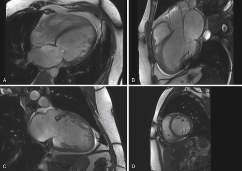
FIGURE 41-1 Examination obtained February 2011. A, Four-chamber view. B, Three-chamber view. C, Two-chamber view. D, Short-axis view.

image

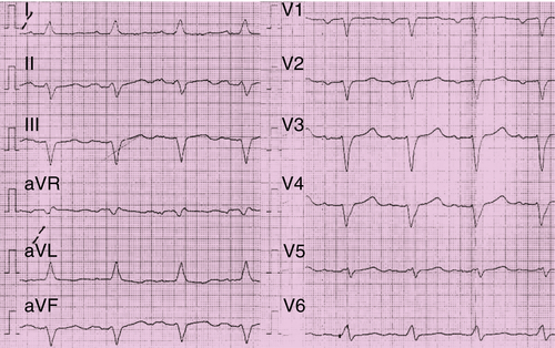
FIGURE 41-2 Electrocardiogram before implantation of the biventricular implantable cardioverter-defibrillator in February 2011.

In August 2011 the patient’s fatigue and dyspnea were worsening. Interrogation of the implantable cardioverter-defibrillator (ICD) revealed abnormalities with the atrial lead. Impedance values and pacing and sensing thresholds with the right and left ventricular leads were stable. Chest radiography revealed Twiddler’s syndrome (Figure 41-3), with the atrial electrode drawn back into the left subclavian vein. The patient admitted that he had touched and rotated the device frequently. During the operative revision the device was observed to have been rotated around its axis 18 times. The device was repositioned, and the right atrial electrode was revised successfully.
In February 2012 the patient was hospitalized again because of cardiac decompensation and dyspnea at rest. He had ankle edema and pleural effusions. Device interrogation did not reveal significant ventricular or supraventricular tachyarrhythmias. The patient again required intravenous diuretic therapy.
Although cardiac compensation was achieved, the patient again developed dyspnea after walking only a few meters. His quality of life was measured with a 21-item scale according to the Minnesota Living with Heart Failure Questionnaire, with a score of 79. The N-terminal probrain natriuretic peptide value was elevated to 12.067 ng/L. An exercise test showed highly decreased VO2 peak value of 10.7 mL/kg/min with maximum exercise capacity of 40 watts.
image

FIGURE 41-3 During the operative revision the device was observed to have been rotated around its axis 18 times.

Current Medications

The patient was taking atorvastatin 40 mg daily, carvedilol 37.5 mg daily, torsemide 40 mg daily, spironolactone 12.5 mg daily, enalapril 7.5 mg daily, opipramol 50 mg daily, pantoprazole 20 mg daily, mixed insulin 30 units daily, and aspirin 100 mg daily.

Current Symptoms

The patient was experiencing dyspnea on minimal effort and fatigue.

Physical Examination
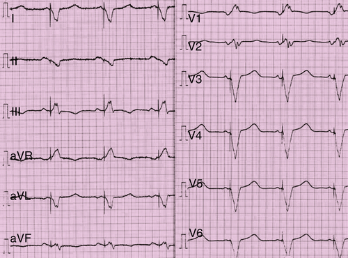
Laboratory Data

Electrocardiogram

Findings

The ECG showed normal rate and normal sinus rhythm, LBBB with a QRS width of 128 ms, and QRS onset to peak R time of 60 ms in V5 and V6 (see Figure 41-2).

Findings

The ECG showed active biventricular stimulation (Figure 41-4).

Findings

The last three QRS complexes are followed by active high-amplitude Optimizer III stimulation (Figure 41-5).

Chest Radiograph

Findings

image

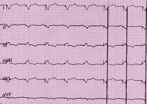
FIGURE 41-5  Electrocardiogram with active stimulation of the Optimizer III system.

image

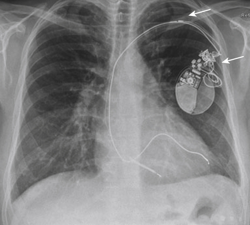
FIGURE 41-6 Chest radiograph from August 2011, 6 months after implantation of the biventricular implantable cardioverter-defibrillator.

Findings

A chest radiograph obtained a few hours after implantation of the Optimizer III device (Figure 41-7, A) shows the enlarged heart. Follow-up chest radiography shows considerable reduction of cardiac dimensions (see Figure 41-7, B).
The following were visualized on chest radiography: the generator of the biventricular ICD (1), Optimizer III device (2), atrial electrode of the CRT-D (3), ventricular electrode of the CRT-D located in the apex of the right ventricle (4), coronary sinus electrode located in a posterolateral vein (5), atrial sensing electrode of the Optimizer III system (6), upper ventricular stimulation electrode of the Optimizer III (7), and lower ventricular stimulation electrode of the Optimizer III (8).

Exercise Testing

Exercise testing in February 2012, before implantation of the Optimizer III, showed a VO2 peak of 10.7 mL/kg/min and exercise capacity of 40 W. Exercise testing had to be stopped because of dyspnea and pain in both lower legs.

Echocardiogram

Findings

Echocardiography in February 2012 (Figure 41-8) showed severely decreased ventricular ejection fraction (10%-14%). Figure 41-8, A, is a parasternal longitudinal axis view with severe dilation of the left ventricle and left atrium. Figure 41-8, B, is a parasternal longitudinal axis view with M-mode Doppler in which septal and posterior wall akinesia are observed.

Magnetic Resonance Imaging

Findings

Cardiac magnetic resonance imaging revealed a severely reduced LVEF (11%), left ventricular end-diastolic volume of 375 mL (184 mL/m2), left ventricular end-systolic volume of 335 mL (164 mL/m2), ejection volume of 41 mL, septal wall thickness of 7 mm, posterior wall thickness of 7 mm, and left ventricular end-diastolic dimension of 81 mm (see Figure 41-1). The contrast agent revealed transmural late enhancement septal midventricularly to apically.

Catheterization

Catheterization was performed in February 2011. Cardiac output was 4.4 L/min; cardiac index was 2.1 L/min/m2; mean right atrial pressure was 14 mm Hg; right ventricular pressure was 61 mm Hg systolic and 21 mm Hg end diastolic; pulmonary artery pressure was 59 mm Hg systolic, 34 mm Hg diastolic, and 44 mm Hg mean; and pulmonary capillary wedge pressure was 30 mm Hg.
image

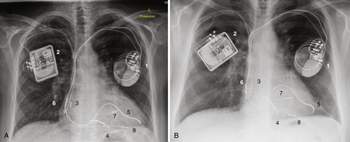
FIGURE 41-7 A, Chest radiograph obtained March 2012 after implantation of the Optimizer III device. B, Follow-up chest radiograph from July 2012, 4.5 months after implantation of the Optimizer III device. The visualized parts of the two devices are marked with numbers 1-8 to make the identification clearer.

image

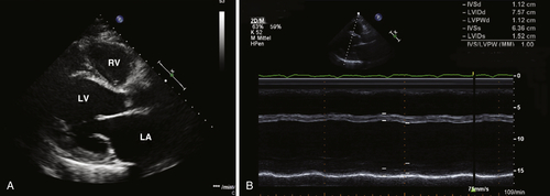
FIGURE 41-8 Echocardiogram.

Findings

The findings on catheterization were two-vessel coronary artery disease with a peripheral occlusion of the left anterior descending artery (100%), outlet stenosis of the first diagonal branch (80%) and small right coronary artery (50%), and left ventriculogram with an LVEF of 10%.

Focused Clinical Questions and Discussion Points

Question

What are indications for CRT? Which patients receive the best clinical results?

Discussion

Cardiac resynchronization therapy has become a standard therapy in patients with heart failure and interventricular and intraventricular conduction disturbances.5
CRT with biventricular pacing is an effective adjunctive therapy to pharmacologic management in reducing the rate of hospitalization and death in symptomatic patients with advanced heart failure symptoms (NYHA class III or IV), an ejection fraction of 35% or less, and an intraventricular conduction delay of 120 msec or more.3,5 Newer data also show favorable outcomes for patients with less advanced heart failure status, for example, patients with NYHA Class II symptoms.7
Unfortunately, approximately 40% of the patients are considered nonresponders to CRT. Analyses of prespecified subgroups of patients have demonstrated the best clinical results in patients with a QRS duration of 150 msec or more.7 Patients with a narrower QRS width are more likely to be nonresponders.
The patient’s clinical situation did not improve after CRT implantation. In February 2012, he deteriorated with persistent NYHA class IV symptoms despite successful (95%-97% capture) biventricular pacing. Therefore he was clearly a nonresponder to CRT.

Question

What therapeutic options are available for nonresponders to CRT?

Discussion

The current European Society of Cardiology guidelines for the treatment of patients with persistent severe heart failure symptoms indicate consideration for heart transplantation (if eligible) and left ventricular assist device (LVAD) implantation. The guidelines also suggest that digoxin therapy be considered. However, wait times for heart transplantation are increasingly long and LVAD therapy is not a therapy desired or recommended for all patients. Beyond this, the only other approved (in the European Union) and available therapy for heart failure is cardiac contractility modulation (CCM).

Question

What clinical data exist on the efficacy of treatment with CCM?

Discussion

To date, two multicenter, randomized controlled clinical trials have been conducted.2,6

FIX-CHF-4

The FIX-CHF-4 double-blind, double-crossover study was conducted in Europe and included 164 patients with heart failure with ejection fractions of 35% or less and NYHA class II or III.2 Co-primary end points in the trial were changes in peak oxygen consumption and changes in quality of life assessed by the Minnesota Living with Heart Failure Questionnaire. Secondary efficacy end points consisted of NYHA class and 6-Minute Walk Test. Each of the co-primary end points was significantly improved during the phase with active CCM. The treatment also was shown to be safe.

FIX-CHF-5

The FIX-CHF-5 prospective, randomized, parallel-group, controlled trial was conducted in the United States.6 It tested the longer term (1 year) safety and efficacy of CCM treatment. A total of 428 patients with NYHA class III to IV, ejection fraction of 35% or less, and narrow QRS were randomized to either CCM or no CCM therapy. This study also showed CCM therapy to be safe in this patient population. In the overall population, CCM therapy significantly improved peak VO2, Minnesota Living with Heart Failure Questionnaire results, and NYHA class, but did not improve the ventilatory anaerobic threshold, which was the declared the primary end point. However, in a prespecified subgroup analysis consisting of approximately 50% of the overall population characterized by baseline ejection fraction of 25% or greater and NYHA class III, the primary end point was reached.1

Consideration of Both Studies

All patients included in the FIX-CHF-4 and FIX-CHF-5 studies had narrow QRS complexes and were therefore ineligible for CRT. Nevertheless, the patient cohort in the FIX-CHF-4 study had NYHA classes II and III and included a small cohort with QRS duration greater than 130 ms. Our patient was in NYHA class IV, had a wide QRS duration, and already had a CRT-D device in place. Therefore the data of these two studies cannot be used for direct comparison with the situation of the patient in this case.

Question

Are there clinical data on the outcome of CRT nonresponders treated with CCM therapy?

Discussion

To date, little is known about the outcome of CRT nonresponders treated with additional CCM therapy. No randomized clinical data have been published about this very ill group of patients. Case reports and a case series have described first experiences with the technique.4,8 Näegele and colleagues reported a case series of 16 patients with NYHA class III and IV symptoms in which they showed the feasibility of the method as a useful adjunct in CRT nonresponders when no other options are available. No electrical interference was observed between the CCM therapy and CRT systems, and, in particular, at no time was the CRT-D device found to be delivering inadequate or inappropriate shocks. However, the mortality rate and number of clinical events remained high.
Long-term clinical outcome studies need to be performed to clarify the impact of CCM therapy in nonresponders to CRT. To date, the FIX-CHF-12 study is under way to evaluate clinical follow-up of patients with combined CRT and CCM therapy.

Final Diagnosis

The final diagnosis in this case was that the patient was a nonresponder to CRT.

Plan of Action

The plan for this patient was implantation of an additional Opimizer III device.

Intervention

Implantation of the Optimizer III device was performed in March 2012. Ventricular tachycardia detection algorithms of the CRT-D device were turned off during CCM implantation to prevent inappropriate shocks. A submuscular pocket was made in the right subclavian region, and three standard screw-in pacemaker electrodes were introduced into the subclavian vein after venipuncture. One electrode was positioned in the right atrium and used for sensing atrial activity (Tendril ST, 58 cm, St. Jude Medical, St. Paul, Minn.). The other two electrodes (Tendril ST 1888, 65 cm, St. Jude Medical) were positioned on the right ventricular septum. Figure 41-7, A, shows the postoperative chest radiograph with electrode positions, and Figure 41-5 shows the ECG with combined CRT and CCM therapy.
During implantation, left ventricular dP/dtmax testing was measured using a 5-French Millar micromanometer catheter placed in the left ventricle. Initially, changes in dP/dtmax from baseline with different VV delays (40 to 40 ms) programmed in the CRT-D device were tested. Changes of VV delay did not induce significant changes from baseline in contrast to a left ventricular VV delay of 0 ms. Subsequently, exclusive left ventricular stimulation was tested (stimulation vector left ventricular ring to right ventricular coil), which showed a mild increase of dP/dtmax of 6%. After activation of the Optimizer III device, an increase of dP/dtmax from baseline of 18% was achieved.
No cross talk between the devices occurred up to a train delay of 80 ms. The Optimizer III device was programmed to provide therapy for 7 hours per day. Both septal stimulation electrodes were programmed “on.” The CCM signal amplitude was programmed to 5.5 V. At the last follow-up examination, device interrogation showed a CCM signal delivery rate of 98%.

Outcome

The most recent clinical follow-up examination occurred 4 months after Optimizer III implantation. The patient reported significant improvement of physical ability. He had no dyspnea during activities of daily living, and his formerly constant fatigue has been eliminated. He reported dyspnea only on strong exertion, corresponding to an improvement of overall symptoms from NYHA III to IV to NYHA II. His mental status also was overtly improved. The repeat Minnesota Living with Heart Failure Questionnaire score had decreased to 29 points, a dramatic 50-point reduction. Echocardiography showed increased LVEF to 25% and a reduction in left ventricular end-diastolic and end-systolic dimensions (to 69 and 58 mm, respectively, measured at the level of the mitral leaflet tips in the parasternal long axis).
The patient’s N-terminal probrain natriuretic peptide (NT-proBNP) decreased to 3028 ng/L in May 2012. Follow-up exercise testing was also performed at that time, demonstrating an increase in peak VO2 to 13 mL/kg/min at 70 W; the patient was without dyspnea, and the test had to be ended early because of orthopedic problems. Figure 41-7, B, shows the follow-up chest radiograph with obvious reduction of the cardiac dimensions.
In the context of a case report, concern always exists that improvements are related to placebo effect. In this case the dramatic clinical improvements observed from CCM therapy were observed after the lack of any clinical effects achieved by CRT implant; had the patient been susceptible to placebo, expectations would have been that these would be observed after the CRT implant. Furthermore, the multiple objective findings (i.e., decreased NT-proBNP, reduced left ventricular dimensions, and improved LVEF) in the face of constant medical therapy also speak against a placebo effect. Although further systematic randomized study needs to be done, the current findings point to the promise of CCM to help at least some patients who are nonresponders to CRT.

Selected References

1. Abraham W.T., Nadamanee K., Volosin K. et al. Subgroup analysis of a randomized controlled trial evaluation the safety and efficacy of cardiac contractility modulation in advanced heart failure. J Card Fail. 2011;17:710–717.

2. Borggrefe M.M., Lawo T., Butter C. et al. Randomized, double blind study of non-excitatory, cardiac contractility modulation electrical impulses for symptomatic heart failure. Eur Heart J. 2008;29:1019–1028.

3. Bristow M.R., Saxon L.A., Boehmer J. et al. Cardiac-resynchronization therapy with or without an implantable defibrillator in advanced chronic heart failure. N Engl J Med. 2004;350:2140–2150.

4. Butter C., Meyhofer J., Seifert M. et al. First use of cardiac contractility modulation (CCM) in a patient failing CRT therapy: clinical and technical aspects of combined therapies. Eur J Heart Fail. 2007;9:955–958.

5. Cleland J.G., Daubert J.C., Erdmann E. et al. The effect of cardiac resynchronization on morbidity and mortality in heart failure. N Engl J Med. 2005;352:1539–1549.

6. Kadish A., Nademanee K., Volosin K. et al. A randomized controlled trial evaluating the safety and efficacy of cardiac contractility modulation in advanced heart failure. Am Heart J. 2011;161:329–337.

7. Moss A.J., Hall W.J., Cannom D.S. et al. Cardiac resynchronization therapy for the prevention of heart failure events. N Engl J Med. 2009;14:1329–1338.

8. Nägele H., Behrens S., Eisermann C. et al. Cardiac contractility modulation in non-responders to cardiac resynchronization therapy. Europace. 2008;10:1375–1380.

Share this: