34. Extraction of a Biventricular Defibrillator with a Starfix 4195 Coronary Venous Lead

Published on 02/03/2015 by admin

Filed under Cardiovascular

Last modified 22/04/2025

Print this page

rate 1 star rate 2 star rate 3 star rate 4 star rate 5 star
Your rating: none, Average: 0 (0 votes)

This article have been viewed 1627 times

History

A 58-year-old man with ischemic cardiomyopathy with an ejection fraction of 20%, a history of coronary artery bypass graft surgery in 1993, and placement of a biventricular defibrillator (Medtronic Concerto; Guidant 0185 RV ICD lead; Attain Starfix 4195 coronary venous lead) on October 25, 2007 for primary prevention, a history of a left ventricular thrombus, and persistent atrial fibrillation sought treatment at a hospital with fevers, chills, and worsening dyspnea on exertion occurring over the previous 2 months. On admission, blood cultures were drawn that were positive for methicillin-resistant Staphylococus aureus (MRSA). A transesophageal echocardiogram suggested a vegetation on the implantable cardioverter-defibrillator (ICD) lead, prompting transfer to a tertiary referral center for complete system extraction.

Comments

Cardiac implantable electronic device (CIED) infection is typically defined as the presence of local warmth, erythema, swelling, edema, pain, or discharge from the device pocket, along with a positive culture from the device, device pocket, blood, or lead. Device-associated endocarditis is defined as the presence of a lead or valvular vegetation on echocardiogram. Prompt removal of the CIED and all leads and administration of a prolonged course of antibiotics is the appropriate management for CIED-related infections. Lead extraction procedures must be performed only at facilities with cardiac surgery programs with experienced cardiac surgeons on site and ready to initiate emergency cardiac surgery. The facility to which this patient presented lacked such capability, making the transfer to a tertiary referral center with significant experience in lead extraction appropriate.

Current Medications

The patient was receiving vancomycin 1 g intravenously twice daily and heparin according to a drip rate nomogram; he was also taking carvedilol 6.25 mg orally twice daily, lisinopril 2.5 mg orally daily, omeprazole 20 mg orally daily, atorvastatin 20 mg orally daily at bedtime, and aspirin 81 mg orally daily.

Comments

Vancomycin is a first-line therapy for MRSA-related bloodstream infections. Staphylococcal species are responsible for 60% to 80% of CIED infections. The optimal duration of antimicrobial therapy for CIED infection is unclear. At least 2 weeks of intravenous antimicrobial therapy is recommended after extraction of an infected device for patients with infected CIED systems, but with positive blood cultures or vegetations the recommendation is usually 4 to 6 weeks. The patient’s heparin drip was stopped the night before the planned extraction.

Current Symptoms

The patient was experiencing fatigue, shortness of breath, and nausea.
image

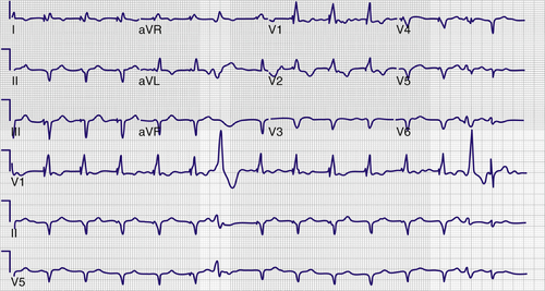
FIGURE 34-1 Preextraction electrocardiogram.

Comments

In patients with a CIED infection, fevers and chills are often absent.

Physical Examination

Laboratory Data

Comments

The patient’s white blood cell count was within normal limits. Many patients with CIED infection fail to present with significant leukocytosis.

Electrocardiogram

Findings

The preextraction electrocardiogram shows atrial fibrillation with a biventricular paced rhythm and occasional premature ventricular contractions (Figure 34-1).

Chest Radiograph

Findings

Portable chest radiography demonstrated the presence of a Medtronic 4195 Starfix coronary sinus lead in a midmyocardial position (Figure 34-2).

Echocardiogram

Findings

image

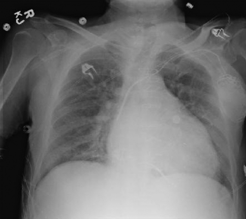
FIGURE 34-2 Portable chest radiograph showing the Starfix coronary venous lead in the midventricular position.

image

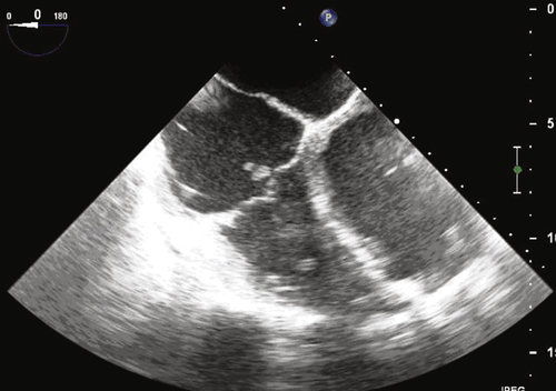
FIGURE 34-3 Transesophageal echocardiogram depicting a vegetation on the implantable cardioverter-defibrillator lead as it crosses the tricuspid valve.

Comments

The size of the vegetation is important in extraction planning. Some operators will opt for a surgical procedure in patients with large vegetations (>2-3 cm) to limit the risk for septic pulmonary emboli. At our institution, vegetations larger than 3 cm have been extracted. In making the decision on whether to extract leads with large vegetations percutaneously, the comorbidities of the patient need to be thoroughly assessed, the risks of both procedures carefully balanced, and timing and route for reimplantation anticipated in this plan. The 1.8-cm lesion in this case is within reason to undergo percutaneous extraction.

Focused Clinical Questions and Discussion Points

Question

How are CIED infections diagnosed and treated?

Discussion

Patients with CIED infections often present with localized inflammatory changes at the device site, occasionally accompanied by cutaneous erosion. Such a presentation, however, is not uniform, as demonstrated by the current case. Some patients simply initially seek treatment with vague systemic symptoms such as fatigue, anorexia, or decreased functional capacity. On presentation, two sets of blood cultures should be obtained before the initiation of antibiotics. Bacteremia caused by staphylococcal species, in particular, increases the likelihood of CIED involvement. Often, the original imaging study to search for lead or valve infection is a transthoracic echocardiogram. Because of low sensitivity and specificity for picking up vegetations, however, a negative transthoracic echocardiogram cannot rule out the presence of vegetations. A transesophageal echocardiogram is significantly more sensitive than a transthoracic echocardiogram to find lead and valvular vegetations. Even in patients in whom a transthoracic echocardiogram has demonstrated a lead-adherent mass, a transesophageal echocardiogram is still indicated to rule out left-sided valvular involvement. Once a diagnosis of a CIED infection has been made, all components of the CIED system must be removed in a timely fashion followed by a prolonged course of antibiotics.1,5

Question

What are the challenges involved with removal of a traditional non–active fixation coronary venous pacing lead?

Discussion

Because of the thin walls of the coronary venous system, coronary venous lead extraction would seem to pose substantial risk for percutaneous extraction.2 In studies in small animals a high incidence of hemopericardium after coronary venous lead extraction has been noted. In humans, however, an increased complication rate with coronary venous lead extraction has not been borne out. Bongiorni and colleagues3 reported on a cohort of 37 coronary venous lead extractions. The age of the coronary venous leads in this study was 19.5 ± 16.5 months. All leads were successfully extracted, with 27 extracted with simple traction alone and the remaining 10 with mechanical dilation. The areas of adhesion for the coronary venous leads were most commonly found in the subclavian vein (60%). In a separate cohort of 173 patients undergoing coronary venous lead extraction from our institution, a total of 76.9% of coronary venous leads were removed using simple traction alone, with the remaining leads requiring the use of a laser-powered sheath. A total of 3.5% of leads required intervention (manual dissection or laser-powered dissection) within the coronary sinus. Major complications were rare, occurring in 1.2% of patients, and minor complications occurred in 7.5% of patients. In a separate cohort of 114 leads from the Mayo Clinic, 91.2% were removed with simple traction alone.9 Minor complications were reported in 7.2% of patients and major complications in 1.6% of cases. Given the thin-walled nature of the coronary sinus, advancement of dissection sheaths into the coronary sinus itself in difficult cases is discouraged and should be reserved for experienced operators in select patients. Overall, extraction of traditional non–active-fixation coronary venous leads has been shown to be a safe, effective procedure with a low incidence of complications.4,6-9

Question

What are the technical challenges involved with removal of the Medtronic Attain Starfix 4195 active fixation coronary venous pacing lead?

Discussion

The Medtronic Attain Starfix 4195 coronary venous lead was approved by the United States Food and Drug Administration in June 2008 as the first active fixation coronary venous lead. The active fixation mechanism consists of three polyurethane lobes that are deployed by advancing tubing around the lead. When deployed, the lobes range from 5 to 24 French in diameter. Acutely, the locking mechanism can be deployed and undeployed. Over time, however, fibrotic tissue ingrowth between the lobes may preclude undeployment. Extraction of Starfix leads can be very challenging. Unlike the case with conventional coronary venous lead extraction, advancement of an extraction sheath into the coronary sinus is often needed with Starfix leads and, in approximately 40% of cases, a sheath must be advanced distally into the coronary sinus tributary itself. Given the high incidence of fibrotic ingrowth and the resultant inability to undeploy the lobes, Starfix lead extraction should be reserved for highly experienced operators at high-volume centers.

Question

Discussion

Successful reimplantation of a coronary venous lead after extraction of a biventricular device is achievable in 80% to 85% of cases, a rate slightly lower than for de novo implants. This slightly lower success rate is likely due to the increased procedural difficulty involved with right-sided procedures (which most reimplants are). In addition, most operators have significantly less experience with right-sided implants. Finally, occlusion of the original coronary venous tributary may be present in up to 50% of cases. Therefore venous tributaries that are difficult to access are occasionally the only available target.

Final Diagnosis

The final diagnosis for this patient was CIED-related endocarditis.

Plan of Action

The plan for this patient was full system extraction.10

Intervention

Under general anesthesia, right radial arterial and right femoral venous sheaths were inserted for hemodynamic monitoring and vascular access, respectively. A left infraclavicular incision yielded access for removal of the CIED and leads. The pocket was grossly infected, with purulent material noted. The entire fibrotic capsule and all infected-appearing tissue were debrided. Deep tissue cultures of the pocket were sent for culture. The ICD lead was prepared with an LLD-EZ locking stylet (Spectranetics, Colorado Springs, Colo.). Although the helical screw did not retract, torque was able to be transmitted to the lead tip. The ICD lead was manually unscrewed from the myocardium, and the lead was removed with simple traction. The coronary venous lead was then prepared with an LLD-EZ locking stylet. An attempt was made to undeploy the lobes of the lead locking mechanism. After prolonged efforts, the lobes would not undeploy. Fortunately, after application of gentle traction, the lead was withdrawn into the the brachiocephalic vein, where it became lodged. A 14-French laser sheath was then advanced over the lead, and the lead was successfully extracted from the body. On inspection of the lead tip, significant fibrotic growth between the lobes of the lead was noted, with a cast of the cardiac vein trailing from the fibrosis (Figure 34-4). The pocket was irrigated and closed. The patient was treated with a 5-week course of vancomycin.
image

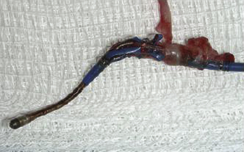
FIGURE 34-4 Tissue ingrowth into the lobes of the Starfix coronary venous lead preventing undeployment.

Outcome

After extraction and 10 days of antibiotics with multiple negative blood cultures, the patient was cleared for reimplantation on the right side. A Medtronic 4193 lead was placed in a lateral branch. The patient was discharged to home on 4 weeks of antibiotics.

Findings

A venogram was performed at the time of reimplantation demonstrating a lack of opacification of the high lateral vein from which the Starfix lead was extracted, suggesting complete occlusion of this vein (Figure 34-5).

Comments

Given the significant tissue ingrowth into the locking mechanism, extraction of the Starfix lead is traumatic to the coronary venous system. Although data are lacking, occlusion of the original vessel at time of reimplantation is likely to be significantly higher than that for traditional non–active-fixation coronary venous leads.
image

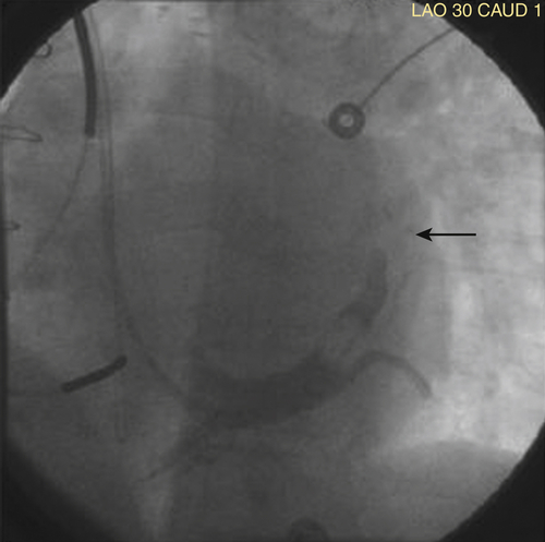
FIGURE 34-5 Occlusion of venous branch from which the Starfix coronary venous lead was extracted.

Selected References

1. Baddour L.M., Epstein A.E., Erickson C.C. et al. Update on cardiovascular implantable electronic device infections and their management: a scientific statement from the American Heart Association. Circulation. 2010;3:458–477.

2. Baranowski B., Yerkey M., Dresing T. et al. Fibrotic tissue growth into the extendable lobes of an active fixation coronary sinus lead can complicate extraction. Pacing Clin Electrophysiol. 2011;7:e64–e65.

3. Bongiorni M.G., Zucchelli G., Soldati E. et al. Usefulness of mechanical transvenous dilation and location of areas of adherence in patients undergoing coronary sinus lead extraction. Europace. 2007;1:69–73.

4. Burke M.C., Morton J., Lin A.C. et al. Implications and outcome of permanent coronary sinus lead extraction and reimplantation. J Cardiovasc Electrophysiol. 2005;8:830–837.

5. Chua J.D., Wilkoff B.L., Lee I. et al. Dagnosis and management of infections involving implantable electrophysiologic cardiac devices. Ann Intern Med. 2000;8:604–648.

6. Maytin M., Carrillo R.G., Baltodano P. et al. Multicenter experience with transvenous lead extraction of active fixation coronary sinus leads. Pacing Clin Electrophysiol. 2012;6:641–647.

7. Rickard J., Tarakji K., Cronin E. et al. Cardiac venous left ventricular lead removal and reimplantation following device infection: a large single-center experience. J Cardiovasc Electrophysiol. 2012;23:1213–1216.

8. Rickard J., Wilkoff B.L. Extraction of implantable cardiac electronic devices. Curr Cardiol Rep. 2011;13:407–414.

9. Sheldon S., Friedman P.A., Hayes D.L. et al. Outcomes and predictors of difficulty with coronary sinus lead removal. J Interv Card Electrophysiol. 2012;35:93–100.

10. Wilkoff B.L., Love C.J., Byrd C.L. et al. Transvenous lead extraction: Heart Rhythm Society expert consensus on facilities, training, indications, and patient management. Heart Rhythm. 2009;7:1085–1104.

Share this: