33. A Difficult Case of Diaphragmatic Stimulation

Published on 26/02/2015 by admin

Filed under Cardiovascular

Last modified 22/04/2025

Print this page

rate 1 star rate 2 star rate 3 star rate 4 star rate 5 star
Your rating: none, Average: 0 (0 votes)

This article have been viewed 1535 times

History

A 70-year-old woman with nonischemic cardiomyopathy, with New York Heart Association (NYHA) class III symptoms, a left ventricular ejection fraction (LVEF) of 22%, left ventricular end-diastolic dimension of 72 mm, QRS duration of 170 ms, and a left bundle branch block pattern (Figure 33-1) was referred for possible cardiac resynchronization therapy defibrillator (CRT-D) implantation.
Comorbidities included hypertension, type 2 diabetes, dyslipidemia, obesity, and paroxysmal atrial fibrillation well controlled with amiodarone. Workup revealed moderate diffuse coronary artery disease (<40% narrowing), a 6-Minute Walk Test distance of 342 m, and a peak VO2 of 13.2 mL/kg/min.

Current Medications

Medical therapy was optimized with carvedilol 6.25 mg twice daily, fosinopril 20 mg daily, furosemide 60 mg daily, spironolactone 25 mg daily, amiodarone 200 mg daily, acetylsalicylic acid 80 mg daily, and dose-adjusted warfarin for a target INR 2.0–3.0.

First Intervention

Implantation of the CRT-D device was uneventful. However, the angiogram was of poor quality because of obesity and thus provided little guidance. It was not repeated given the patient’s underlying renal dysfunction (i.e., glomerular filtration rate ≤40 mL/min). Three coronary sinus branches were blindly identified. The anterior and posterior branches had septal courses. The bipolar left ventricular lead (1056K, St. Jude Medical, St. Paul, Minn.) was positioned in a long, large-caliber midlateral branch after confirming the absence of phrenic nerve capture despite high-output (10 V) pacing. Left ventricular pacing thresholds were 3.4 V for the distal electrode and 2.2 V for the proximal ring, both with the right ventricular coil as anode.

Outcome

The following morning, the patient reported symptoms consistent with diaphragmatic stimulation. On interrogation, the best left ventricular capture threshold was 3.75 V (proximal ring to right ventricular coil) and both pacing electrodes elicited phrenic nerve capture at voltages down to 3.25 V. It was impossible to identify a pacing configuration with a left ventricular threshold lower than that of the phrenic nerve capture threshold. The lead appeared to be well positioned on chest radiograph.

Second Intervention

A second intervention was therefore performed to rectify the situation. Other potential coronary sinus branches could not be identified. The anterior branch was of small caliber and too far septal and the proximal posterior branch (middle cardiac vein) was deemed unsuitable. A new 1056K left ventricular lead was implanted in the same venous branch. Phrenic nerve capture occurred from both electrodes, but only on deep inspiration. The problem was far less pronounced in a very proximal position. The lead was not wedged into place in the hope that its spiral design would ensure stability.

Outcome

Five years later, she remained with NYHA class III symptoms and had four heart failure–related hospitalizations within 8 months. Her left ventricular systolic function remained stable (LVEF 20%; left ventricular end-diastolic dimension 70 mm). Meanwhile, she developed new-onset complete atrioventricular block and became dependent on right ventricular pacing.
Two months previously, she presented with recurrent presyncope. Intermittent ventricular oversensing (Figure 33-2) resulting from electrical failure of a Riata lead (St. Jude Medical) was documented. The left ventricular lead remained nonfunctional. In addition, a high atrial lead threshold was noted, with a P wave of approximately 1.0 mV.

Third Intervention

A backup temporary pacemaker was immediately implanted. Lead extraction was subsequently performed, with all three leads removed by simple traction. Three new leads were implanted, including a quadripolar deeply inserted left ventricular lead (Quartet, St. Jude Medical) in the same lateral branch of the coronary sinus (Figure 33-3). Phrenic nerve capture was present (down to 3.5 V) with the three proximal electrodes but not the distal electrode.
image

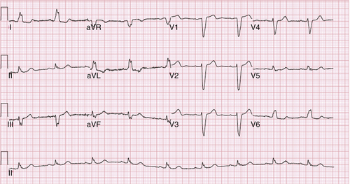
FIGURE 33-1 Patient with left bundle branch block pattern in electrocardiogram.

image

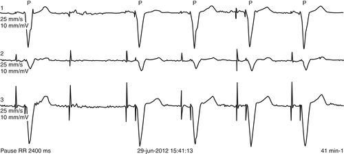
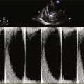
FIGURE 33-2 Patient with intermittent ventricular oversensing resulting from electrical failure of a Riata lead (St. Jude Medical).

Outcome

Despite a lack of improvement in LVEF, the patient has not been hospitalized for heart failure during 12 months of follow-up. Effective biventricular pacing is shown in Figure 33-4. On interrogation, a left ventricular pacing threshold of 2.25 V was obtained from the distal electrode in a unipolar configuration (D1 to right ventricular coil). No phrenic nerve stimulation occurred with up to 7.5 V. For the three other electrodes, the left ventricular pacing threshold was either too high (>4.0 V for the P4 ring) or higher than the phrenic nerve capture threshold (M2 and M3).
image

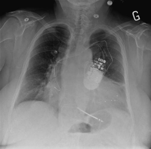
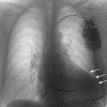
FIGURE 33-3 A posteroanterior chest radiograph is shown depicting the position of the three implanted leads, including a quadripolar left ventricular lead (Quartet, St. Jude Medical) deeply inserted in the lateral branch of the coronary sinus.

Focused Discussion Points

Diaphragmatic contraction resulting from phrenic nerve capture is observed in up to 20% to 30% of CRT implants.13 Unfortunately, it is not always apparent at the time of implantation and may develop in the hours or days that follow. In most instances, the problem may be overcome by reprogramming pacing configurations, pacing output, or both. In a minority of patients, the only options are to reintervene or inactivate left ventricular pacing.4,6
Several options are available for reintervention. The new quadripolar left ventricular leads present an attractive solution, offering greater flexibility in programming options.5,10,11 As this case demonstrates, quadripolar leads also may be considered when standard bipolar left ventricular leads fail to deliver effective CRT.9 Additional options include implanting a bipolar active fixation lead more proximally within the coronary sinus (e.g., right ventricular lead or Attain Starfix left ventricular lead [Medtronic, Minneapolis, Minn.]). However, such an option may complicate future extraction interventions.7 Finally, alternative approaches to implanting a left ventricular lead may be considered, either endocardially by a transseptal approach or epicardially by surgical access. In weighing potential options, results from the REPLACE study should be considered, which indicate that the highest risk for device infection occurs in the context of CRT system revision.8 In this case, a second procedure was initially attempted but failed. The system was entirely revised 6 years later, when reintervention was required for right ventricular lead dysfunction.
image

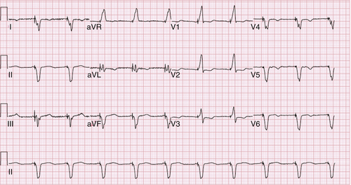
FIGURE 33-4 Patient with effective biventricular pacing shown in electrocardiogram.

Selected References

1. Biffi M., Boriani G. Phrenic stimulation management in CRT patients: are we there yet? Curr Opin Cardiol. 2011;26:12–16.

2. Biffi M., Exner D.V., Crossley G.H. et al. Occurrence of phrenic nerve stimulation in cardiac resynchronization therapy patients: the role of left ventricular lead type and placement site. Europace. 2013;15:77–82.

3. Biffi M., Moschini C., Bertini M. et al. Phrenic stimulation: a challenge for cardiac resynchronization therapy. Circ Arrhythm Electrophysiol. 2009;2:402–410.

4. Champagne J., Healey J.S., Krahn A.D. et al. The effect of electronic repositioning on left ventricular pacing and phrenic nerve stimulation. Europace. 2011;13:409–415.

5. Forleo G.B., Mantica M., Di Biase L. et al. Clinical and procedural outcome of patients implanted with a quadripolar left ventricular lead: early results of a prospective multicenter study. Heart Rhythm. 2012;11:1822–1828.

6. Klein N., Klein M., Weglage H. et al. Clinical efficacy of left ventricular pacing vector programmability in cardiac resynchronization therapy defibrillator patients for management of phrenic nerve stimulation and/or elevated left ventricular pacing thresholds: insights from the efface phrenic stim study. Europace. 2012;14:826–832.

7. Maytin M., Carrillo R.G., Baltodano P. et al. Multicenter experience with transvenous lead extraction of active fixation coronary sinus leads. Pacing Clin Electrophysiol. 2012;35:641–647.

8. Poole J.E., Gleva M.J., Mela T. et al. Complication rates associated with pacemaker or implantable cardioverter-defibrillator generator replacements and upgrade procedures: results from the replace registry. Circulation. 2010;122:1553–1561.

9. Shetty A.K., Duckett S.G., Bostock J. et al. Use of a quadripolar left ventricular lead to achieve successful implantation in patients with previous failed attempts at cardiac resynchronization therapy. Europace. 2011;13:992–996.

10. Sperzel J., Danschel W., Gutleben K.J. et al. First prospective, multi-centre clinical experience with a novel left ventricular quadripolar lead. Europace. 2012;14:365–372.

11. Thibault B., Karst E., Ryu K. et al. Pacing electrode selection in a quadripolar left heart lead determines presence or absence of phrenic nerve stimulation. Europace. 2010;12:751–753.