

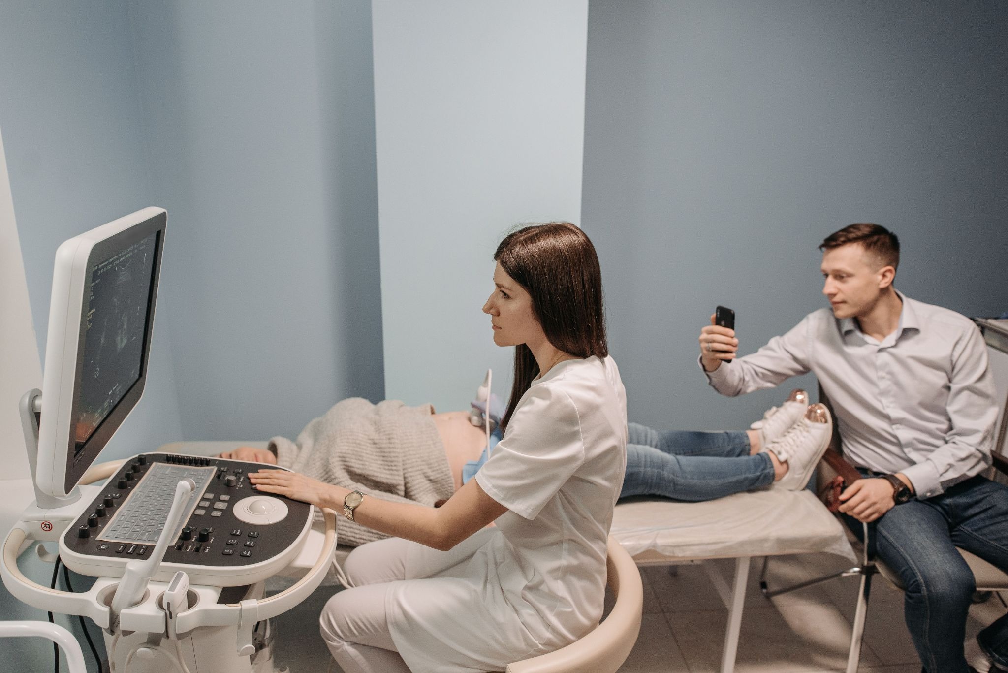
Minutes count when a patient crashes in the Emergency Department. No one can have a radiology slot to wait in. Point-of-Care Ultrasound, or POCUS, solves some of that: The physician carries a compact scanner in his pocket, pushes it into the skin, and peeks inside the body as it happens.
It is not a substitute for a complete radiology examination. It is more of a flashlight rather than a floodlight. It gives a concise answer quickly (has there been blood in the belly, is the heart pumping, is there a blocked vein in the leg?): and then the team acts.
Where It Earns Its Place
Trauma came first. The FAST exam involves identifying free fluid around the heart and abdomen and is one of the most basic skills which new emergency trainees learn. Today’s toolbox is much larger: heart and lung views, screening of the aorta, checking the gall bladder, early pregnancy scans, and even needle guiding for the central line.
A number of these tests only require minutes. This could alter the plan immediately, such as calling the surgeon to inform him or her of the change, initiation of blood thinners, or direct referral to the operating room.
Spotting Blood Clots in the Leg
A 2024 systematic review of studies published between 2011 and 2023 in the Western Journal of Emergency Medicine combined the results of research on emergency physicians doing scans for deep vein thrombosis. Pooled sensitivity was 90% and specificity, 95%. That’s a fast way to a decision, versus waiting for a formal scan.
Technique matters too. The three-point compression test proved more sensitive than the two-point configuration test, and there was a statistically significant difference only when the test was conducted by specialist emergency physicians.
Sorting Out Shortness of Breath
There are many reasons for being out of breath and the clock is ticking. In one study performed by novice POCUS users about acute dyspnea, asthma and COPD were detected with a 88.1% sensitivity and a 96.2% specificity. Acute heart failure was found to be 81.3% sensitive and had a high specificity of 99.1%. Pneumonia reached 80% sensitivity.
Pulmonary embolism had a different tale. Sensitivity fell to 60%. While that was from a small number of cases, it is not to be taken as gospel but it does point to the fact that a normal lung scan doesn’t exclude a clot.
Children, Appendicitis, and No Radiation
In a study conducted in the UK, 226 children between the ages of 1 and 16 had POCUS performed by pediatric emergency doctors. Sensitivity was 89%, specificity was 96% and negative predictive value was as high as 98%. There was an extremely high agreement with radiology performed ultrasound (kappa 0.87).
Kids are small, so images tend to come out clean, and nothing in the exam involves radiation. Parents like that. Doctors do too.
Broken Bones Without the X-Ray Room
For long bone fractures, a prospective study at two emergency departments reported 90.2% sensitivity and 96.1% specificity. That matters in places where an X-ray machine is far away, such as rural clinics or remote sports events.
The Limits Nobody Should Ignore
Not all questions have an unambiguous answer. A multicentre study of adult appendicitis in a general ED reported that the sensitivity of POCUS was 85% while the specificity was 63%. The authors concluded that it was too weak to be used as a definitive test in undifferentiated patients. Deep structures make images more difficult to read than bowel gas or obesity.
There’s the human element too. There is an operator dependency in ultrasound: Two different operators, two different hands on the probe, two different results. If a patient doesn’t receive a diagnosis, he or she may feel assured but later receive unnecessary tests if the diagnosis was miscalled. A good POCUS exam provides one focused question. It should never be made into a complete diagnosis.
How Many Scans Make a Competent Operator?
The American College of Emergency Physicians suggests 25-50 scans per core application and 150-300 per month. The advice for FAST alone varies from 10 exams to 50 exams, depending on the society’s opinion. A team of researchers, who administered a hands-on test known as ACEP range to 29 senior residents, found that they had a broad target area.
Now, a good question, does a number on a checklist equal skill? Not by itself. A person who has 300 bad scans can possibly be worse than a person who has 100 good scans.
Training Late-Career Doctors
Not all people had probes as a part of their childhood. One health system developed a residency-based path to incorporate POCUS. 60 scans were needed for basic privileges in three exam types. On average, doctors worked only 31. Twenty-eight (17.7%) of the FAST examinations had image quality issues.
Therefore, a short course program might not be sufficient. The initial workshop is likely to be the least impactful — mentoring and regular feedback are likely more important.
Learning Together, Even from Far Away
Ultrasound is a visual craft, and it grows faster in company. Residents in different hospitals now swap teaching clips, compare probe angles, and talk through tricky cases over video chat platforms like CallMeChat. Some set up study groups on chat platforms online, where a quick face-to-face chat can replace a long email thread about one confusing image. Others stick to hospital conferencing tools.
What counts is the habit of discussing real scans with someone who can push back. Patient identifiers stay out of it, of course.
Staying Sharp After Certification
Emergency ultrasound fellowships, following ACEP guidance, ask for at least 1,000 exams, plus quality review and teaching duties. Most working doctors will never go that far, yet the same logic applies:
- Keep scanning, regularly.
- Have someone review your saved images.
- Track your misses, not only your hits.
The Bottom Line
POCUS has become a companion to the stethoscope. The numbers are robust in the case of clots, fractures, free fluid, and numerous respiratory issues. They are not for adult appendicitis or pulmonary embolus. The decision of which side of that line a scan falls into is determined by the training. Proper teaching, protected practice time, and image review are the elements to be funded to receive real value from hospitals. You will get noise from those that give out probes and hope for the best.
