GENITOURINARY PATHOLOGY

Published on 07/03/2015 by admin

Filed under Pathology

Last modified 22/04/2025

Print this page

rate 1 star rate 2 star rate 3 star rate 4 star rate 5 star
Your rating: none, Average: 0 (0 votes)

This article have been viewed 1783 times

[level-membership-for-pathology-category]

CHAPTER 2 GENITOURINARY PATHOLOGY

BLADDER AND PROSTATE PATHOLOGY

INTRODUCTION

• Prostate / seminal vesicle pathology is rare in childhood other than involvement by rhabdomyosarcoma
• Bladder specimens predominantly comprise:

image biopsies for assessment of tumor

– usually rhabdomyosarcoma (specific issues detailed in the relevant tumor section)
image specimens with non-specific reactive changes following surgery for congenital bladder anomalies such as bladder extrophy repair
• Other bladder lesions biopsied are rare and include the following

Bladder paraganglioma

• Rare, but reported in children
• Paroxysmal episodes of hypertension, sweating, headache, hematuria
• Histopathological features of paraganglioma at other sites (see tumor section)

Bladder neurofibroma

• Children with NF
• See neurofibroma section

UROTHELIAL PAPILLOMA

Epidemiology

• Mainly adults but reported in childhood

Clinical features

• Hematuria

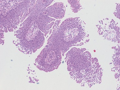
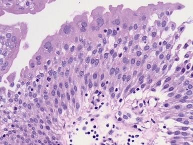
Histopathological features (Figs 2.1, 2.2)

• Papillary structures with no urothelial atypia
• Stromal edema
• Low proliferation rate and rare basal mitoses only
image

Fig. 2.1

image

Figs 2.1–2.2 Photomicrographs of a bladder transitional cell papilloma, demonstrating papillary architecture and no significance cytological atypia.

Immunohistochemical staining

• No p53 abnormalities

image cf papillary carcinomas

Differential diagnoses and pitfalls

• Inverted papilloma

image bland urothelium in nests / cords within lamina propria

TESTICULAR AND PARATESTICULAR PATHOLOGY

TESTICULAR GERM CELL TUMORS

Epidemiology

• Most germ cell tumors are seminoma in young adulthood
• In younger childhood, classical seminoma rare
• Childhood peak in infancy

image teratomas
image yolk sac tumors

Genetics

• Familial cases reported but usually tumors develop in adulthood
• Adult cases i12p associated, in contrast to childhood cases

Clinical features

• Painless testicular mass

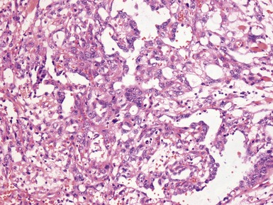
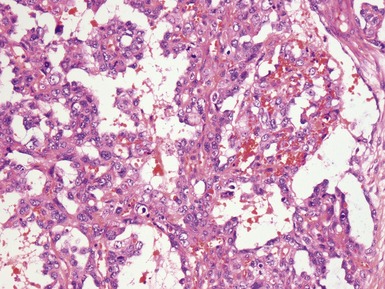
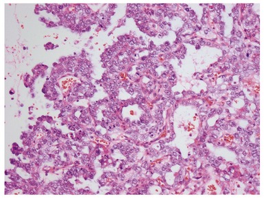
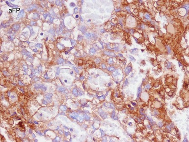
Histopathological features (Figs 2.3–2.18)

SEMINOMA

• Mainly affects adolescents when occurs in childhood
• Sheets of uniform round to ovoid cells
• Minimal pleomorphism in most cases
• Fine trabeculae
• Associated lymphoid infiltrate
• PLAP and CD117 + in almost all
image

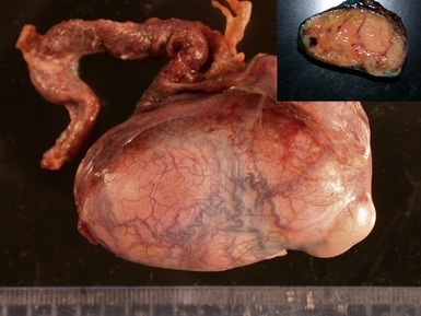
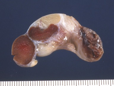
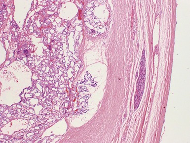
Fig 2.3 Photograph of a testis with a malignant yolk sac tumor. The capsule is stretched and the cut surface (inset) is solid with areas of hemorrhage.
image

Fig. 2.4

image

Fig. 2.5

image

Fig. 2.6

image

Fig. 2.7

image

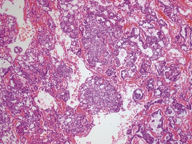
Figs 2.4–2.8 Photomicrographs of testicular malignant yolk sac tumors, demonstrating the range of morphological appearances including microcystic, reticular, solid and papillary patterns.
image

Fig. 2.9

image

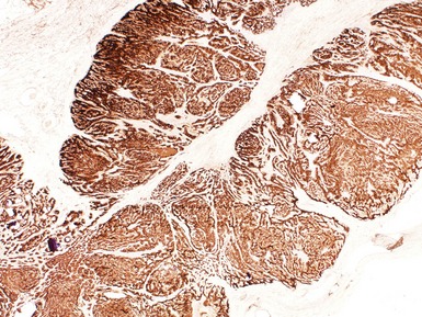
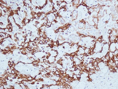
Figs 2.9–2.10 Photomicrographs of testicular malignant yolk sac tumors, demonstrating the tumor cells highlighted by cytokeratin (Fig 2.9) and alphafetoprotein (Fig 2.10) immunostaining.
image

Fig. 2.11

image

Fig. 2.12

image

Fig. 2.13

image

Fig. 2.14

image

Fig. 2.15

image

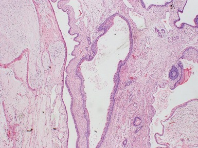
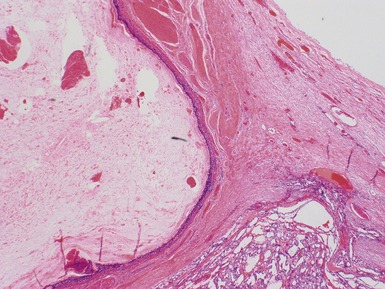
Figs 2.11–2.16 Photomicrographs of testicular teratomas, demonstrating a wide range of mature tissue elements (mature) in addition to immature neuroepithelial tubular structures (immature).
image

Fig. 2.17

image

Figs 2.17–2.18 Photomicrographs of testicular teratomas in which there are additional focal areas of malignant yolk sac tumor. The identification of such malignant areas is important and highlights the need for adequate specimen sampling.

EMBRYONAL CARCINOMA

• Rare
• Sheets, papillae and cysts with poorly differentiated cells
• Polygonal with vesicular nuclei

image prominent nucleoli
image overlapping nuclei
• CD30+
• CK+
• Focal PLAP+

YOLK SAC TUMOR

• Commonest malignant GCT in infancy
• 90% associated with elevated serum aFP levels
• Ovoid vacuolated cells with scattered hyaline globules
• Mitotically active
• Numerous patterns, most cases show features of many admixed

image microcystic / reticular
image macrocytic
image solid
image alveolar
image endodermal sinus (Schiller–Duval bodies)
image papillary
image myxomatous
image polyvesicular / vitelline
image hepatoid
image enteric
• Cytokeratin+
• aFP focally+

CHORIOCARCINOMA

• Very rare in childhood even as component of mixed malignant GCT
• Cytokeratin+, inhibin+, hCG+

TERATOMA

• Infancy
• Composed of tissues recapitulating all three germ layers
• Well demarcated
• Wide range of tissue differentiation
• Sample carefully for malignant areas including focal PNET

MIXED GERM CELL TUMOR

• Multiple combinations of above components

Differential diagnoses and pitfalls

• Normal immature fetal germ cells express PLAP and CD117

image expression reduces during early childhood
image do not overdiagnose ITGCN in childhood testes on the basis of immunostaining only
• Infantile / congenital testicular tumors are most likely to be juvenile granulosa cell tumor rather than GCT

TESTICULAR SEX-CORD STROMAL TUMORS

LEYDIG CELL TUMORS

Epidemiology

• <5% pediatric testicular tumors
• 3–9 years in childhood

Clinical features

• Painless unilateral testicular enlargement
• Endocrine features may be present

image gynecomastia
image precocious puberty

Macroscopic features

• Well circumscribed yellow brown nodule

Histopathological features

• Diffuse sheets of large polygonal cells

image eosinophilic cytoplasm
image Reinke crystals <40%
image regular nuclei, prominent nucleoli
• Spindle cell variant
• Malignant behavior (10%) associated with:

image >5cm diameter
image cytological atypia
image mitoses
image necrosis
image vascular invasion

Immunohistochemical staining

• Vimentin+
• Inhibin+

Differential diagnoses and pitfalls

• Leydig cell hyperplasia is diffuse and interstitial not expansile / destructive
• Adrenogenital syndrome-associated tumors are multiple nodules and occur bilaterally

SERTOLI CELL TUMOR

Epidemiology

• Generally very rare in childhood

Histopathological features

• Bland uniform sheets of ovoid cells
• Tubular formations
• Malignancy associated with

image pleomorphism
image mitoses
image invasion

Immunohistochemical staining

• Vimentin+
• Variably inhibin+

Large cell calcifying Sertoli cell tumor (LCCST) (Figs 2.19–2.21)

• Variant occurring in childhood

image average age 16 years
image sporadic and associated with Carney and Peutz–Jeghers syndromes
• 20% malignant
• 40% bilateral
• Nests / cords of polygonal cells with eosinophilic cytoplasm
• Myxoid / hyaline stroma
• Foci of calcification

image Note: calcification may be absent even though called LCCST
image

Fig. 2.19

image

Fig. 2.20

image

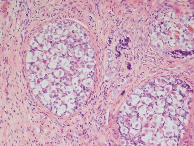
Figs 2.19–2.21 Photomicrographs of a large cell calcifying Sertoli cell tumor, demonstrating nests and cords of polygonal cells with eosinophilic cytoplasm in a hyaline stroma with foci of calcification. Inhibin immunostaining is positive.

JUVENILE GRANULOSA CELL TUMOR

• Adult type granulosa cell tumor of testis not reported in childhood
• JGCT is the most frequent congenital testicular neoplasm

Clinical features

• Testicular mass
• 20% associated with ambiguous genitalia

Histopathological features (Figs 2.22, 2.23)

• Cystic and solid mass with focal areas of hemorrhage
• Cysts with inner granulosa like cells

image outer theca like cells (may be spindled)
• Intervening solid areas with plump ovoid-spindle cells
image

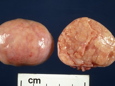
Fig 2.22 Photograph of a testis from an infant, almost completely replaced by a predominantly solid juvenile granulosa cell tumor.

image

Fig 2.23 Photomicrograph of a testicular juvenile granulosa cell tumor, demonstrating cystic areas surrounded by granulosa-type cells and solid areas composed of plump, ovoid-spindled cells.

Immunohistochemical staining

• Vimentin+, cytokeratin+, S100+
• Inhibin+
• Note: Mixed forms of SCSTs with elements of specific lesions may also occur

GONADOBLASTOMA

• Tumor containing mixture of germ cells and sex cord stromal cells

Epidemiology

• Rare but also reported in otherwise normal males

Clinical features

• Painless gonadal mass / incidental

Histopathological features (Figs 2.24, 2.25)

• Nested architecture

image Sertoli-like cells surrounding seminoma-like germ cells
image

Fig. 2.24

image

Figs 2.24–2.25 Photomicrographs of a testicular gonadoblastoma, demonstrating a nested architecture with admixed Sertoli-like cells surrounding seminoma-like germ cells and focal calcifications.

Differential diagnoses and pitfalls

• May be associated with foci of seminoma or other malignant germ cell tumor elements, requires adequate sampling to exclude

SECONDARY TESTICULAR INVOLVEMENT
• In lymphoma relatively common (Figs 2.26, 2.27)

image especially ALL (acute lymphoblastic leukemia)

– interstitial infiltrate of monomorphic cells surrounding and separating tubules
– now not usually biopsied
image

Fig. 2.26

image

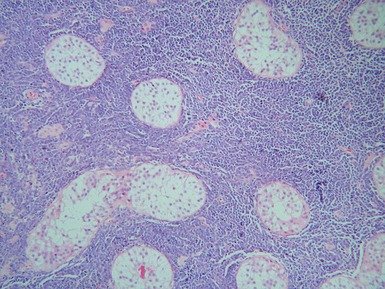
Figs 2.26–2.27 Photomicrographs of testicular biopsies demonstrating involvement by acute lymphoblastic leukemia, with an interstitial infiltrate of monomorphic cells surrounding and separating tubules.

VANISHED TESTIS / TESTICULAR REGRESSION SYNDROME (TRS)

• Nodule / tissue identified on surgical exploration for an impalpable testis

Epidemiology

• 46 XY genetically normal males
• Usually submitted from birth to early childhood

image median 2 years at exploration
• Probably due to intrauterine torsion of developmentally normal testis

Clinical features

• Usually unilateral, impalpable testis
• Phenotypically normal male

Macroscopic features

• Cord-like tissue removed on inguinal exploration

image macroscopically identifiable nodule in about half

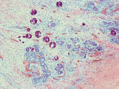
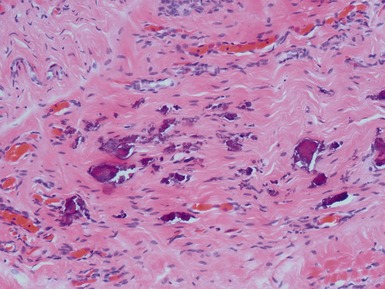
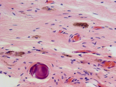
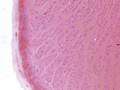
Histopathological features (Figs 2.28–2.32)

• Rudimentary spermatic cord with absence of macroscopically identifiable normal testicular tissue
• Fibrovascular nodule with associated spermatic cord remnants

image epididymis (30%)
image vas deferens (70%)
• Areas containing scattered hemosiderin-laden macrophages (70%) and/or calcification (40%)

image prevalence of these findings decreases with increasing patient age (Law et al 2006)
• Testicular tissue with identifiable tubules in <10%

image fibrosed
image scattered clusters of tubules
image usually no germ cells present

– when present show no atypia
image

Fig. 2.28

image

Fig. 2.29

image

Fig. 2.30

image

Fig. 2.31

image

Figs 2.28–2.32 Photomicrographs of cases of testicular regression syndrome, demonstrating fibrous nodules, within which residual identifiable tubules may be seen in a minority, in association with hemosiderin deposition and dystrophic calcification.

CRYPTORCHIDISM

• One or both testis not palpable in normal scrotal position but present inguinally or intra-abdominally

Epidemiology

• 2–4% term males at birth
• 1% at 3+ months of age

Clinical features

• Usually isolated but may be associated with underlying syndromes and/or other genitourinary defects
• Increased risk of development of testicular germ cell tumor

image especially seminoma and gonadoblastoma in intersex states with Y chromosome
• Testicular germ cell malignancy six times more likely in men in whom orchidopexy is delayed or not performed, compared to those with orchidopexy before 10 years of age (Walsh et al 2007)

image optimal timing of orchidopexy still uncertain, <1 year of age may be best to also preserve fertility (Leung & Robson 2004)

Histopathological features

• Histological changes related to patient age and hence length of cryptorchid period prior to surgery
• If operated in infancy, may show minimal abnormalities, but histological changes increase with age

image reduced germ cell number
image germ cell maturational arrest
image reduced tubular diameter
image interstitial fibrosis
image tubular sclerosis and thickened tubular basement membrane
image Sertoli only tubules
image possible ITGN and Leydig cell hyperplasia development in adolescents

Immunohistochemical stains

• ITGN cells express CD117 and PLAP but

image caution in using these markers in prepubertal children since normal immature germ cells also express CD117 and PLAP (Law et al 2006)
• ITGN diagnosis required additional characteristic morphological changes and abnormal central germ cell intratubular location

TESTICULAR AND TESTICULAR APPENDAGE TORSION

Epidemiology

• All ages of childhood affected but:

image true testicular torsion is much more common in adolescents / early adulthood
image adnexal torsion more common in younger boys
• Around 50% of cases of acute scrotum in children represent torsion of an appendage rather than testis (Murphy et al 2007)

Clinical features

• Acute scrotal pain / swelling

Histopathological features (Fig 2.33)

• Testis torsion

image congestion
image edema
image interstitial hemorrhage
image progressing to frank hemorrhagic infarction
image always examine even apparently necrotic tissue to exclude underlying tumor
• Appendage torsion

image marked congestion and interstitial hemorrhage
image rarely frank necrosis
image

Fig 2.33 Photomicrograph of a case of testicular torsion in a child, demonstrating hemorrhagic infarction with scattered residual tubules.

Differential diagnoses and pitfalls

• Testicular appendage cysts (eg Hydatid of Morgagni) without torsion may also be removed when found incidentally during scrotal exploration, or rarely, due to a palpable cyst

PARATESTICULAR / ADNEXAL TUMORS

Paratesticular spindle cell rhabdomyosarcoma

• see RMS section

Adenomatoid tumor

• Mesothelial lesion
• Mainly affects adults but reported in teenagers
• Smooth well-circumscribed lesion arising from / near epididymis
• Histologically composed of:

image cords and tubules of mesothelial cells

– bland, no atypia
– vacuolated cytoplasm
image fibrous stroma
• AE1/3+, EMA+

Nodular mesothelial hyperplasia

• May occur in hernia sacs of children
• Nodules of polygonal cells (probably histiocytes) with overlying mesothelial proliferation
• Admixed fibrin and inflammatory cells

Melanotic neuroectodermal tumor of infancy

• May arise in epididymis and paratesticular region (see tumor section)
• Composed of:

image smaller neuroblast-like cells

Desmoplastic small round cell tumor

• May arise in paratesticular location (see tumor section)

Other rare paratesticular tumors

• Include lipoma, hemangioma, neurofibroma, granular cell tumor, fibrous hamartoma of infancy

NONTUMOROUS PARATESTICULAR MASSES

• May be due to

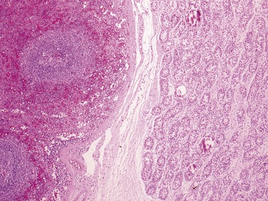
image splenogonadal fusion (Figs 2.34, 2.35)

– splenic and gonadal tissue
image ectopic renal or adrenal tissue

– renal or adrenal tissue
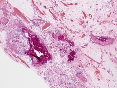
image meconium periorchitis (Fig 2.36)

– secondary to in-utero gastrointestinal perforation, with or without cystic fibrosis
– fibrosis, chronic inflammation with giant cells, pigmented macrophages
image

Fig 2.34 Photograph of a case of splenogonadal fusion, demonstrating juxtaposed but not admixed, splenic and gonadal elements.
image

Fig 2.35 Photomicrograph of a case of splenogonadal fusion, demonstrating juxtaposed but not admixed, splenic and gonadal elements.

image

Fig 2.36 Photomicrograph of a case of intrauterine bowel perforation with meconium periorchitis, demonstrating areas of fibrosis, chronic inflammation and pigmented macrophages with dystrophic calcification.

CYSTIC DYSPLASIA OF THE RETE TESTIS (CDRT)

• Congenital malformation of rete testis

Epidemiology

• Presents in childhood through to adulthood

Clinical features

• Apparent testicular enlargement or scrotal mass
• Many ipsilateral genitourinary associated lesions described (embryological ductal malformation) (Bouron-Dal Soglio et al 2006, Park et al 2008)

Macroscopic features

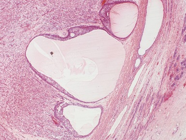
• Multiple dilated and slit-like cystic spaces adjacent to testis at hilum

Histopathological features

• Anastomosing cystic spaces lined by flattened simple cuboidal epithelium
• Connective tissue septae
• Adjacent testis may be compressed / displaced but is otherwise normal

INTERSEX / DISORDERS OF SEXUAL DIFFERENTIATION (DSD)

• A new classification system for patients with DSD proposed

image associated modification of risk for germ cell tumors in specific diagnostic groups (Cools et al 2006)
• No longer use terms ‘intersex’ or ‘hermaphroditism’ (Lee et al 2006)

Sex chromosome DSD

• 45,XO
• 47,XXY
• 45XO/46XY

image MGD, ovotesticular DSD
• 46XX/46XY

image chimeric, ovotesticular DSD

46XY DSD

• Disorders of testicular development

image complete gonadal dysgenesis
image partial gonadal dysgenesis
image testicular regression
image ovotesticular DSD
• Disorders of androgen synthesis or action

image androgen synthesis defects
image androgen insensitivity syndrome
image this group may show:

– hyalinized nodule of smooth muscle at one pole
– hamartomatous Sertoli cell adenomas
– development of seminoma

· usually in adulthood (partial AIS higher risk than complete AIS since germ cells remain)
• LH receptor defects
• Anti-Mullerian hormone (and receptor) defects
• Only 50% of 46XY DSD have a specific diagnosis

46XX DSD

• Disorders of ovarian development

image ovotesticular DSD
image testicular DSD
image gonadal dysgenesis
• Androgen excess

image eg 21-hydroxylase deficiency
image most virilized XX infants are due to congenital adrenal hyperplasia

Other

• Most surgical pathology specimens in this setting require (Figs 2.37, 2.38):

image descriptive report of structures present
image comment on germ cell neoplasia

– increased risk of germ cell neoplasia development if Y chromosome present
– overall risk GCN in GD is around 10–15%
– <5% in undervirilization syndromes
– androgen insensitivity

· risk around 5–10% overall
· <1% with CAIS
· 10–15% with PAIS
– most tumors develop after puberty in adulthood

· usually carcinoma in situ or seminoma
· gonadoblastoma rare
image possible previous increased risk of CIS was overestimated due to misinterpretation of maturational delay in immature germ cells

– gonadal dysgenesis with Y chromosome present

· 20–40% risk of gonadoblastoma / seminoma / dysgerminoma
· in this group GCTs may be present in childhood
· note some Turner syndrome (XO) may also have part of Y chromosome present with associated risk
• Detection of abnormal germ cells

image PLAP+
image CD117+
image OCT3/4+

– strong nuclear staining
image maturational delay (MD) may be precursor for CIS and tumor development

– not in itself premalignant
– common in GD
• Widespread, luminal OCT3/4+ germ cells in an infant represents MD
• CIS germ cells

image on the basal lamina not luminal
image patients >1 year old
image focal clusters not diffuse
image

Fig 2.37 Low power photomicrograph of a case of ovotestis, with adjacent ovarian and testicular elements.

image

Fig 2.38 Photomicrograph of a case of ovotestis, with adjacent ovarian and testicular elements.

REFERENCES

Bouron-Dal Soglio D, Harvey I, Jovanovic M, Oligny LL, Fournet JC. Bilateral cystic dysplasia of the rete testis with renal adysplasia. Pediatr Dev Pathol. 2006;9:157-160.

Cools M, Drop SL, Wolffenbuttel KP, Oosterhuis JW, Looijenga LH. Germ cell tumors in the intersex gonad: old paths, new directions, moving frontiers. Endocr Rev. 2006;27:468-484.

Law H, Mushtaq I, Wingrove K, Malone M, Sebire NJ. Histopathological features of testicular regression syndrome: relation to patient age and implications for management. Fetal Pediatr Pathol. 2006;25:119-129.

Lee PA, Houk CP, Ahmed SF, Hughes IA. International Consensus Conference on Intersex organized by the Lawson Wilkins Pediatric Endocrine Society and the European Society for Pediatric Endocrinology. Consensus statement on management of intersex disorders. International Consensus Conference on Intersex. Pediatrics.. 2006;118:e488-e500.

Leung AK, Robson WL. Current status of cryptorchidism. Adv Pediatr. 2004;51:351-377.

Murphy FL, Law H, Mushtaq I, Sebire NJ. Testicular and paratesticular pathology in infants and children: the histopathological experience of a tertiary pediatric unit over a 17-year period. Pediatr Surg Int. 2007;23:867-872.

Park E, Morrison S. Cystic dysplasia of the rete testis in an adolescent with VATER association. Pediatr Radiol. 2008;38:123.

Walsh TJ, Dall’Era MA, Croughan MS, Carroll PR, Turek PJ. Prepubertal orchiopexy for cryptorchidism may be associated with lower risk of testicular cancer. J Urol. 2007;178:1440-1446.

[/level-membership-for-pathology-category][not-level-membership-for-pathology-category]<

Buy Membership for Pathology Category to continue reading. Learn more here

[/not-level-membership-for-pathology-category]