

[level-membership-for-dermatology-category]70
Protozoa and Worms
Leishmaniasis
• Three major forms: (1) cutaneous (Fig. 70.1); (2) mucocutaneous (Fig. 70.2); and (3) visceral (e.g. liver, spleen).
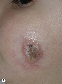
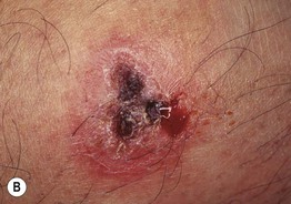
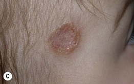
Fig. 70.1 Variable presentations of cutaneous leishmaniasis. Ulcerated plaques with rolled border (A) and central crusting (A, B). Plaque with translucent borders containing telangiectasias and central scarring (C). Cutaneous leishmaniasis is sometimes mistaken for a basal cell carcinoma in adults. A, C, Courtesy, Julie V. Schaffer, MD.
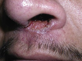
Fig. 70.2 Mucocutaneous leishmaniasis. Ulceration and induration of the nasal vestibule extending onto the cutaneous lip due to Leishmania braziliensis. Courtesy, Kalman Watsky, MD.
• Caused by more than 15 different species of Leishmania (Table 70.1).
Table 70.1
Four major species of Leishmania that cause cutaneous disease.
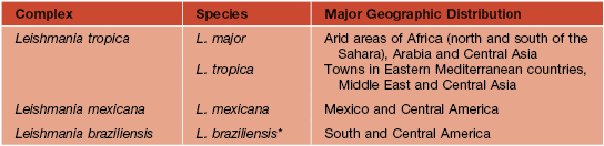
* Can also cause mucosal leishmaniasis or visceral leishmaniasis in immunocompromised individuals.
• Vector = sandfly (Phlebotomus and Lutzomyia spp.) (Fig. 70.3).
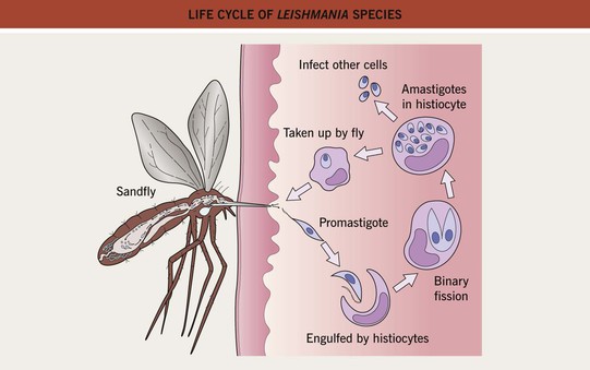
Fig. 70.3 Life cycle of Leishmania species. Promastigotes develop within the gut of the sandfly and then migrate to the proboscis.
• Disease seen worldwide but endemic in areas of Asia, Africa, Latin America, and the Mediterranean basin (Fig. 70.4).
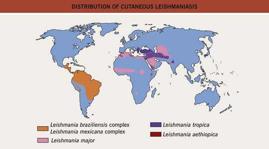
Fig. 70.4 Distribution of cutaneous leishmaniasis. Adapted with permission from Davidson RN, Leishmaniasis. In Cohen J, Powderly WG (Eds.), Infectious Diseases. Edinburgh, UK: Mosby, 2004.
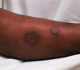
• Cutaneous disease affects skin only and is commonly a papule that expands and ulcerates (Fig. 70.5); pattern may be sporotrichoid (Fig. 70.6); lesion(s) may heal spontaneously (Fig. 70.7).
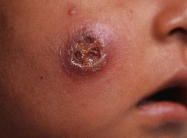
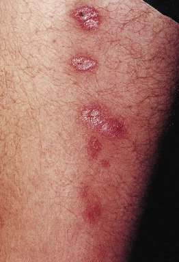
Fig. 70.6 Sporotrichoid form of cutaneous leishmaniasis. With permission from Tyring S, Lupi O, Hengge U (Eds.), Tropical Dermatology. Oxford: Churchill Livingstone, 2005.
Amebiasis
Free-Living Ameba
Trypanosomiasis – American
• Trypanosoma cruzi carried by reduviid (kissing) bugs.
• Endemic in areas of Central and South America.
• Systemic disease that can affect the autonomic nervous system, gastrointestinal tract, and heart.
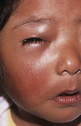
• Primary acute phase: local erythema and edema at inoculation site ± regional lymphadenopathy; when periorbital, termed Romaña sign (Fig. 70.8).
Trypanosomiasis – African
• Found in both West (Trypanosoma brucei gambiense) and East (Trypanosoma brucei rhodesiense) Africa.
• Skin findings: trypanosomal chancre (localized bite reaction; Fig. 70.9) and annular erythematous eruption with fever.
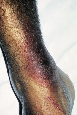
Fig. 70.9 Trypanosomal chancre. The bite reaction, the earliest clinical lesion, is known as a ‘trypanosomal chancre.’ It resembles a boil but is usually painless. Fluid aspirated from the nodule contains actively dividing trypanosomes. This reaction is seen more commonly in T. b. rhodesiense than in T. b. gambiense infection. With permission from Peters W, Pasvol G, Tropical Medicine and Parasitology, 6th ed. London: Mosby, 2007.
Toxoplasmosis
Cutaneous Larva Migrans
• Worldwide, but especially common in tropical/subtropical areas and the southwestern United States.
• Larvae in infected soil, including sand, penetrate the skin.
• Pruritic, inflamed, serpiginous tracks are produced by migrating organisms (Fig. 70.10); migration averages 1–2 cm/day.
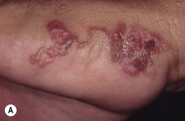
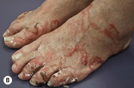
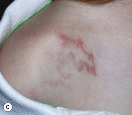
Fig. 70.10 Cutaneous larva migrans. Note the characteristic serpiginous erythematous tracks on the lateral foot (A), both feet (B), and the shoulder (C). Vesiculation and crusting (B) are sometimes seen. B, Courtesy, Peter Klein, MD; C, Courtesy, Julie V. Schaffer, MD.
• Most common locations are the lower extremities, especially the feet, and buttocks, due to walking and sitting at the beach.
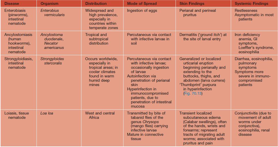
• DDx: not to be confused with larva currens (secondary to Strongyloides; see Table 70.2).
Onchocerciasis
• Secondary to Onchocerca volvulus, a tissue-dwelling nematode.
• Vector = black fly (Simulium).
• Endemic in Africa, Yemen, and some areas of Central and South America; near fast-flowing rivers.
• Skin findings: subcutaneous nodules containing adult worms (onchocercomas), pruritic papular dermatitis, depigmented, lichenified skin, especially over the shins (sowda) (Fig. 70.11).
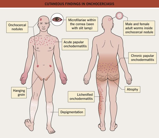
Fig. 70.11 Cutaneous findings in onchocerciasis.
Filariasis
Schistosomiasis
Swimmer’s Itch
• Worldwide; endemic to the Great Lakes.
• Erythematous papules on exposed skin (Fig. 70.12).
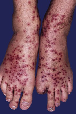
Fig. 70.12 Swimmer’s itch. Numerous edematous dark red papules on the feet and ankles. Courtesy, Kalman Watsky, MD.
• Not to be confused with seabather’s eruption (secondary to Linuche, Edwardsiella spp.), which affects areas under the swimsuit (see Chapter 72).
Cysticercosis
Gnathostomiasis
See Table 70.2 for a summary of other major parasitic worms that cause skin findings.
For further information see Ch. 83. From Dermatology, Third Edition.
[/level-membership-for-dermatology-category][not-level-membership-for-dermatology-category]70
Protozoa and Worms
Leishmaniasis
• Three major forms: (1) cutaneous (Fig. 70.1); (2) mucocutaneous (Fig. 70.2); and (3) visceral (e.g. liver, spleen).
Fig. 70.1 Variable presentations of cutaneous leishmaniasis. Ulcerated plaques with rolled border (A) and central crusting (A, B). Plaque with translucent borders containing telangiectasias and central scarring (C). Cutaneous leishmaniasis is sometimes mistaken for a basal cell carcinoma in adults. A, C, Courtesy, Julie V. Schaffer, MD.
Fig. 70.2 Mucocutaneous leishmaniasis. Ulceration and induration of the nasal vestibule extending onto the cutaneous lip due to Leishmania braziliensis. Courtesy, Kalman Watsky, MD.
• Caused by more than 15 different species of Leishmania (Table 70.1).
Table 70.1
Four major species of Leishmania that cause cutaneous disease.
* Can also cause mucosal leishmaniasis or visceral leishmaniasis in immunocompromised individuals.
• Vector = sandfly (Phlebotomus and Lutzomyia spp.) (Fig. 70.3).
Fig. 70.3 Life cycle of Leishmania species. Promastigotes develop within the gut of the sandfly and then migrate to the proboscis.
• Disease seen worldwide but endemic in areas of Asia, Africa, Latin America, and the Mediterranean basin (Fig. 70.4).
Fig. 70.4 Distribution of cutaneous leishmaniasis. Adapted with permission from Davidson RN, Leishmaniasis. In Cohen J, Powderly WG (Eds.), Infectious Diseases. Edinburgh, UK: Mosby, 2004.
• Cutaneous disease affects skin only and is commonly a papule that expands and ulcerates (Fig. 70.5); pattern may be sporotrichoid (Fig. 70.6); lesion(s) may heal spontaneously (Fig. 70.7).
Fig. 70.6 Sporotrichoid form of cutaneous leishmaniasis. With permission from Tyring S, Lupi O, Hengge U (Eds.), Tropical Dermatology. Oxford: Churchill Livingstone, 2005.
Amebiasis
• Protozoan infection (Entamoeba histolytica) that most commonly causes colitis; fecal–oral spread.
• Rx: for Entamoeba histolytica
[/not-level-membership-for-dermatology-category]
