[level-membership-for-cardiovascular-category]
CASE 44
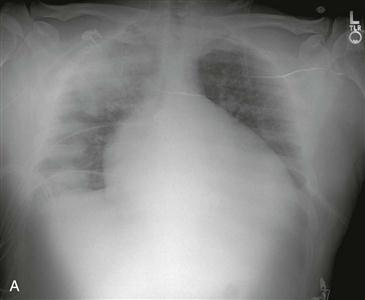
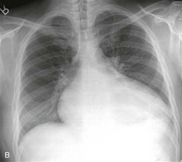
1. The radiograph in Fig. A was performed 1 month after the radiograph in Fig. B. What should be included in the differential diagnosis? (Choose all that apply.)
A. Pneumonia
B. Lung cancer
2. What is the predominant distribution of the lung opacity?
3. If the patient had severe hemoptysis, what would be the most likely diagnosis?
A. Pneumonia
B. Lung cancer
4. If the patient has mitral regurgitation, what is the most likely diagnosis?
A. Pneumonia
B. Lung cancer
ANSWERS
Reference
Walker CM, Reddy GP, Steiner RM. Radiology of the heart. In: Rosendorff C, ed. Essential Cardiology. ed 3 Philadelphia: Saunders; 2012.
Cross-Reference
Cardiac Imaging: The REQUISITES, ed 3, p 23.
Comment
Etiology and Findings
The jet in mitral regurgitation can be directed into the right superior pulmonary vein, which can result in asymmetric right upper lobe pulmonary edema. Asymmetric right upper lobe pulmonary edema (Fig. A) is seen in 9% of adult patients and 22% of pediatric patients with severe mitral regurgitation. In adults, it is usually caused by a flail posterior valve leaflet secondary to myocardial infarction. A flail posterior leaflet causes the mitral regurgitant jet to be preferentially directed into the right superior pulmonary vein. This leads to focal increased hydrostatic pressure and pulmonary edema within the right upper lobe. In the setting of chest pain, acute mitral regurgitation must be considered and can be confirmed with echocardiography.
Differential Diagnosis
The differential diagnosis for right upper lobe opacity varies depending on the patient’s clinical presentation. In a patient with fever and productive cough, the imaging findings are consistent with pneumonia. In a patient with hemoptysis, pulmonary hemorrhage is the most likely diagnosis. In a patient with severe chest pain and acute myocardial infarction, severe mitral regurgitation secondary to a flail posterior valve leaflet should be considered. If the abnormality has persisted over months, adenocarcinoma or organizing pneumonia must be excluded.
[/level-membership-for-cardiovascular-category][not-level-membership-for-cardiovascular-category]
CASE 44
1. The radiograph in Fig. A was performed 1 month after the radiograph in Fig. B. What should be included in the differential diagnosis? (Choose all that apply.)
A. Pneumonia
B. Lung cancer
2. What is the predominant distribution of the lung opacity?
3. If the patient had severe hemoptysis, what would be the most likely diagnosis?
[/not-level-membership-for-cardiovascular-category]
