19. Left Ventricular Endocardial Pacing in a Patient with an Anomalous Left-Sided Superior Vena Cava

Published on 26/02/2015 by admin

Filed under Cardiovascular

Last modified 22/04/2025

Print this page

rate 1 star rate 2 star rate 3 star rate 4 star rate 5 star
Your rating: none, Average: 0 (0 votes)

This article have been viewed 1107 times

History

The patient had a complex past medical history. He underwent bioprosthetic aortic valve replacement in 2006 related to infective endocarditis with consequent severe aortic regurgitation and had moderately impaired left ventricular systolic function (left ventricular ejection fraction [LVEF] 45%-50%). Coronary angiography did not show significant coronary artery disease, and therefore surgical revascularization was not necessary at the time of aortic valve replacement.
The patient then developed persistent (cardioversion on two occasions in 2007) and then permanent atrial fibrillation with fast ventricular rate response despite the use of digoxin, verapamil, and beta blockade (all in maximum tolerated or appropriate doses). He underwent atrioventricular nodal ablation in 2007 with implantation of a single-chamber rate-responsive right ventricular pacemaker.
Over the subsequent 2 years, he developed worsening systolic heart failure (New York Heart Association [NYHA] class III) that was refractory to medical therapy at maximal tolerated dosages (including an angiotensin-converting enzyme [ACE] inhibitor, beta blocker, nitrate, hydralazine, and loop diuretic). His ejection fraction was reduced to 35%. He therefore received an upgrade to a biventricular pacemaker.1 Because of the presence of an anomalous left-sided superior vena cava, left ventricular pacing via an epicardial coronary sinus branch was challenging. Nevertheless pacing was achieved at an anatomically appropriate site, although only one target coronary sinus tributary was available.
The patient responded well to biventricular pacing with improvement in ejection fraction (LVEF 45%) and NYHA status to class II.
Thirty-six months after implantation of the left ventricular pacing lead, he had late infection of the pacing pocket, with raised inflammatory markers and positive blood cultures for Staphylococcus.

Focused Clinical Questions and Discussion Points

Question

What is the indication for device and lead extraction?

Discussion

With evidence for both local and systemic infection, no other option is available but to perform system extraction.

Question

What is the reason for delay between extraction and re-implantation?

Discussion

This patient is pacing-dependent and requires temporary pacing before reimplantation of a new pacing system.
image

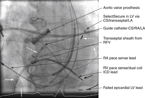
FIGURE 19-1 Left ventricular (LV) endocardial lead on anteroposterior projection. A transseptal sheath was passed from the right femoral vein (RFV) and has dilated the puncture site. Then a steerable guide catheter was passed via the anomalous left superior vena cava, through the enlarged coronary sinus to the right atrium (RA) and maneuvered across the transseptal puncture site into left atrium (LA). Then a Select Secure pacing lead has been passed to the LV via the guide catheter. CS, Coronary sinus; ICD, implantable cardioversion defibrillator.

Question

What are the challenges of pacing via an anomalous left-sided superior vena cava draining into an enlarged coronary sinus.

Discussion

Access is difficult, and manipulation of leads to achieve entry to the appropriate cardiac chamber requires expert handling. However, it is possible to gain access to the right ventricle and right atrium.

Question

Can transseptal access be gained from a superior approach?

Discussion

Transseptal access can be achieved using modified catheters.4 Studies are ongoing to understand the clinical value of this approach and of left ventricular endocardial pacing,2,3 although several studies supported the approach in terms of feasibility, safety, and patient response.

Final Diagnosis

The infected pacing system with all three pacing leads was extracted. A temporary pacing lead was inserted to provide pacing during a period of prolonged intravenous and subsequent oral antibiotic treatment. The patient received vancomycin and intravenous flucloxacillin followed by oral flucloxacillin.

Plan of Action

The decision was made to continue with antibiotic treatment for 6 weeks before reimplantation of a new, permanent biventricular pacing system. An attempt was made to establish LV epicardial pacing5 but the implanted pacing lead failed two days after the surgical procedure and the decision was made to attempt LV endocardial pacing.

Intervention

A new biventricular pacing system was implanted. Patency of the left subclavian vein was confirmed by venography before commencement of the procedure. Access then was gained to the left subclavian vein using a cutdown and after creation of a new pacemaker pocket in the left prepectoral region.
Anterograde coronary sinus angiography via the anomalous left superior vena cava showed occlusion of the previously targeted coronary sinus branch and no alternative target. A permanent right ventricular pacing lead was implanted via the left subclavian vein, coronary sinus, and right atrium.
Right femoral venous access was gained, and a transseptal catheter passed to the right atrium via the inferior vena cava. The procedure’s catheter and lead manipulations are summarized in the fluoroscopic images (Figures 19-1 and 19-2). Transseptal access to left atrium was achieved using a radiofrequency needle. The transseptal puncture site was dilated using a 12-French transseptal guide catheter and an extra support guide wire passed to left atrium. Intravenous heparin was administered to maintain an activated clotting time at greater than 300 seconds. Via the left subclavian vein and coronary sinus access route, a steerable guide catheter was passed to right atrium. Then, using a dilator inserted into this guide catheter and with the inferior approach guide wire as reference, the steerable guide was introduced to the left atrium and orientated to left ventricle. Using this steerable orientated catheter, a polyurethane 4.1-French pacing lead was passed to the left ventricle and secured by an active fixation screw to the lateral left ventricular endocardial wall, just beneath the mitral ring. Pacing assessment demonstrated a pacing impedance of 980 Ω at 0.2 mV amplitude and 0.04 msec pulse width. When the pacing leads were secured, the delivery systems were removed and a biventricular pacemaker (atrial port capped) was attached to the respective leads. The pacemaker was secured in the prepectoral pouch, and the wound was closed in a standard fashion (Figure 19-3).
image

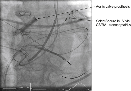
FIGURE 19-2 Anteroposterior projection. The transseptal guide catheter has now been withdrawn, leaving the Select Secure lead in place after fixation. The transseptal catheter remains in place until the end of the procedure to allow for reentry to the left atrium (LA) should there be displacement of the pacing lead as its delivery system is withdrawn. CS, Coronary sinus; LV, left ventricle; RA, right atrium; RV, right ventricle.

Postprocedure assessment demonstrated appropriate pacing parameters and a narrow paced QRS. The patient was established on an anticoagulation regimen using warfarin and maintaining an international normalized ratio between 2 and 3. The patient experienced remarkable clinical improvement within 1 month, with an LVEF of 50% and improvement to NYHA I.

Outcome

The patient received biventricular pacing using left ventricular endocardial pacing4,6 in a clinical situation in which alternative mechanisms for achieving biventricular pacing were not possible. The patient has experienced remarkable clinical benefit from the procedure.

Findings

No periprocedural or postprocedural complications occurred, and to date the patient has received significant clinical benefit from left ventricular endocardial pacing.
image

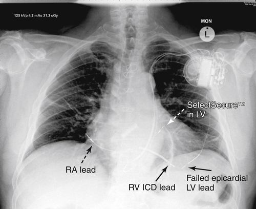
FIGURE 19-3 Posteroanterior chest radiograph obtained at 24 hours after implantation of the left ventricular (LV) lead. Permanent pacing lead positions are indicated. CS, Coronary sinus; LA, left atrium; RA, right atrium.

Selected References

1. Cazeau S., Alonso C., Jauvert G. et al. Cardiac resynchronization therapy. Europace. 2004;5(Suppl 1):S42–S48.

2. Fish J.M., Brugada J., Antzelevitch C. Potential proarrhythmic effects of biventricular pacing. J Am Coll Cardiol. 2005;46:2340–2347.

3. Garrigue S., Jaïs P., Espil G. et al. Comparison of chronic biventricular pacing between epicardial and endocardial left ventricular stimulation using Doppler tissue imaging in patients with heart failure. Am J Cardiol. 2001;88:858–862.

4. Morgan J.M., Scott P.A., Turner N.G. et al. Targeted left ventricular endocardial pacing using a steerable introducing guide catheter and active fixation pacing lead. Europace. 2009;11:502–506.

5. Puglisi A., Lunati M., Marullo A.G. et al. Limited thoracotomy as a second choice alternative to transvenous implant for cardiac resynchronisation therapy delivery. Eur Heart J. 2004;25:1063–1069.

6. van Gelder B.M., Scheffer M.G., Meijer A. et al. Transseptal endocardial left ventricular pacing: an alternative technique for coronary sinus lead placement in cardiac resynchronization therapy. Heart Rhythm. 2007;4:454–460.