ENT Emergencies
Edited by Peter Cameron
18.1 Ears, nose and throat emergencies
Sashi Kumar
The ear
Introduction
Emergency presentations for ear, nose and throat (ENT) problems are common and all emergency physicians need to be familiar with the basic skills required for assessment and management of these problems.
Foreign body
Foreign bodies in the ear are most common in children under the age of 5 and in mentally handicapped adults. Animate objects, such as insects in the ear, can affect all ages, especially adults who enjoy the outdoors, particularly at dusk.
Accidental foreign bodies, such as the end of a cotton bud or a matchstick, occur in people obsessed with cleaning their ears with such objects.
Management
Removal of a live foreign body
This is a true ENT emergency. The insect should be killed as a matter of urgency, as considerable damage is being done to the sensitive skin of the bony meatus and the tympanic membrane by the flapping wings and appendages of the desperate insect trying to escape.
The movement of the insect also causes intense pain and tinnitus, thereby creating further anxiety and distress.
Any liquid used to kill the insect should be carefully chosen so as to avoid damage to the sensitive skin and tympanic membrane: strong corrosive agents, knockdown spray or alcohol should be avoided. The common agents of choice are lignocaine 2%, olive oil, water for injection or normal saline.
One of the preferred methods is to instil some water for injection from a 10 mL plastic ampoule and leave an examination light on the pinna. The insect swims up to surface towards the light and can be helped to safety by holding the tip of the ampoule [1].
Removal of a foreign body in a child or a mentally handicapped adult may be done in one of two ways. The patient is either cooperative and unrestrained or fully restrained. It is vital not to attempt any procedure with partial restraint, as any movement of the patient during the attempt could cause trauma to the ear canal and the tympanic membrane.
There are two techniques used to remove a foreign body. The dry method is by using a Jobson Horne probe for solid objects, such as beads, or alligator forceps for an insect or a cotton bud. The wet method is by syringing the ear canal with tepid water. The water should be close to body temperature to avoid a caloric effect, which produces nystagmus and vertigo.
The key to success is good lighting, preferably through a head lamp, a cooperative or fully restrained patient and a patient, gentle approach by the clinician, who knows when to stop if unsuccessful.
Impacted cerumen
Impacted hard cerumen or wax causes pain and hearing loss.
Sudden onset of hearing loss after a swim is classical of impacted ceruman as the wax swells up when in contact with water.
A 3–5-day course of Waxsol or Cerumol ear drops 3–5 drops three times a day followed by syringing of the ear canal with warm water should clear up the ear canal.
Trauma
Trauma to the ear canal requires the ear to be kept dry for about a week with antibiotic ear drops for 4–5 days in severe cases to avoid progressing into otitis externa.
Penetrating trauma can cause perforation of the eardrum and, occasionally, disruption of the ossicular chain. Dislocation of the footplate of the stapes following such an injury can cause permanent sensorineural hearing loss. Referral to an ENT specialist is essential in all cases of traumatic perforation with suspected ossicular chain disruption.
Blunt trauma
Boxing and other contact sports can lead to blunt trauma to the pinna. Accumulation of blood under the perichondrium, if not treated properly, may progress to cartilage necrosis and the end result is a ‘cauliflower ear’.
A slap on the ear can also produce a ruptured tympanic membrane with or without ossicular chain disruption.
Assessment
Assessment of the injury includes a clinical assessment of the hearing loss. A ruptured eardrum without ossicular chain disruption does not usually cause a significant hearing loss. Any evidence of nystagmus or tinnitus suggests damage to the inner ear.
Management
A simple traumatic perforation of the eardrum is managed by simple analgesics and keeping the ear dry. On no account should any drops or water be allowed into the ear, as this may precipitate otitis media.
If ossicular chain disruption or inner ear trauma is suspected, an urgent ENT opinion is required to assess the need for urgent tympanotomy and repair.
Haematoma of the pinna requires urgent release of the accumulated blood by aseptic incision and drainage and the immediate application of a firm mastoid bandage, to prevent reaccumulation, and this should be left in place for up to a week. The patient should be placed on broad-spectrum antibiotics to prevent infection.
Infection
Otitis externa
Infection of the external ear is common and affects between 3 and 10% of the patient population [2]. It can be localized (furuncle) or diffuse. The symptoms are pain, itching and tenderness to palpation, followed by aural fullness, hearing loss and discharge. The common pathogens responsible are Pseudomonas aeruginosa, Proteus spp. and Staphylococcus aureus[3].
The diagnosis is usually self-evident, but the diagnostic signs of otitis externa are tragal tenderness or pain on pulling the pinna. This is a disease of the cartilaginous ear canal, with swelling and discharge causing occlusion of the meatus. It may be extremely painful to pass the ear speculum and often the tympanic membrane is not able to be visualized.
Management
The most important step in the treatment is thorough and atraumatic cleansing of the ear canal [4]. Tolerance and cooperation between the patient and the clinician is vital. Pope Otowick (Xomed) is very useful in the management of this condition. This is a semirigid foam wick that, when inserted into the ear canal, swells, absorbing moisture to increase the size of the ear canal. Topical otic drops, such as Sofradex (Roussel), are used three to four times a day and the patient is reviewed on a daily basis to change the wick and continue the ear toilet. Occasionally, oral antibiotics, such as ciprofloxacin or flucloxacillin, may be required [5], particularly if there is evidence of cellulitis. The patient is advised to keep the ear clear of any water. Strong analgesics are usually required.
Fungal otitis externa (otomycosis) tends to be not that painful and is treated with ear toilet as described and topical antifungal ear drops, such as Loco corten vioform.
Otitis media
Acute otitis media is a common infection and is due to blocking of the eustachian tube (eustachian catarrh) and negative pressure in the middle ear cavity. Although viral in origin, secondary bacterial infection often supervenes. The most frequently isolated pathogens are Streptococcus pneumoniae, Haemophilus influenzae and Moraxella catarrhalis[6]. The symptoms are earache, fullness, hearing loss and fever, with ear discharge if the drum has perforated. The development of discharge usually marks an improvement in the pain and fever.
The clinical findings vary from a retracted dull eardrum to a congested bulging drum or a white eardrum with pus behind and a perforated tympanic membrane with discharge in the ear canal. A perforated eardrum without much pain is usually a sign of chronic otitis media.
Management
Treatment is almost always empiric and amoxicillin is a good first-line therapy. Cephalosporins and trimethoprim/sulpha are also used with considerable success. The newer macrolides, such as azithromycin and clarithromycin, are rational alternatives [6].
In otitis media with a perforated eardrum, the mainstay of treatment should be toilet by dry mopping followed by antibiotic drops, such as Sofradex. The ear should be kept dry and regular follow up arranged until the perforation has healed.
Labyrinthitis
Acute labyrinthitis usually has cochlear symptoms, such as hearing loss and tinnitus, which should be referred for audiometry and urgent ENT evaluation. If the symptoms are limited to vertigo and nystagmus, it is more likely to be due to acute vestibular neuronitis.
Management
The management of labyrinthitis includes bed rest, antiemetics, e.g. prochlorperazine, benzodiazepine, e.g. diazepam, and admission if severely debilitating. In the presence of hearing loss, a course of oral steroids or intratympanic dexamethasone may be started after discussions with the ENT surgeon.
Otitis media with effusion (glue ear)
This is most common in children in developed countries. The symptoms are fullness and hearing loss and, occasionally, pain. Management includes the diagnosis based on history and examination, which reveals a dull, retracted drum or fluid behind the drum without redness. The most reliable sign of a glue ear is an immobile eardrum on Valsalva manoeuvre or pneumatic otoscopy. Repeated attacks of glue ear are an indication for the insertion of tympanostomy tubes.
Mastoiditis
Acute mastoiditis is a complication of acute or chronic otitis media. It is a rare condition in the developed world, although still quite prevalent in the developing world and the Aboriginal population of Australia. Otitis externa with a painful and tender postauricular lymph node is usually mistaken for acute mastoiditis due to the postauricular tenderness. Extension of infection can cause meningitis or temporal lobe abscess, with life-threatening complications if untreated.
Examination reveals infection in the middle ear cavity by way of an injected drum or a perforated drum with discharge. The cardinal sign of acute mastoiditis is tenderness at the base of the mastoid on digital pressure. The diagnosis is confirmed by computed tomography (CT) scan.
Management
Admission, intravenous antibiotics and surgical intervention, such as mastoidectomy, to remove the infected mastoid air cells and drain any abscess collection.
The nose
Foreign body
A foreign body in the nose is common in preschool children and adults with mental retardation. The most common types of foreign body are beads, buttons and pieces of paper.
The diagnostic sign of a neglected nasal foreign body is a unilateral foul-smelling nasal discharge. The patient or the parent usually provides the history as to the type of foreign body and for how long present.
Management
The removal of the foreign body follows the same rules as for a foreign body in the ear. An additional method is to blow forcefully through the patient’s mouth while occluding the unaffected nostril. This could be done by the parent with instruction.
The suggested method of removal is to pass the ring end of a Jobson–Horne probe above and behind the foreign body and to roll it along the floor of the nose. The patient should be cooperative and unrestrained, or fully restrained. At the first sign of trauma or bleeding, removal should be organized under general anaesthesia as soon as practically possible.
Trauma
Fractured nose
This is a common presentation in the emergency department. The history is often quite clear and the findings include pain and tenderness over the nasal bones with or without crepitus and swelling at the bridge of the nose with or without epistaxis.
Careful examination will usually rule out CSF rhinorrhoea due to cribriform plate fracture and any external deformity. Active bleeding from the nostril should be controlled by direct pressure by pinching the nostril; if it does not settle it may require nasal packing.
Radiographs are not indicated for nasal bone fracture as this is a clinical diagnosis. It is often difficult to visualize the fracture line on the X-rays and radiographs do not help in the management. If associated facial fractures are suspected, X-ray facial views or CT scan should be taken.
Management
Acute intervention is required in the following circumstances:
Sinusitis
Approximately 90% of upper respiratory infections have associated sinus cavity disease [7]. Viral rhinosinusitis is the most common cause and is associated with the common cold. Approximately 0.5–2% of these cases progress to bacterial sinusitis.
Clinical features
Symptoms of viral sinusitis are rhinorrhoea, nasal obstruction and sneezing and facial pressure with or without headache. With bacterial superinfection, a purulent or coloured nasal discharge and fever of 38°C or higher develop. Significant facial pain and maxillary toothache with no obvious dental cause also occurs. The common organisms involved are Streptococcus pneumoniae and H. influenzae. Patients with allergic sinusitis typically have sneezing and itching, with watery eyes, as a leading symptom.
Radiographs of the sinus are not very helpful unless they demonstrate a distinct air–fluid level, as this increases with the likelihood of bacterial sinusitis. CT can indicate the presence of sinus abnormalities and evidence of infection. A raised white cell count is neither sensitive nor specific in the diagnosis of bacterial sinusitis.
Management of viral rhinosinusitis is symptomatic and it is generally self-limiting. Bacterial sinusitis must be treated with antibiotics: amoxicillin, augmentin or keflex could be used as first-line drugs. Although of unproven value, an oral decongestant or antihistamine is commonly used. Complications of sinus disease include meningitis, orbital extension and brain abscess. Diagnosis is by CT scan and treatment is intravenous antibiotics with surgical intervention by an otolaryngologist.
Epistaxis
Nose bleeding is the most common ENT emergency: a Scottish study reported an incidence of 30/100 000 people [8] in which the cause could only be found in 15% [9]. The common identified causes are trauma, blood dyscrasias, anticoagulation therapy and, occasionally, hereditary haemorrhagic telangiectasia [10]. Although hypertension has been traditionally labelled as a cause of epistaxis, studies have shown that blood pressure in these patients is no higher than in the control population [11,12].
The history is vital and all patients should be asked where the blood appeared first – anteriorly in the nose or in the back of the throat. Anterior epistaxis can usually be controlled in the emergency department and the patient safely discharged home without a nasal pack after cautery.
Management
The control of epistaxis due to a general cause, such as uncontrolled warfarin therapy or a bleeding disorder, is to reverse the cause. Local measures can still be used to stem the flow.
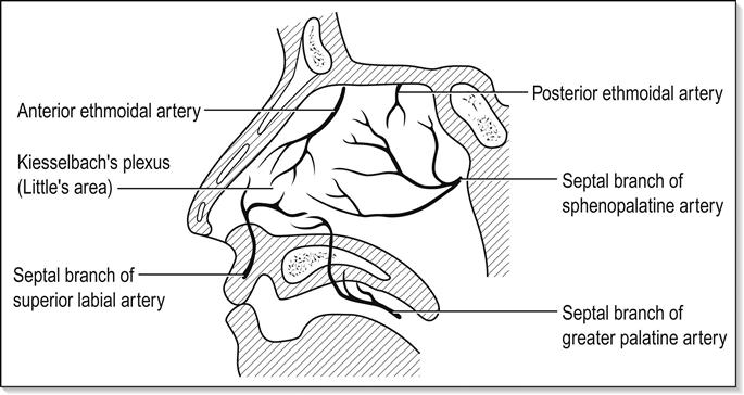
Idiopathic epistaxis, or that due to a local cause such as trauma, can be dealt with using local measures. The most common cause of anterior epistaxis is bleeding from Kiesselbach’s plexus in the Little’s area of the septum [13] (Fig. 18.1.1), which can easily be controlled by simple measures in the emergency department. Careful examination of a seated patient applying direct pressure to the bleeding vessel by pinching the anterior nares with the thumb and forefinger for up to 10 min will usually slow or cease the bleeding. At this point, it is essential to remove all the blood clots from the nasal cavity and the postnasal space using suction.
Following this, the application of cotton pledgets soaked in 5 or 10% cocaine or lignocaine with adrenaline or cophenylcaine (phenylephrine and lignocaine) will provide analgesia and vasoconstriction to the septum and the anterior part of the lateral wall.
Examination may reveal the bleeding vessel on the septum, which can be cauterized under direct vision using silver nitrate sticks. Following this, the patient is observed for a short time and can be discharged from the emergency department. The patient is advised not to pick, rub or blow the nose for 10 days and is advised to keep the cauterized area moist by applying chloromycetin eye ointment or Vaseline twice a day.
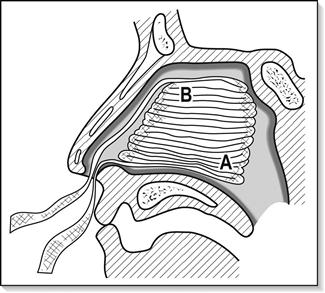
If the bleeding cannot be controlled by the above measures, or the bleeding point is posteriorly placed, the nasal cavity should be packed. There are several ways to pack the nose, the most traditional being to use ribbon gauze to fill the entire nasal cavity in layers (Fig. 18.1.2). A Foley urinary catheter can be used to control the posterior bleed, but it can be better controlled with a specifically designed epistaxis catheter, such as a Brighton’s epistaxis catheter, which has a double balloon for anterior and posterior tamponade. A Merocel nasal pack (Xomed) can be used as a nasal tampon for anterior bleeds. Posterior bleeds require a Rapid Rhino which has inflatable anterior and posterior balloons with hydrocolloid fabric on the outside surface, that promotes clotting when soaked in sterile water for a few minutes prior to insertion to activate the ingredient. Almost all patients with nasal packing need admission and observation. When the above measures are unsuccessful, further invasive procedures, such as postnasal packing, examination under anaesthesia and septal surgery or arterial ligation may be required under general anaesthesia.
Summary
Patients with anterior nasal bleeds can usually be managed by chemical cautery with silver nitrate and then be discharged. Posterior bleeding or failure to control by simple measures may require nasal packing and admission for further invasive procedures.
The throat
Foreign body
Coins are a common oesophageal foreign body in children. In adults, the foreign body is usually a fish, chicken or meat bone and, occasionally, objects such as partial dentures, safety pins, etc.
The common lodgement sites include the cricopharynx, the oesophagus at the level of the aortic arch and the gastro-oesophageal junction. Fish bones can lodge in the tonsil, the posterior third of the tongue or the vallecula prior to entering the oesophagus.
Management
Careful examination of the oropharynx initially, especially if the patient localizes the foreign body above the level of the hyoid and to one side. If the foreign body is found it should be removed under direct vision.
A foreign body at or below the cricopharynx requires general anaesthesia and endoscopy.
Lateral X-ray of the neck for soft tissues is useful in identifying and localizing radiopaque foreign bodies, such as coins and bones, including large fish bones.
Patients presenting with odynophagia but no dysphagia following the ingestion of a fish bone and a negative physical examination, can be discharged safely for review in 48 h. Symptoms of increasing odynophagia, fever, haematemesis or dysphagia warrant admission for endoscopy. Patients with a confirmed foreign body should be admitted for endoscopy and removal.
ENT surgeons use a rigid scope for visualizing and removal of foreign bodies at the cricopharynx and the proximal one-third of the oesophagus and gastroenterologists use a flexible scope for distal oesophageal foreign bodies which can be removed or more commonly pushed distally into the stomach.
A food bolus not containing bone causing obstruction should have a trial of IV glucagon 1 mg, IV buscopan 20–40 mg and sips of a fizzy drink before arranging for an endoscopy, as one of these may dislodge the obstruction, thereby avoiding an urgent endoscopy. Recurrent food bolus obstructions require an endoscopy to rule out stricture or malignancy.
Button batteries
A button battery lodged in the oesophagus can cause liquefaction necrosis and perforation so urgent endoscopy and removal is recommended [14].
If the battery is lodged past the oesophagus in the stomach most, if not all, will pass in the next 48–72 h and the patient can be safely discharged following reassurance [15].
Inhaled foreign body
History of choking, coughing or gagging while eating.
Immediate treatment is firm back blows to dislodge the foreign body. Examination may reveal unilateral wheeze. Inspiratory and expiratory chest X-ray may reveal the foreign body or air trapping distal to it.
Urgent referral for a bronchoscopy and removal of the object, especially if the foreign body is organic material, such as nuts, which can cause excessive tissue reaction and pneumonitis if there is excessive delay.
Infection
Tonsillitis
Patients with acute tonsillitis present to the general practitioner and, occasionally, to the emergency department. The emergency department patients usually have severe symptoms or are not responding to oral antibiotics. They are often dehydrated, toxic, with a high temperature and unable to take adequate oral fluids. Treatment includes intravenous penicillin in high doses (e.g. 2 g 4-hourly), intravenous fluids, one off dose of 8 mg of intravenous dexamethasone and adequate analgesia.
Infectious mononucleosis (glandular fever)
The symptoms are similar to severe tonsillitis with odynophagia, fever and a hot potato voice. The tonsils are quite swollen but have a smooth diffuse swelling with not much exudate on the fossa. The cervical lymph nodes are enlarged and tender and there may also be axillary and inguinal lymphadenopathy with or without hepatosplenomegaly.
The diagnosis is made by abnormal lymphocytes in the blood film and monospot test.
Treatment is symptomatic. Amoxicillin should not be used as it produces a rash.
Quinsy
Peritonsillar abscess or quinsy is a condition which occurs mostly in young adults in which the infection in the tonsil has breached the capsule and caused cellulitis in the adjacent soft palate (peritonsillitis) and, eventually, a collection of pus (quinsy).
Examination reveals a congested tonsil being pushed medially and downwards by a diffuse swelling of the soft palate. The opposite tonsil may look injected. There are often unilateral or bilateral enlarged and tender jugulodigastric lymph nodes in the neck. The patient is usually febrile, toxic and dehydrated. There is marked trismus due to masseteric spasm and referred otalgia.
Management includes admission to hospital for intravenous fluids, penicillin or cephalosporin and adequate analgesia. Aspiration of the pus using a large-bore needle or incision and drainage of the quinsy can be done in the emergency department under local anaesthesia in a conscious patient sitting up. Sometimes this needs general anaesthesia. Intubation of such patients should be performed by a skilled anaesthetist preferably by awake fibreoptic technique and every effort must be made to avoid rupturing the abscess to avoid aspiration of pus.
A second attack of quinsy usually requires tonsillectomy.
Retropharyngeal abscess
This is predominantly a disease of young children, as the retropharyngeal lymph nodes atrophy after the age of 5. In older patients, it could be secondary to trauma or lodgement of a foreign body, such as a fish bone. Diagnosis is made on symptoms of fever, swelling of the neck due to cervical lymphadenopathy and, especially in young children, stridor. Clinical suspicion leads to imaging procedures, such as CT, which is diagnostic.
Management includes admission to hospital, intravenous antibiotics and urgent ENT consultation. Treatment is incision and drainage of the abscess under general anaesthesia.
Epiglottitis
Childhood epiglottitis is rare these days due to a highly successive immunization programme against Haemophilus influenzae. Adults with epiglottitis are still occasionally seen in emergency departments. The classical symptoms are acutely painful throat, drooling, odynophagia and increased pain to speech with enlarged tender bilateral cervical lymph nodes.
A quick bedside test is to ask the patient to poke his or her tongue out and wiggle it from side to side as this will increase the pain substantially.
Management is mainly intravenous antibiotics and analgesia. If the patient is showing signs of imminent upper airway obstruction, an attempt should be made to intubate by the most skilled emergency doctor available with the surgical airway kit readily available in case of failure.
All other patients should be taken to the operating theatre for a gaseous induction by a skilled anaesthetist with an ENT surgeon standing by for immediate surgical airway if unable to intubate.
Post-tonsillectomy bleed
Haemorrhage from the tonsillar fossa that occurs 24 h after tonsillectomy is termed ‘secondary haemorrhage’. This differs from primary, which happens during surgery, and reactionary haemorrhage, which occurs within 24 h of surgery while the patient is still in the hospital. The cause of secondary haemorrhage is usually infection and this occurs classically 10 days postoperatively. This incidence is about 1% and is usually not very severe. The management is intravenous antibiotics, usually penicillin. The patient should be admitted and bloods taken for estimation of haemoglobin and cross-matching. Application of a swab soaked in 1 in 1000 adrenaline (epinephrine) to the tonsillar fossa after removal of the clot may help to stop the bleeding. Rarely, the patient may need to have a general anaesthetic to cauterize/ligate the bleeder [16].


