16. Video-Assisted Thoracotomy Surgery for Implantation of an Epicardial Left Ventricular Lead

Published on 02/03/2015 by admin

Filed under Cardiovascular

Last modified 22/04/2025

Print this page

rate 1 star rate 2 star rate 3 star rate 4 star rate 5 star
Your rating: none, Average: 0 (0 votes)

This article have been viewed 1385 times

History

Nonischemic cardiomyopathy with a left ventricular ejection fraction of 15% to 20%, down from 30% in a span of 6 months, and New York Heart Association (NYHA) class III congestive systolic heart failure was diagnosed 5 months before the current admission. The patient also had a long history of chronic anemia that predates the decompensated heart failure. The patient was on optimal medical therapy. She underwent placement of an implantable cardioverter-defibrillator (ICD) 2 months earlier with the intent to place a cardiac resynchronization therapy device, but a left ventricular lead could not be placed through the coronary sinus. During the procedure, coronary sinus venography showed one high lateral branch with a proximal right-angle curve and a tight stenosis at the tip of the angle, a midcardiac vein with no posterior or lateral branches, and an anterior branch. The high lateral branch was cannulated with a Whisper wire, and using the over-the-wire technique an attempt was made to advance the lead into the branch. However, the lead could not be advanced beyond the stenosis. Repeat venography to better assess the severity of the stenosis demonstrated the vein to be occluded beyond the stenosis. Because no other vein suitable for transvenous left ventricular lead placement was available, the patient was referred for surgical epicardial lead implantation.

Current Medications

The patient was taking aspirin 81 mg daily, carvedilol 6.25 mg twice daily, furosemide 20 mg twice daily, potassium chloride 10 mEq daily, spironolactone 25 mg daily, and valsartan 40 mg daily.

Current Symptoms

The patient was experiencing shortness of breath when walking short distances (NYHA class III).

Physical Examination

Laboratory Data

Electrocardiogram

Findings

The findings on the electrocardiogram were ventricular rate 81 bpm, atrial rate 81 bpm, pulse rate 146 ms, QRS 140 ms, QT 420 ms, QTc 487 ms, and P-R-T axes 039, 51, and 127 degrees (Figure 16-1).
image

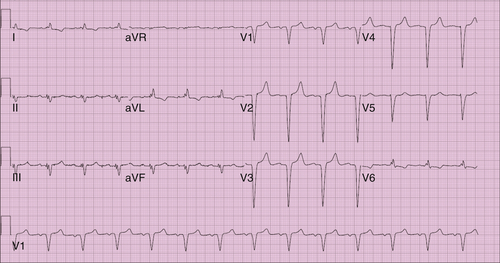
FIGURE 16-1 Electrocardiogram 3 days before surgery.

image

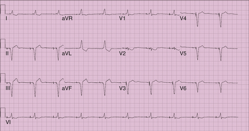
FIGURE 16-2 Electrocardiogram 20 days after surgery.

Comments

Normal sinus rhythm, left axis deviation, anterior wall myocardial infarction (age indeterminate) were noted.

Findings

Repeat electrocardiogram findings were ventricular rate 60 bpm, atrial rate 60 bpm, pulse rate 128 ms, QRS 130 ms, QT 400 ms, QTc 400 ms, and P-R-T axes 0, 80, and 105 degrees (Figure 16-2).

Comments

Atrioventricular pacing with biventricular system.

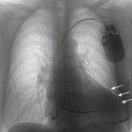
Chest Radiographs Findings

Figure 16-3 shows the heart is mildly enlarged, with left ventricular dominance. Calcification is seen within the aortic knob. An ICD is satisfactorily oriented with right atrial and ventricular leads. The hilar structures are mildly prominent. Small bilateral pleural effusions are seen.
In Figure 16-4, the cardiac silhouette is marginal in size, with intracardiac defibrillator leads directed into the right atrium and both ventricles. Also, a left chest tube had been placed. An abnormal pleural-based opacity is visible in the right lower thorax. Parenchymal consolidation or pleural fluid is not otherwise demonstrated.
Immediately postoperatively there was a patchy right basilar infiltrate and/or a right pleural effusion present with small residual left pneumothorax, see white arrows (see Figure 16-4).
On postoperative day 20 all the previous changes noted on Figure 16-5 have disappeared with no evidence of focal pulmonary consolidation, pleural effusion, or active vascular congestion (Figure 16-6).
image

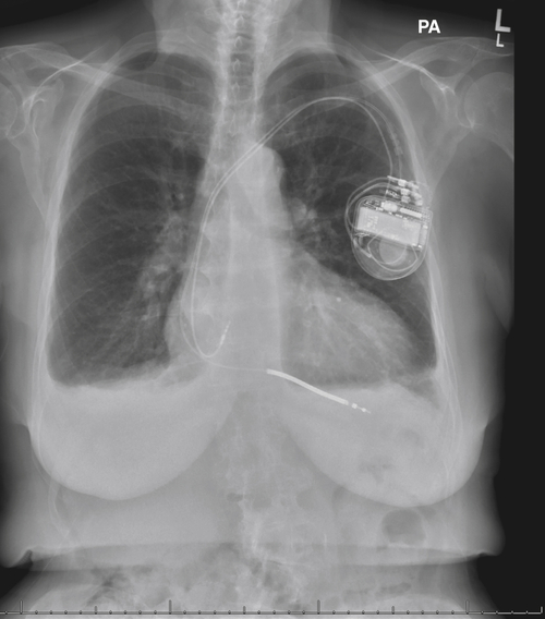
FIGURE 16-3 Anteroposterior chest radiograph obtained 3 days before surgery.

image

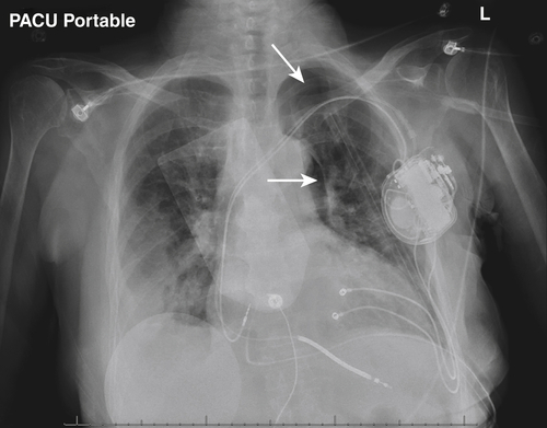
FIGURE 16-4 Postoperative anteroposterior chest radiograph. Arrows denote a very small postoperative pneumothorax.

Echocardiogram

5 Months Before Surgery

Findings

The echocardiogram revealed severely reduced systolic function of the left ventricle, with an ejection fraction of 15% to 20%. The left ventricular cavity was moderately increased. Both atria were moderately to severely dilated. Mild pulmonic regurgitation was seen, and the aortic valve was moderately calcified. Moderate-to-severe regurgitation was noted in both mitral and tricuspid valves. There was mitral valve sclerosis with a valve area of 1.36 cm2 and the pulmonary artery systolic pressure was mildly elevated at 40 to 50 mm Hg.
image

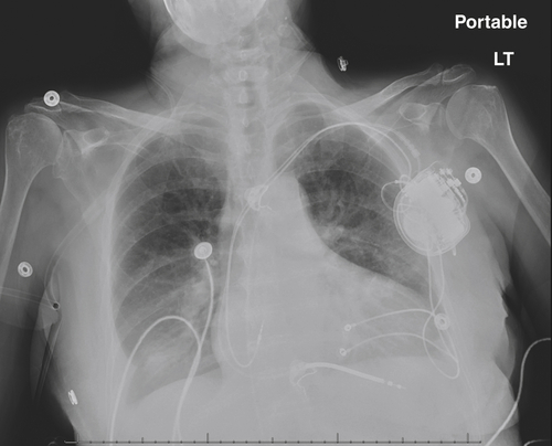
FIGURE 16-5 Anteroposterior chest radiograph obtained 3 days postoperatively. The small pneumothorax is no longer seen.

image

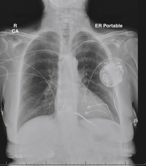
FIGURE 16-6 Anteroposterior chest radiograph obtained 20 days postoperatively.

image

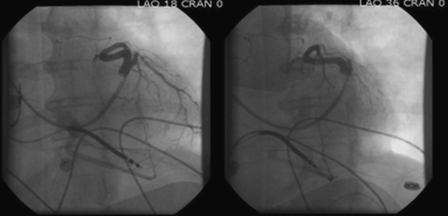
FIGURE 16-7 Venogram 2 months before surgery.

Postoperative Day 3

Findings

The postoperative echocardiogram revealed severely reduced systolic function of the left ventricle and ejection fraction increased to 20% to 25%. Moderate paradoxical motion of the septum was seen. The left ventricular cavity size was moderately increased and the left atrium mildly dilated. A pacemaker wire was identified in the right ventricle. Mild-to-moderate regurgitation was noted in both mitral and tricuspid valves. The pulmonary artery systolic pressure was mildly elevated, at 35 to 45 mm Hg.

Venogram

Findings

Using the balloon occlusion technique a coronary sinus venogram was obtained in the left anterior oblique (LAO) projection using ioversol (Optiray) contrast agent (Figure 16-7). Venography showed one high lateral branch with a proximal right-angle curve and tight stenosis at the tip of the angle, a midcardiac vein with no posterior or lateral branches, and an anterior branch. A Whisper wire with extra distal support was used to cannulate the high lateral branch. Using an over-the-wire technique, a bipolar Attain Ability Plus steerable lead was advanced to the proximal portion of the lateral branch. However, the lead was unable to advance beyond the stenosis point and was removed so a smaller lead could be used. A repeat venogram in right anterior oblique and LAO projections to better assess the degree of stenosis showed that the vein was occluded beyond the stenosis. Left ventricular lead placement was abandoned because no other vein could accommodate a left ventricular lead on the lateral or posterior wall.

Focused Clinical Questions and Discussion Points

Question

Why was this patient recommended for biventricular pacing?

Discussion

The patient had NYHA class III congestive heart failure and severely impaired left ventricular systolic function, as demonstrated by the extremely low ejection fraction. In addition, the patient had impairment despite traditional right atrial–right ventricular pacing. The lack of response is likely related to the nonspecific intraventricular block, indicating the potential benefit of the addition of left ventricular pacing.

Question

Why was this patient considered for surgical left ventricular lead placement?

Discussion

The surgical platform allows for direct epicardial access, thereby overcoming the limitations of coronary sinus anatomy in situations in which no branches or inadequate venous branches in the preferred target zone are available. In this patient, venography of the heart showed only one high lateral branch suitable for left ventricular lead placement, but it had a proximal right-angle curve and a tight stenosis at the tip of the angle that occluded beyond the stenosis during the lead placement attempt. The other coronary sinus branches, a midcardiac vein with no posterior or lateral branches, and an anterior branch were not desirable alternatives. Direct visualization of scar tissue and phrenic nerve position avoids left ventricular lead placement at sites of a previous myocardial infarction and diaphragmatic capture, respectively. Transvenous left ventricular placement should be the first option, but epicardial lead placement should be considered in cases of aberrant coronary sinus anatomy, venous branching that precludes stable lead placement or placement in a suitable target zone (as in this patient), phrenic nerve stimulation, or excessive pacing thresholds via transvenous route.

Question

What are the advantages and disadvantages of performing video-assisted thoracotomy surgery over other surgical platforms?

Discussion

Video-assisted thoracotomy surgery is a minimally invasive technique that produces less pain and has a shorter recovery time while still allowing for visualization of the epicardium. However, because the procedure is minimally invasive, the operating field is smaller than that with a limited thoracotomy. In addition, the lack of tactile feedback can make the operation difficult for an inexperienced surgeon. However, these obstacles can be overcome as the surgeon gains more experience with the procedure.

Final Diagnosis

The final diagnosis was severe congestive heart failure after placement of an ICD.

Plan of Action

The plan of action for this patient was video-assisted thoracotomy for implantation of an epicardial left ventricular lead.

Intervention

The patient was positioned in the supine position on the operating room table. Intravenous cefazolin was given less than 1 hour before skin incision (discontinued within 24 hours). The patient was then intubated with a double-lumen endotracheal tube. The lines were placed by the anesthesia team. She was then prepped and draped in the usual sterile manner, and a beanbag was used to keep her in the right lateral decubitus position. To avoid interfering with the procedure, the left arm was supported and retracted to allow access to the pacemaker pocket.
Three small incisions were made in the third, fifth, and eighth intercostal spaces, and three ports were inserted. A camera was passed through the middle port, and the pericardium was visualized. The pericardium was opened and the first marginal branch of the circumflex artery visualized. The left atrial appendage was also visualized. At this time, an attempt to deploy one of the leads through the superior port failed; the lead would not deploy properly from its holder because of the small intercostal space. A small, 2-cm incision was made at the level of the camera and the leads deployed through the opening. Two leads were placed at the base of the heart in between the obtuse marginal branch and branches of the circumflex artery.
The proximal ends of the pacing wires were then guided submuscularly through the third intercostal space to the pacemaker pocket. One of the leads was connected to the ICD generator, and the backup lead was capped and placed posterior to the ICD generator. At this time, a Blake drain was placed in the pericardial sac and the pericardium was closed. A posterior chest tube was placed in the pleural space. The working port, generator pocket, and port site incisions were all closed in the usual manner. The patient was then extubated in the operating room and taken to the recovery room in stable condition. No complications occurred during the operation.
The new leads were tested and were found to be working very well. The left ventricular lead connected to the generator had a pacing threshold of 0.5 V with an impedance of 532 Ω, and the backup left ventricular lead had a threshold of 2.5 V with an impedance of 794 Ω. The pacemaker was programmed to DDD and set to an atrioventricular delay of 130 msec and a V-V delay of 20 ms, with left ventricular stimulation preceding right ventricular stimulation by 20 ms.

Outcome

Left ventricular lead implantation carries a small failure rate because of variant coronary sinus and venous anatomy, as demonstrated in this patient.1,2 Furthermore, lead dislodgement can result in an additional 5% to 10% late failure rate of left ventricular lead capture and sensing.2,7 In cases in which transvenous left ventricular lead insertion is not feasible, the patient should be considered for surgical implantation of an epicardial lead. Surgical left ventricular lead placement has been shown to be highly successful in nearly 100% of attempts and offers the additional advantage of direct access to the left ventricular surface, leading to the possibility of detailed left ventricular mapping and precise site-directed resynchronization.10
The development of video-assisted thoracotomy surgery began with the thoracotomy, which was an attempt to avoid the complications associated with a full sternotomy while still allowing for excellent exposure of the posterolateral wall. However, the traditional thoracotomy involved a large incision and formal rib spreading and therefore was quite invasive; thus the limited thoracotomy was created. The limited thoracotomy approach allows for minimally invasive access to the anterior or lateral walls without rib spreading. In both cases, the incisions are kept to a minimum and rib spreading is avoided. Surgeons have gained significant experience in using these methods because they are commonly used in coronary revascularization procedures such as in minimally invasive direct coronary artery bypass in which the left internal mammary artery is anastomosed to the left anterior descending artery by a small anterior thoracotomy. Reports on the outcomes of limited left thoracotomy are positive, showing an average length of stay in the intensive care unit of 2.1 days, with no reports of significant complications, morbidity, or mortality rates79 with a 5-day hospital stay.8
Video-assisted thoracotomy developed as an extension of the limited thoracotomy. Videoscopic guidance enhances exposure to the posterolateral surface of the left ventricle, limiting incision size and the need for rib spreading. Postoperative pain and discomfort are decreased with these approaches, and significant improvement of visualization of the entire area of interest allows more precise left ventricular lead placement. The proper site of lead implantation is paramount to the success of complete resynchronization.3 More specifically, posterolateral pacing on the left ventricle has been shown to result in optimal resynchronization, whereas some evidence suggests that more anterior sites may actually hinder resynchronization.3 The ideal lead for this approach is the screw-in lead. The introduction of new tools for the secure and controlled deployment of the lead has made minimally invasive approaches for left ventricular lead placement more feasible. Video-assisted thoracotomy has been shown to be comparable to the limited thoracotomy in terms of complication and mortality rates.46
The average length of hospital stay for a patient who has undergone video-assisted thoracotomy is approximately 4 days.4,5 In the patient with prior cardiac surgery, fine and careful dissection of all surrounding structures can be difficult because the surgeon works without tactile feedback. Nonetheless, enhanced left ventricular access and left ventricular mapping have been facilitated by the aid of the videoscope and can be easily accomplished in most cases.

Selected References

1. Abraham W.T. Cardiac resynchronization therapy for heart failure: biventricular pacing and beyond. Curr Opin Cardiol. 2002;17:346–352.

2. Alonso C., Leclercq C., d’Allonnes F.R. et al. Six year experience of transvenous left ventricular lead implantation for permanent biventricular pacing in patients with advanced heart failure: technical aspects. Heart. 2001;86:405–410.

3. Ansalone G., Giannantoni P., Ricci R. et al. Doppler myocardial imaging to evaluate the effectiveness of pacing sites in patients receiving biventricular pacing. J Am Coll Cardiol. 2002;39:489–499.

4. Fernandez A.L., Garcia-Bengochea J.B., Ledo R. et al. Minimally invasive surgical implantation of left ventricular epicardial leads for ventricular resynchronization using video-assisted thoracoscopy. Rev Esp Cardiol. 2004;57:313–319.

5. Gabor S., Prenner G., Wasler A. et al. A simplified technique for implantation of left ventricular epicardial leads for biventricular re-synchronization using video-assisted thoracoscopy (VATS). Eur J Cardiothorac Surg. 2005;28:797–800.

6. Mair H., Jansens J.L., Lattouf O.M. et al. Epicardial lead implantation techniques for biventricular pacing via left lateral mini-thoracotomy, video-assisted thoracoscopy, and robotic approach. Heart Surg Forum. 2003;6:412–417.

7. Mair H., Sachweh J., Meuris B. et al. Surgical epicardial left ventricular lead versus coronary sinus lead placement in biventricular pacing. Eur J Cardiothorac Surg. 2005;27:235–242.

8. Puglisi A., Lunati M., Marullo A.G. et al. Limited thoracotomy as a second choice alternative to transvenous implant for cardiac resynchronisation therapy delivery. Eur Heart J. 2004;25:1063–1069.

9. Shah R.V., Lewis E.F., Givertz M.M. Epicardial left ventricular lead placement for cardiac resynchronization therapy following failed coronary sinus approach. Congest Heart Fail. 2006;12:312–316.

10. Steinberg J.S., Derose J.J. The rationale for nontransvenous leads and cardiac resynchronization devices. Pacing Clin Electrophysiol. 2003;26:2211–2212.

Share this: