15. Persistent Left Superior Vena Cava

Published on 26/02/2015 by admin

Filed under Cardiovascular

Last modified 22/04/2025

Print this page

rate 1 star rate 2 star rate 3 star rate 4 star rate 5 star
Your rating: none, Average: 0 (0 votes)

This article have been viewed 1023 times

History

This patient had complete heart block, with 30% left ventricular ejection fraction (LVEF) visualized on echocardiography. A temporary pacemaker wire was inserted into the right ventricle via femoral venous access. Coronary artery angiography was performed, ruling out coronary artery disease. The patient was scheduled for a biventricular pacemaker.

Current Medications

The patient was taking carvedilol 6.25 mg twice daily, digoxin (Lanoxin) 0.25 mg daily, furosemide plus spironolactone (Lasilactone) 25 mg twice daily, and enalapril 2.5 mg twice daily.

Current Symptoms

The patient was experiencing symptoms consistent with New York Heart Association (NYHA) class III symptoms.

Physical Examination

Laboratory Data

Comments

The patient’s hematologic and chemistry values were normal.

Electrocardiogram

Findings

The patient had a left bundle branch block and a QRS duration of 140 ms.

Chest Radiograph

Findings

Chest radiography revealed cardiomegaly, with a cardiothoracic ratio of 60%.
image

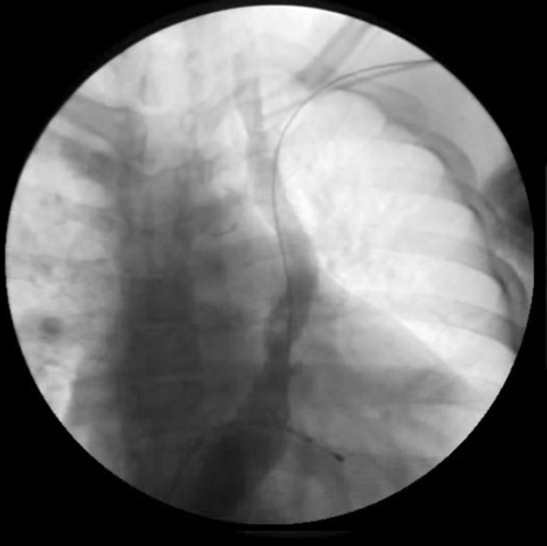
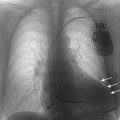
FIGURE 15-1 Posterior-anterior fluoroscopic view showing the guide wire in serted via left axillary vein puncture, tracking into the persistent left superior vena cava. Contrast injection did not show an innominate vein or posterior lateral tributaries suitable for coronary sinus lead implantation.
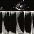
Echocardiogram

Findings

Echocardiolography revealed dysynergic septal motion with an LVEF of 30%.

Final Diagnosis

The final diagnosis in this patient was idiopathic cardiomyopathy with persistent left superior vena cava and NYHA class III disease.

Intervention

Left axillary vein access was obtained, revealing a persistent left superior vena cava (PLSVC). A venogram was performed via the introducer sheath, showing absence of an innominate vein; it did not show any postero-lateral branches (Figure 15-1). The levophase of the coronary artery angiogram was then retrieved, revealing a posterolateral vein (Figure 15-2). A Judkins right 4 diagnostic catheter was inserted via an Attain MB2 coronary sinus guiding sheath (Medtronic, Minneapolis, Minn.) to subselect the posterolateral vein (Figure 15-3). A Medtronic Starfix lead was implanted over a 0.014-inch balanced middle-weight guide wire (Figure 15-4). This lead was chosen to ensure stability. A right atrial screw-in lead was placed in the right atrial appendage, and a right ventricular lead on the interventricular septum using a J-stylet that allowed easy positioning of the lead directly at the desired site (Figure 15-5).
image

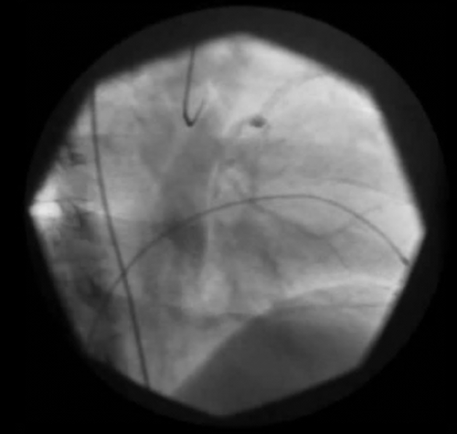
FIGURE 15-2 Levophase of the left coronary angiogram in the right anterior oblique 30-degree projection showing a dilated coronary sinus (the persistent left superior vena cava is also visible) and a posterior lateral vein.

Outcome

Comments

As mentioned in the case in Chapter 14, coronary sinus lead implantation is often easier via a right-sided venous access with retrograde cannulation of the coronary sinus. However, in rare cases, the right superior vena cava is absent1 or coronary sinus atresia2 may be present, with drainage to the subclavian vein. In these cases, the only option is to implant the coronary sinus lead via the PLSVC.
In our patient, the levophase of the coronary arteriogram was useful in revealing the presence of a lateral vein that was accessible via the PLSVC and allowed successful coronary sinus lead implantation without having to cross over to a right-sided venous access. Positioning of the right atrial lead is easy via the PLSVC, but the right ventricular lead may be challenging. A useful technique is to use a J-shaped stylet that allows positioning of the lead directly on the interventricular septum.
image

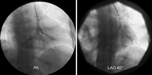
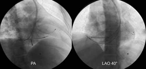
FIGURE 15-3 Subselection of the lateral vein with a Judkins right 4 diagnostic catheter in the posterior-anterior (left) and left anterior oblique 40-degree (right) views.

image

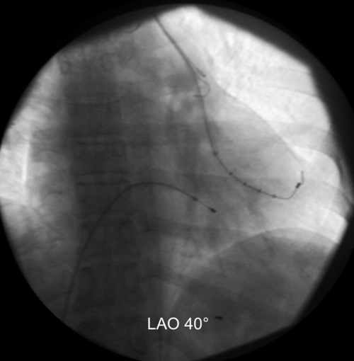
FIGURE 15-4 Positioning of the left ventricular lead in the lateral branch.

image

FIGURE 15-5 Final position of the lead in the posterior-anterior (left) and left anterior oblique 40-degree (right) views.

Selected References

1. Ratliff H.L., Yousufuddin M., Lieving W.R. et al. Persistent left superior vena cava: case reports and clinical implications. Int J Cardiol. 2006;113:242–246.

2. Gasparini M., Mantica M., Galimberti P. et al. Biventricular pacing via a persistent left superior vena cava: report of four cases. Pacing Clin Electrophysiol. 2003;26:192–196.