[level-membership-for-emergency-medicine-category]
Genitourinary Emergencies
Edited by George Jelinek
10.1 Acute kidney injury
Nicholas Adams and Linas Dziukas
Introduction
The basic process in acute kidney injury (AKI) is a rapid (hours to days) reduction in the glomerular filtration rate (GFR) due to renal hypoperfusion, damage to glomeruli, tubules, interstitium or blood vessels, or obstruction to urine flow. The GFR is inversely related to the serum creatinine (SCr) concentration and the diagnosis of AKI is made when there is an acute increase in the SCr concentration, with or without a decrease in the urine output. A simple definition of AKI is an acute and sustained (lasting for 48 hours or more) increase in the SCr of 44 μmol/L if the baseline is less than 221 μmol/L, or an increase in the SCr of more than 20% if the baseline is more than 221 μmol/L. A more comprehensive definition (the RIFLE system) is used to classify persons with acute impairment of renal function (Table 10.1.1) [1].
Table 10.1.1
RIFLE classification of acute renal failure
| Stage | Serum creatinine (SCr) concentration | Urine output |
| RISK | Increase of 1.5 times the baseline | <0.5 mL/kg/h for 6 h |
| INJURY | Increase of 2.0 times the baseline | <0.5 mL/kg/h for 12 h |
| FAILURE | Increase of 3.0 times the baseline or SCr is 355 μmol/L or more when there has been an acute rise of greater than 44 μmol/L for 24 h or anuria for 12 h | <0.3 mL/kg/h |
| LOSS | Persistent acute renal failure; complete loss of kidney function for longer than 4 weeks | |
| END-STAGE RENAL DISEASE | End-stage renal disease for longer than 3 months |
Aetiology and pathogenesis
The causes of AKI are grouped according to the source of renal injury: prerenal (hypoperfusion), renal (parenchymal) and post-renal (obstructive). More than one cause can be present simultaneously.
Prerenal acute kidney injury
Prerenal AKI is initially an adaptive response to severe volume depletion and hypotension in structurally intact nephrons. Prerenal AKI that is prolonged or inadequately treated can be followed by parenchymal renal damage (acute tubular necrosis). Prerenal AKI is a potentially reversible cause of acute renal failure (ARF).
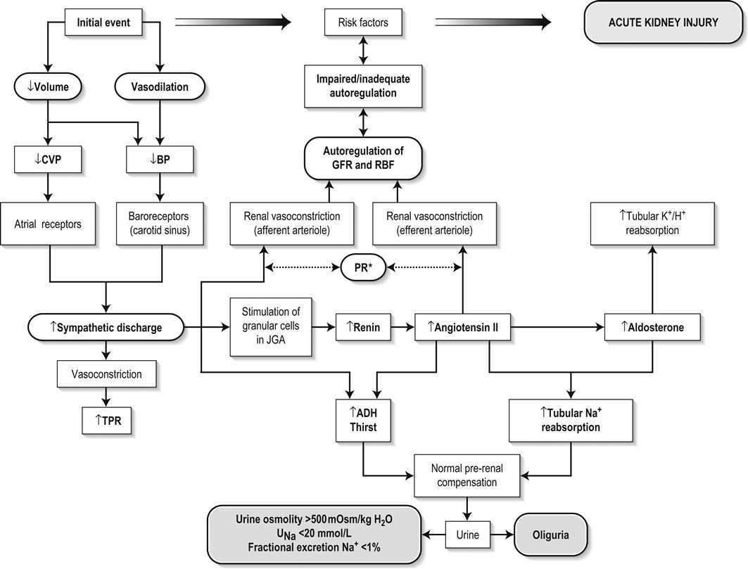
Reductions in renal blood flow (RBF) and GFR occur in the setting(s) of hypovolaemia, hypotension, oedematous states with a reduced ‘effective’ circulating volume (cardiac failure, hepatic cirrhosis, nephrotic syndrome) or impaired renal perfusion (renal artery stenosis, hepatorenal syndrome). Drugs that interfere with renal autoregulation (e.g. prostaglandin inhibitors, angiotensin-converting enzyme [ACE] inhibitors or angiotensin II receptor antagonists) can reduce glomerular perfusion [2]. The physiological responses to volume depletion and hypotension, and the link to prerenal AKI are shown in Figure 10.1.1.
Renal acute kidney injury
Ischaemic, cytotoxic or inflammatory processes may damage the renal parenchyma. The causes of the damage can be grouped according to the major structures that are damaged: vessels, glomeruli, renal tubules or renal interstitial tissue.
Vascular causes involving the larger vessels include acute thrombosis of the renal artery, embolism of the renal arteries, renal artery dissection and renal vein thrombosis. Microvascular causes include vasculitis, malignant hypertension and thrombotic microangiopathy.
The glomeruli are the site of injury in acute glomerulonephritis which can cause proteinuria, haematuria, nephrotic syndrome or nephritic syndrome. A number of different forms of glomerulonephritis have been described, generally diagnosed by the histological changes seen on renal biopsy. The distinction between these forms is not of direct concern for the emergency practitioner.
Acute tubular necrosis (ATN) is the most common pathological process that causes AKI. While the terminology suggests that the main cause is tubular damage, the actual pathophysiology is more complex: impaired autoregulation and marked intrarenal vasoconstriction (the main mechanism for the greatly reduced GFR), tubular damage (with cytoskeleton breakdown), increased tubuloglomerular feedback, endothelial cell injury, fibrin deposition in the microcirculation, release of cytokines, activation of inflammation and activation of the immune system [3].
ATN is often classified as ischaemic ATN or cytotoxic ATN but both processes may be present in some patients. Ischaemic ATN represents an advanced form of prerenal AKI, but the distinction between these two entities is based on histopathological changes and is of little use to the clinician. Important causes of cytotoxic ATN are listed in Table 10.1.2. Non-steroidal anti-inflammatory drugs (NSAIDs), ACE inhibitors and angiotensin receptor blockers (ARBs) often cause a gradual and asymptomatic decrease in the GFR, but can also cause AKI. NSAIDs do not impair renal function in healthy persons, but can reduce the GFR in the elderly with atherosclerotic cardiovascular disease, in persons with chronic renal failure, when chronic prerenal hypoperfusion is present (e.g. cardiac failure, cirrhosis) or in persons using diuretics and calcium channel blockers [4]. AKI may occur after the administration of intravenous or intra-arterial radiocontrast agents. A number of risk factors have been identified for this, the most important being pre-existing renal impairment, hypovolaemia, a large contrast load and the use of hyperosmolar contrast agents [5]. Drugs that alter angiotensin levels (ACE inhibitors and ARBs) reduce renal perfusion by their antihypertensive effects or by impairing vasoconstriction of the efferent arteriole when renal perfusion is reduced by renal artery stenosis. The nephrotoxicity of haem pigments (myoglobin and haemoglobin) is enhanced by volume depletion, low urine flow rates and possibly low urine pH.
Table 10.1.2
Causes of toxic acute tubular necrosis
Exogenous agents
Radiocontrast
Non-steroidal anti-inflammatory drugs
Antibiotics: aminoglycosides, amphotericin B
Antiviral drugs: acyclovir, foscarnet
Immunosuppressive drugs: ciclosporin
Organic solvents: ethylene glycol
Poisons: snake venom, paraquat, paracetamol
Chemotherapeutic drugs: cisplatin
Herbal remedies
Heavy metals
Endogenous agents
Haem pigments: haemoglobin, myoglobin
Uric acid
Myeloma proteins
Correct intravascular volume depletion
Maintain perfusion pressure
Choice of resuscitation fluid
Diuresis in rhabdomyolysis
Avoid nephrotoxins
Use derived GFR or creatinine clearance when calculating drug doses
Abnormalities of renal interstitial structure and function are only one feature of ATN, but represent the primary abnormality in acute tubulointerstitial nephritis (ATIN). The damage in ATIN is due to immunological mechanisms, the most important involving cell-mediated immunity. ATIN is usually due to an allergic reaction to a drug, commonly antibiotics (β-lactam antibiotics, sulphonamides, fluoroquinolones), NSAIDs, cyclooxygenase-2 inhibitors, proton pump inhibitors, diuretics, phenytoin, carbamazepine and allopurinol.
Post-renal acute kidney injury
Obstructive uropathy refers to the functional or structural processes in the urinary tract that impede the normal flow of urine and obstructive nephropathy is the renal damage caused by the obstruction. Hydronephrosis and hydroureter refer to dilatation of the renal urinary collecting system and the ureters, respectively. They may occur in the absence of obstruction and, conversely, may be absent in some patients with obstruction.
Casts or crystals within the renal tubular lumen can cause intrarenal obstruction. Extrarenal obstruction can develop in the urethra, bladder, ureter or the pelvi-ureteric junction. Obstructive uropathy in adults is commonly caused by prostate disease or retroperitoneal neoplasm (cancer of the cervix, uterus, bladder, ovary or colon). Metastatic cancer, lymphomas or inflammatory processes in the retroperitoneum (appendicitis, diverticulitis, Crohn’s disease) or a neurogenic bladder can also cause obstructive uropathy. Bilateral renal stones are an uncommon cause of obstructive uropathy.
Obstructive nephropathy usually develops gradually and can cause chronic renal failure if the obstruction involves the urethra, the bladder or both ureters. Unilateral ureteric obstruction will cause AKI only if it involves a single functioning kidney.
Epidemiology
Studies of the pathogenesis of community-acquired ARF have produced conflicting results. In one study, the major processes were identified as prerenal in 70% of cases, renal in 11% of cases and post-renal in 17% of cases [6]. There are geographical differences in the causes of ATN. In Africa, India, Asia and Latin America, ATN is usually caused by infections (e.g. diarrhoeal illnesses, malaria, leptospirosis), ingestion of plants or medicinal herbs, envenomation, intravascular haemolysis due to glucose-6-phosphate dehydrogenase deficiency or poisoning.
Prevention
Maintaining intravascular volume and renal perfusion
The rate and volume of intravenous fluid given to hypovolaemic persons depends on the nature of the intravascular depletion, the blood pressure and heart rate, the (estimated) volume of fluid lost, cardiac function and ongoing circulatory losses. The response to treatment is evaluated by simple bedside measurements (heart rate, blood pressure, urine output).
Rhabdomyolysis
Most studies on the prevention of ATN after rhabdomyolysis have been in persons with crush injury after earthquakes, where the incidence of AKI is about 50%. In this situation, fluid resuscitation should, if possible, begin before the crush is relieved. These patients may require massive amounts of fluid because of fluid sequestration in the injured muscles. The goal of intravenous fluid treatment is to produce a urine output of 200–300 mL/h while myoglobinuria (discoloured urine) persists. There is no evidence to support this rate of fluid replacement in persons who have rhabdomyolysis and AKI without crush injury, although a urine output of 100 mL/h would be reasonable while the urine is discoloured. The intravenous administration of mannitol and sodium bicarbonate to produce an alkaline diuresis as a means of preventing ATN in severe rhabdomyolysis has not been shown to be effective [7].
Radiocontrast nephropathy
The incidence of radiocontrast nephropathy can be reduced by saline infusion to produce intravascular volume expansion and by using low osmolar contrast agents. N-acetyl cysteine administration before and after radiocontrast administration does not appear to be effective [5].
Clinical features
The diagnosis of AKI should be considered when there is a decrease in urine output or an elevated SCr concentration. The clinical features depend on the pre-existing conditions that increase the risk of developing AKI, the initiating factor(s) and the effects of AKI (Fig. 10.1.2). The history should include a detailed drug history, enquiry about recent invasive vascular or radiological procedures and any family history of renal disease. This is followed by clinical examination and evaluation of investigations. A number of key issues then need to be resolved (Table 10.1.3).
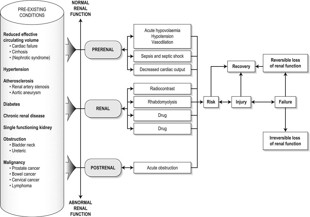
Table 10.1.3
Evaluation of acute kidney injury
Assess the intravascular volume
Look for renovascular disease
Look for symptoms or signs of obstruction to urine flow
Systematic search for presence of infection or sepsis
Evaluate for pre-existing renal disease or chronic renal failure
Obtain a detailed history of medication or drug use
Consider possibility of glomerulonephritis
Evaluation of prerenal (intravascular volume) status
Imprecise terminology, such as ‘dry’ or ‘dehydrated’, should be avoided. ‘Dehydration’ refers to situations where more water than electrolyte(s) has been lost, shrinking body cells and increasing the serum sodium concentration and osmolality. In other words, ‘dehydration’ means water depletion. Hypovolaemia is a decrease in the intravascular volume due to loss of blood (haemorrhage, trauma) or loss of sodium and water (e.g. vomiting, diarrhoea, sequestration of fluid in the bowel, etc.).
The (bedside) assessment of the (extracellular) volume status determines the initial resuscitation strategy. This involves evaluation of heart rate and blood pressure, the state of the skin and mucous membranes and the jugular venous pulse. The examination also includes auscultation of the lungs (for pulmonary crackles), abdominal examination (for ascites or masses) and examination of the legs (for peripheral oedema).
The ‘typical’ features of intravascular volume depletion (tachycardia or hypotension or both, in the supine position, or postural hypotension) are not as consistent or reliable as implied by many textbook descriptions. The presence of (supine) tachycardia has low sensitivity as a diagnostic feature of increasing hypovolaemia in healthy persons. An increase in the pulse rate of 30 beats per minute or more between the supine and standing positions is a highly sensitive and specific sign of hypovolaemia after phlebotomy of large volumes (600–1100 mL) of blood, but the sensitivity is much less after phlebotomy of smaller volumes. The inability to stand long enough for vital signs to be measured because of severe dizziness is a sensitive and specific feature of acute large blood loss. A systolic blood pressure of 95 mmHg or less in the supine position has high specificity but low sensitivity for hypovolaemia. Postural hypotension is present in 10% of normovolaemic people younger than 65 years and in up to 30% of normovolaemic people older than 65 years [8].
The textbook descriptions of the signs of saline depletion in adults (dry mucous membranes, shrivelled tongue, sunken eyes, decreased skin turgor, weakness, confusion) are neither specific nor sensitive compared to laboratory tests for hypovolaemia. The presence of a dry axilla argues somewhat for the presence of saline depletion; the absence of tongue furrows and the presence of moist mucous membranes argue against the presence of saline depletion.
The central venous pressure (CVP) is an indicator of the vena caval or right atrial pressure. A vertical distance greater than 3 cm between the top of the jugular venous pulsation (using the external jugular vein or internal jugular vein) and the sternal angle indicates that the CVP is elevated. An elevated venous pressure in persons with pulmonary crackles or peripheral oedema means that the intravascular volume is greater than normal.
The absence of visible venous pulsation in the neck veins when the patient is supine or in a head down position indicates significant intravascular volume depletion. The presence of visible venous pulsations in the neck at or below the level of the sternal angle that is seen only when the patient is supine indicates that the intravascular volume is below normal.
Evaluation of the renovascular state
Acute renal infarction is caused by dissection of the aorta or renal artery, embolism, renal artery thrombosis, renal vein thrombosis or renal artery aneurysm. Acute arterial occlusion is usually symptomatic, with the development of pain (loin, abdominal or back pain), haematuria, proteinuria, nausea and vomiting. Vascular occlusion of a single functioning kidney produces anuria.
Atheromatous disease of the renal arteries is common in persons older than 50 years with widespread atherosclerosis. Persons with stenosis or occlusion of one or both renal arteries can develop an elevation in SCr concentration after starting treatment with ACE or ARB drugs or develop acute on chronic renal failure.
Exclusion of thrombotic microangiopathy (TMA)
TMA is a syndrome of microangiopathic haemolytic anaemia, thrombocytopaenia and varying degrees of organ injury caused by platelet thrombosis in the microcirculation. There are two clinically distinct entities: haemolytic uraemic syndrome (HUS) and thrombotic thrombocytopaenic purpura (TTP). HUS affects young children and causes AKI with absent or minimal neurological abnormalities. TTP occurs in adults and causes severe neurological involvement in most cases and variable degrees of renal damage. Both conditions are rare.
Pre-existing renal disease or chronic renal failure
It can be difficult to distinguish between chronic and acute renal impairment. The following features suggest the presence of chronic renal failure: documented renal impairment in the past, family history of renal disease, polyuria or nocturia, uraemic pigmentation, normochromic and normocytic anaemia or small kidneys on ultrasound or computed tomography (CT) scans. Renal size may be normal or increased in chronic renal failure associated with diabetes, polycystic kidney disease or amyloidosis.
Exclusion of urinary obstruction
The symptoms and signs of urinary tract obstruction depend upon the site, the cause and the rapidity with which it develops. Pain is more common in acute obstruction and is felt in the lower back, flank or suprapubic region, depending on the level of the obstruction. Chronic obstruction is usually painless. Symptoms of prostatic obstruction include frequency, nocturia, hesitancy, post-void dribbling, poor urinary stream and incontinence. Bladder neck obstruction usually results in an enlarged (and palpable) bladder.
Recognition of rhabdomyolysis
Muscle necrosis releases intracellular contents into the circulation. This causes red-brown urine (that tests positive for haem in the absence of visible red cells on microscopy or tests positive for myoglobin with specific tests), pigmented granular casts in the urine, elevated serum creatine kinase (CK) levels that are five times or more above the upper limit of normal and clear serum (serum is reddish in haemolysis). The severity of the rhabdomyolysis ranges from asymptomatic elevations of muscle enzymes in the serum to AKI and life-threatening electrolyte imbalances.
Urine dipstick findings may be normal because myoglobin is renally cleared from the serum more rapidly than CK, thus myoglobinuria may be absent in patients with renal failure or those who present later in the illness. Muscle pain is absent in about 50% of cases and muscle swelling is an uncommon finding. Muscle weakness occurs in those with severe muscle damage. Fluid sequestration in muscles can cause hypovolaemia. Marked muscle swelling can cause a compartment syndrome.
Other blood test abnormalities include hyperkalaemia, AKI with rapid and marked elevation in SCr (e.g. 220 μmol/L per day), hypocalcaemia (which occurs early and is usually asymptomatic), hyperuricaemia, hyperphosphataemia, metabolic acidosis and disseminated intravascular coagulopathy [7].
Acute kidney injury and acute renal failure
The early stages of AKI are usually asymptomatic and the diagnosis is based on an elevated SCr concentration. It may take 24 hours or more for an initially normal SCr concentration to show a definite increase and up to 48 hours after the event(s) that caused the AKI to distinguish between the early stages of AKI (risk and injury) and the development of renal failure.
The urine output usually decreases and the patient may be oliguric (urine output less than 400 mL per day) or anuric (urine output less than 100 mL per day). Persons with AKI and oliguria have more severe kidney impairment than those without oliguria. Only a few conditions cause complete anuria: total obstruction, vascular lesions, severe ATN or rapidly progressive glomerulonephritis. The clinical features caused by ARF are shown in Table 10.1.4.
Table 10.1.4
Clinical features of acute renal failure
1. Anorexia, fatigue, confusion, drowsiness, nausea and vomiting, and pruritus
2. Signs of salt and water retention in the intravascular and interstitial spaces: an elevated jugular venous pressure, peripheral oedema, pulmonary congestion, acute pulmonary oedema
3. Abnormal plasma electrolyte concentrations, particularly hyperkalaemia
4. Metabolic acidosis
5. Anaemia
6. Uraemic syndrome: ileus, asterixis, psychosis, myoclonus, seizures, pericardial disease (pericarditis, pericardial effusion, tamponade)
Differential diagnosis
The diagnosis of AKI requires synthesis of data from the patient’s history, physical examination, laboratory studies and urine output. The category of AKI (Risk, Injury or Failure) may be difficult to determine in the emergency department (ED) if the baseline SCr is unknown. The reversibility of the AKI may be inferred if there is a marked increase in urine output after correction of prerenal problems, but a reduction in SCr (due to an increase in GFR) may not be seen for 12–24 hours.
Criteria for diagnosis
Serum biochemistry
The following are measured: serum concentration of electrolytes (sodium, potassium, bicarbonate, chloride, calcium, phosphate), serum urea and SCr concentrations, random blood glucose, liver function tests, coagulation tests and CK concentration.
AKI causes acute elevation in the SCr concentration or serum urea concentrations or both. In prerenal AKI, the low urine flow rate favours urea reabsorption out of proportion to decreases in GFR, resulting in a disproportionate rise of serum urea concentration or blood urea nitrogen (BUN) concentration relative to the SCr concentration. However, serum urea concentrations depend on nitrogen balance, liver function and renal function. Severe liver disease and protein malnutrition reduce urea production, resulting in a low serum urea concentration. Increased dietary protein, gastrointestinal haemorrhage, catabolic states (e.g. infection, trauma) and some medications (corticosteroids) increase urea production and increase serum urea concentration without any change in GFR.
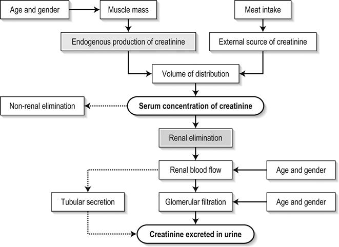
The SCr concentration is the best available guide to the GFR. Acute reductions in GFR produce an increase in the SCr concentration. The changes in SCr concentration lag behind the change in GFR and can be affected by the dilutional effect of intravenous fluid. Correct interpretation of the SCr concentration extends beyond just knowing the normal values (Fig. 10.1.3). Creatinine is a metabolic product of creatine and phosphocreatine, which are found almost exclusively in skeletal muscle. The SCr concentration is affected by the muscle mass, meat intake, GFR, tubular secretion (which can vary in the same individual and increases as the GFR decreases) and breakdown of creatinine in the bowel (which increases in chronic renal failure). The GFR decreases by 1% per year after 40 years of age, yet the SCr concentration remains unchanged because the decrease in muscle mass with age reduces the production of creatinine. The GFR (corrected for body surface area) is 10% greater in males than females, but men have a higher muscle mass per kilogram of body weight. The SCr concentration in men is thus greater than in women.
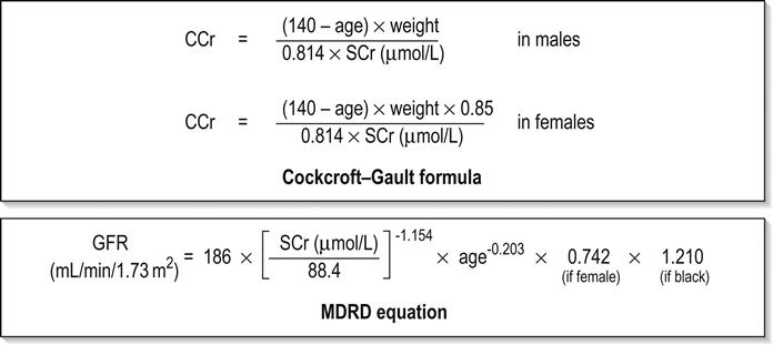
The creatinine clearance (CCr) or GFR are estimated indirectly using formulae (Cockcroft–Gault formula or the modification of diet in renal disease (MDRD) study equation) based on the SCr concentration (Fig. 10.1.4) [9]. These equations assume a steady-state SCr concentration and are inaccurate if the GFR is changing rapidly. They will also be less accurate in amputees, very small or very large persons or persons with muscle-wasting diseases.
Knowledge of a patient’s baseline SCr concentration is important in assessing the severity and progression of AKI. Small changes when the baseline SCr concentration is low are more important than larger changes when the baseline SCr concentration is high. Major decreases in GFR can occur in the normal range of SCr concentration. If the previous SCr concentration is not known, the MDRD equation can estimate the expected (normal) SCr concentration (using a value for the GFR at the lower range of normal).
Hyperkalaemia is a common complication, with the serum K+ usually rising by 0.5 mmol/L/day in ARF. The serum Ca2+ concentration may be normal or reduced in ARF. Both hypocalcaemia and hypercalcaemia may occur at different stages of ARF in rhabdomyolysis. Rhabdomyolysis is characterized by a very high blood CK concentration. Abnormal liver function tests invariably accompany the hepatorenal syndrome associated with hepatic cirrhosis.
Full blood examination
Anaemia develops rapidly in ARF, but its presence or the degree of anaemia does not reliably distinguish between acute and chronic renal failure. Leucocytosis is usually seen if sepsis is the cause of ARF. Eosinophilia is often present in acute interstitial nephritis, polyarteritis nodosa and atheroembolic disease. Anaemia and rouleaux formation suggest a plasma cell dyscrasia. Disseminated intravascular coagulation can complicate ARF due to rhabdomyolysis. A microangiopathic blood film associated with ARF occurs in vasculitis or thrombotic thrombocytopenic purpura.
Serological tests
Tests for the detection of antinuclear antibody (ANA) or antineutrophil cytoplasmic antibody (ANCA) or measurement of complement concentration are indicated in suspected cases of vasculitis or glomerulonephritis.
Urine tests
The results of urine analysis may be normal in AKI. A positive test for leucocytes, nitrates or both is found in urinary tract infections. A positive test for blood, protein or both suggests a renal inflammatory process. The presence of red cell casts on microscopy is diagnostic of glomerulonephritis.
The measurement of the concentration of electrolytes in the urine and the calculation of their fractional excretion is of intellectual interest in understanding the pathophysiological responses of the nephron to different types of AKI. The calculations are cumbersome, the results are inconsistent and the information obtained does not alter the patient’s immediate treatment.
Imaging
A chest X-ray is taken to assess the heart size and the presence of cardiac failure, infection, malignancy or other abnormalities. Ultrasound can define renal size and demonstrate calyceal dilation and hydronephrosis, but the findings depend on the expertise of the operator. Obtaining adequate images is difficult in obese patients, in ascites or where there is a large quantity of gas within the bowel. Ultrasound also provides information about bladder size and can detect prostamegaly.
A normal ultrasound examination can occur in the very early stages of obstruction or if ureteric obstruction is due to retroperitoneal fibrosis or to infiltration by tumour. Hydronephrosis not due to obstruction occurs in pregnancy, vesicoureteric reflux or in diabetes insipidus.
Doppler scans are useful for detecting the presence and nature of renal blood flow in thromboembolism or renovascular disease; however, because renal blood flow is reduced in prerenal or intrarenal AKI, test findings are of little use in the diagnosis of AKI. CT scans of the urinary tract evaluate renal size and renal position, renal masses, renal calculi, the collecting system and the bladder. Non-contrast CT is the examination of choice in persons with suspected renal calculi and can be used to assess the urinary tract in persons at risk of radiocontrast AKI. Injection of intravenous contrast is used for CT urography, CT angiography and CT venography, which may be necessary in some circumstances. Radionuclide can be used to assess renal blood flow and tubular function.
Renal biopsy
A renal biopsy provides a tissue diagnosis of the intrarenal cause of AKI and is indicated if the findings will identify a treatable condition. A renal biopsy is also valuable when renal function does not recover after several weeks of ARF and a prognosis is required for long-term management.
Treatment
The basis of emergency management is recognizing that AKI is present, correcting reversible factors, providing haemodynamic support and treating life-threatening complications. This is followed by treatment (if available) of the specific cause of AKI and management of ARF by supportive measures and (if required) renal replacement treatment.
Correction of hypovolaemia
Hypovolaemia not only causes AKI but also worsens all forms of AKI. The clinical diagnosis of hypovolaemia can be difficult if the jugular venous pressure is not easily seen or if there is pre-existing cardiac failure. When there are definite signs of hypovolaemia, the patient is resuscitated with rapid infusion of crystalloid. If hypovolaemia is a possibility, or if the person’s urine output has decreased markedly, the patient should have 250–500 mL of crystalloid infused rapidly (fluid challenge) and the response (urine output, vital signs, jugular venous pressure) evaluated. An increase in urine output or an increase in blood pressure following a fluid challenge suggests that hypovolaemia was present.
Invasive measurement of volume status using central venous and pulmonary artery catheters can increase mortality, lengthen hospital stay and increase the cost of care. There is no evidence to justify the routine use of these invasive measures in patients with AKI. The main indications for central venous cannulation in AKI in the ED are difficulties obtaining intravascular access in the limbs or the need to give drugs which can only be given into a large central vein (e.g. noradrenaline).
Haemodynamic support
AKI impairs autoregulation of GFR and renal blood flow throughout all ranges of mean arterial pressure. Renal perfusion in ATN is linearly dependent on mean arterial pressure even in the normal range of blood pressure. Episodes of mild or severe decrease in blood pressure lead to recurrent ischaemic injury. Inotrope/vasopressor drugs (noradrenaline or adrenaline) should be commenced if hypotension persists after correction of hypovolaemia. Dopamine appears to have no clinical advantage compared to other agents and has, in fact, resulted in increased mortality in some studies.
Monitoring and maintaining urine output
Urinary Catheter
Accurate measurement of urine output requires insertion of a urinary catheter, but this is not needed in the less severe forms of AKI if there is frequent spontaneous voiding. A catheter is required initially in persons with oliguria or (apparent) anuria, shock or obstruction to bladder outflow.
Diuretics
Frusemide is used to produce a diuresis in the treatment of AKI due to hypercalcaemia and in the treatment of severe rhabdomyolysis. A trial of high-dose frusemide (80–120 mg intravenously) can be used in persons with AKI who have acute pulmonary oedema if dialysis is not readily available. Persons with less severe forms of AKI (e.g. Risk or Injury) who have a low urine output (less than 0.5 mL/kg/h) that does not increase after correction of hypovolaemia are often given low doses of frusemide (e.g. 20–40 mg intravenously). A subsequent increase in urine output is not necessarily associated with a decrease in the SCr concentration. There is no evidence that the use of diuretics to convert the less severe forms of AKI from a (presumed) oliguric to a non-oliguric stage affects outcome [11].
Electrolyte abnormalities
Potassium
The serum potassium concentration may be low, normal or high. AKI due to diarrhoea causes hypokalaemia and metabolic acidosis, while AKI due to vomiting or diuretics causes hypokalaemia with metabolic alkalosis. A serum [K+] less than 3.0 mmol/L is treated with oral or intravenous potassium. Diabetic ketoacidosis (DKA) causes renal loss of K+, depleting the body of potassium. Persons with AKI due to DKA who have a normal or low serum [K+] need intravenous potassium during treatment with intravenous fluids and insulin.
Hyperkalaemia is due to an imbalance between potassium intake and renal potassium excretion or follows redistribution of potassium from the intracellular to the extracellular space. Hyperkalaemia in AKI can be asymptomatic, produce electrocardiogram (ECG) changes or cause potentially fatal changes in cardiac rhythm.
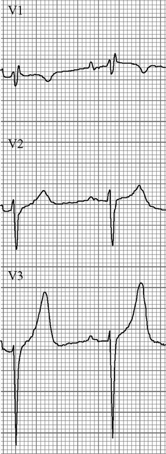
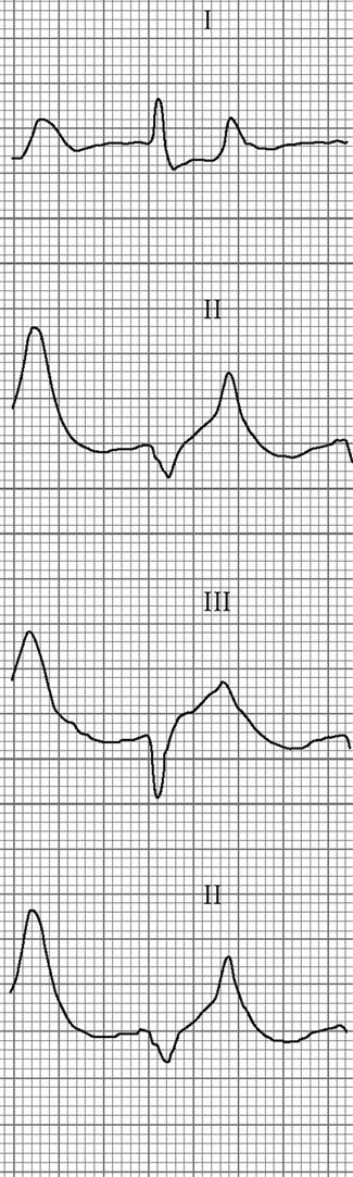
The initial ECG changes in hyperkalaemia are shortening of the PR and QT interval, followed by peaked T waves that are most prominent in leads II, III and V2 through V4 (Fig. 10.1.5). Marked ST-T segment elevation (pseudomyocardial infarction pattern) may occur. Bradycardia with sinoatrial (SA) block or atrioventricular block (including complete heart block) can develop and progress to periods of cardiac standstill or asystole. More commonly, the PR interval is prolonged and the QRS complex is widened, with the QRS complex having a left or right bundle branch block configuration (Fig. 10.1.6). At high serum [K+] (8–9 mmol/L), the sinoatrial (SA) node may stimulate the ventricles without ECG evidence of atrial activity (sinoventricular rhythm). When the serum [K+] is 10 mmol/L or greater, SA conduction no longer occurs and junctional rhythms are seen. The QRS complex width continues to increase and, eventually, the QRS complexes and the T wave blend, producing a sine wave ECG. At this stage ventricular fibrillation or asystole are imminent [10].
The higher the serum [K+] concentration, the more likely is the occurrence of ECG changes and life-threatening arrhythmias. However, nearly half of persons with a serum [K+] greater than 6.8 mmol/L do not have ECG changes of hyperkalaemia. Physicians predict the presence of hyperkalaemia solely on the basis of ECG changes with a sensitivity of less than 50%.
Drugs, such as oral potassium tablets, ACE inhibitors and aldosterone antagonists, should be ceased in AKI. Hyperkalaemia is treated when the serum [K+] is greater than 6.5 mmol/L (even if there are no ECG changes) or when there are ECG changes of hyperkalaemia. The emergency treatment of hyperkalaemia is covered in Chapter 12.2 Electrolyte disturbances.
Sodium
The sodium concentration in AKI may be normal, low (when water excess is present) or high (when water depletion is present). Patients with AKI and symptomatic hyponatraemia should be treated with haemofiltration or dialysis. Hypernatraemia is treated by slow intravenous infusion of hypotonic saline or 5% dextrose.
Calcium, phosphate, uric acid and magnesium
The serum calcium concentration is normal or slightly reduced in the Risk and Injury stages of AKI and is moderately reduced in later stages. Hypocalcaemia does not require therapy unless tetany is present. Hyperphosphataemia is present in nearly all persons with ARF, but does not need treatment in the ED.
Hyperuricaemia is common in AKI, but also occurs in chronic renal failure and in persons without AKI. Episodes of acute gout are very uncommon in AKI and the hyperuricaemia does not need treatment. Hypermagnesaemia is common in AKI, but is usually asymptomatic. Severe symptomatic hypermagnesaemia can occur if magnesium is administered to persons with AKI.
Acid–base abnormalities
Increased loss of bicarbonate-rich intestinal secretions (diarrhoea or an ileal conduit) can cause AKI with a normal anion-gap metabolic acidosis. AKI accompanied by acid loss from the stomach (vomiting or nasogastric suction) or caused by diuretics can result in a hypochloraemic metabolic alkalosis. Persons with the Risk and Injury stages of AKI often have a decrease in the serum bicarbonate concentration. More severe AKI causes a mild-to-moderate metabolic acidosis with an increased anion gap. This acidosis does not usually require specific treatment. Severe acidosis occurs in rhabdomyolysis and in lactic acidosis. The presence of a very severe metabolic acidosis in AKI is an indication for dialysis.
Fluid overload
The management of AKI in patients with peripheral oedema or pulmonary congestion due to cardiac failure is challenging. The clinical diagnosis of hypovolaemia in these patients is difficult and rapid intravenous administration of large volumes of fluid can worsen the pulmonary congestion or heart failure. Hypovolaemia is treated (or excluded) in these cases by assessing the response to small volume (200 mL) fluid challenges.
Patients with acute pulmonary oedema may have a raised SCr, which can be due to chronic renal failure, AKI or acute on chronic renal failure. These patients usually improve following treatment with vasodilators, continuous positive airway pressure (CPAP) ventilation and loop diuretics (40–80 mg frusemide intravenously). Patients with AKI and acute pulmonary oedema who do not respond to these measures need haemofiltration or haemodialysis.
Hypertension
Persons with AKI may have an elevated blood pressure that predated the renal injury or AKI itself may cause hypertension. A markedly elevated blood pressure reading (greater than 180/120 mmHg) in a person with AKI can be treated with glyceryl trinitrate applied as a skin patch (at a dose of 25–50 mg), sublingual nifedipine (5–10 mg) or oral hydralazine (20 mg). Intravenous drugs (glyceryl trinitrate or hydralazine) are used if AKI is associated with a hypertensive emergency, such as acute pulmonary oedema, hypertensive retinopathy or hypertensive encephalopathy.
Specific causes of AKI
Obstruction
Obstruction is relieved by decompression or diversion of the urinary tract. The site of the obstruction determines the technique used: placement of a Foley catheter or insertion of a suprapubic catheter, ureteral catheters (stents) or nephrostomy tubes. Relief of obstruction is often followed by a post-obstructive diuresis. Fluid replacement after relief of obstruction is based on frequent measurements of urine volume and urinary electrolytes.
Other causes
Specific treatments include immunosuppressive agents (glomerulonephritis, vasculitis), plasma exchange (thrombotic microangiopathy), systemic anticoagulation or revascularization (renovascular disease).
Management of ATN
Reduction of damage/accelerating recovery
Despite much experimental laboratory work and numerous clinical trials, no therapeutic intervention has hastened the recovery of renal function in established ATN. Therapeutic trials of dopamine, atrial natriuretic peptide and various growth factors have been ineffective. The use of high-dose loop diuretics to convert oliguric ATN to non-oliguric ATN was based on the observation that patients with non-oliguric ATN had a lower mortality and better renal recovery rates than those with oliguric ATN. The use of high-dose loop diuretics does not affect the duration of ATN, the need for dialysis or the outcome.
Supportive treatment
This includes monitoring fluid input and fluid output, measuring serum electrolyte values frequently, preventing sepsis by reducing the number of intravenous lines and removing urinary catheters if possible, culturing periodically and using antibiotics when clinically indicated. The fluid intake is restricted to insensible water loss (about 500 mL per day in the absence of fever) plus all measured fluid losses (urine output, gastrointestinal losses, chest tube drainage). Nephrotoxic agents should be avoided and the dosage of renally excreted drugs reduced. Because the increase in SCr lags behind the decrease in GFR, drug doses should be calculated based on a GFR of less than 10 mL/min per 1.73 m2 rather than on the SCr value.
Renal replacement treatment
Renal replacement treatment (RRT) is required in most patients with oliguric ARF and one-third of patients with nonoliguric ARF. The indications for RRT are summarized in Table 10.1.5.
Table 10.1.5
Indications for renal replacement treatment in acute kidney injury*
Oliguria (urine output<200 mL/12 hours) or anuria (urine output 0–50 mL/12 hours)
Serum urea concentration>35 mmol/L
Serum creatinine concentration>400 μmol/L
Serum potassium concentration>6.5 mmol/L or rapidly rising
Serum sodium concentration<100 mmol/L or>160 mmol/L
Pulmonary oedema not responding to diuretics
Severe (uncompensated) metabolic acidosis with pH<7.1
Uraemic syndrome (asterixis, psychosis, myoclonus, seizures, pericarditis)
Overdose with a toxin that is dialyzable
*Presence of two or more indications in a patient means that renal replacement will be needed.
Prognosis
The prognosis of ARF is largely dependent on the underlying cause and the presence of co-morbidities. Mortality varies from about 40% in those with no co-morbidity to more than 80% in those who have three or more failed organ systems.
10.2 The acute scrotum
Gino Toncich
Torsion of the spermatic cord (testicle)
Torsion is a twisting, not of the testicle but of the spermatic cord, which then interferes with the vascularity of the testicle, ultimately leading to infarction.
Aetiology
Torsion is due to a powerful contraction of the cremaster muscles in an abnormally attached testis. A normal testis is anchored posterolaterally to the scrotal sac and is therefore fixed in place. The main abnormality found in patients with torsion is an enlarged tunica vaginalis, which surrounds the whole of the testes and epididymis, preventing the testis from creating any attachment to the scrotal wall. The testis, therefore, floats freely like a clapper inside a bell. The contraction of the cremaster causes the testes and adnexae to rotate, thereby twisting the cord [1].
Pathology
The twisting of the cord causes obstruction of the lymphatic and venous outflows, but allows arterial inflow, leading to venous engorgement. Eventually, the pressure rises and occludes the arterial inflow.
The extent and rapidity of the damage depends on the degree of torsion, that is the number of turns. An incomplete rotation (<360°) may not completely occlude arterial flow, while a complete turn (360°) causes necrosis in 12–24 hours. Two or more turns (>720°) cause necrosis in less than 2 hours because arterial flow is obstructed [1,2].
Clinical presentation
Classically, there is a sudden onset of severe scrotal or abdominal pain. There are no irritative voiding symptoms. Between one-third and one-half of patients have had previous episodes of acute scrotal pain [3,4].
The patient looks pale and may vomit. The testicle is tender and rides high in the scrotum. Other signs include loss of cremasteric reflex, scrotal oedema, testicular swelling and retraction. These clinical signs are unreliable with sensitivities of 60–91% and specificities of 27–68% [5].
Systemic signs, such as fever, are classically absent. Urinalysis is normal.
Intermittent torsion of the testis
This is a syndrome of recurrent acute scrotal pain, usually lasting less than 2 hours, which resolves spontaneously. Creagh et al. [4] describe a series of 27 patients who underwent elective orchidopexy for these symptoms. Three patients developed acute torsion while on the waiting list; of those coming to operation, one had an atrophic testis and four had evidence of torsion of the appendages of the testis. One patient subsequently had torsion after surgery because absorbable sutures were used [4].
Differential diagnosis of acute testicular pain
Differential diagnoses to consider in acute testicular pain are listed in Table 10.2.1.
Table 10.2.1
Differential diagnosis of acute testicular pain
Epididymo-orchitis
Strangulated hernia
Haematocoele
Hydrocoele
Testicular tumour
Henoch–Schonlein purpura in children
Idiopathic scrotal oedema
Traps in the clinical diagnosis
There are many potential pitfalls in the clinical diagnosis of the acute scrotum:
 Age: the abnormality is present for life, so the torsion could potentially occur at any age. In those under 18 years of age, an acutely painful scrotum should always be considered to be torsion [3]. Most of the literature concerns itself with the under 18-year-old population. Less than 4% of torsions occur in patients over 30 years. It is most common in adolescence (12–18 years) [6]. In teenagers, sexually transmitted disease may confuse the diagnosis. There is an old surgical aphorism: ‘Question: When do you diagnose epididymo-orchitis in a teenager? Answer: After you have fixed the torsion.’
 Pain: in 25% of cases there is no sudden onset of pain, nor is it necessarily severe. Some patients with epididymo-orchitis (EDO) have severe pain [1,3].

 Abnormal position of testis: this is only seen if 360° or greater rotation occurs [3].
 Previous repair: torsion can occur in a testis that has previously been fixed, especially if absorbable sutures have been used [4].
 Dysuria: irritative voiding symptoms rarely occur with torsion and suggest infection [3].
 Fever: temperatures>38.9°C have been noted in up to 15% of torsion patients [1].
Clinical findings remain misleading and none can reliably exclude the diagnosis of torsion [7].
Investigations
Surgical exploration of the scrotum
This is the investigation of choice where the diagnosis of torsion is likely and maximizes the chance of saving the testis.
Delaying the diagnosis has been termed ‘castration by neglect’ [3,7]. Surgical exploration requires only a skin incision and has no major complications [5,7].
Low rates of torsion diagnosed at operation have led to interest in other tests to predict torsion preoperatively.
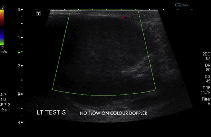
Colour Doppler imaging of the testis
It is useful in diagnosing torsion but also in elucidating other scrotal pathology. Comparison of blood flow to the asymptomatic side is crucial. If there is reduced flow to one side then some degree of torsion must be suspected (E-Fig. 10.2.1 and E-Fig. 10.2.2). If the testis has untwisted, hyperaemic flow may be noted. The sensitivity of colour Doppler imaging (CDI) for torsion can be as low as 82%, missing one in five cases, and is affected by:


Role of investigations in suspected testicular torsion
When the diagnosis of torsion remains probable, then the investigation is surgical exploration of the scrotum; any other investigations that delay theatre are unnecessary. If torsion is unlikely clinically and a surgical opinion concurs, then CDI can be used, provided it is available on an urgent basis [1,3,7]. It is important that there is early communication and discussion with the responsible surgeon to avoid delays between the suspicion of torsion and surgical intervention [1–3].
Treatment
Manual untwisting
This manoeuvre is not universally recommended and should be done only as a temporizing measure or when surgical exploration cannot be performed. The spermatic cord is infiltrated with local anaesthetic and the testis is untwisted. Untwisting is done by turning the left testis anticlockwise (outward) and the right one clockwise, like opening the pages of a book [8].
Surgery
The testes is untwisted and inspected for return of colour and bleeding. An obviously infarcted testis is removed at the initial surgery. A viable testis is sutured into place on the scrotal wall. The tunica should be inverted and also sutured to the scrotal wall. It is vital that the normal side is also explored and fixed to the scrotal wall, as the abnormality is bilateral in most cases. Retorsion following orchidopexy has occurred when absorbable sutures have been used [1,3–6].
Prognosis
Viability depends on the number of twists and the time taken to untwist the testis. There is 100% salvage if the testis is untwisted in less than 4 hours. Up to 24 hours, the rate falls to 50%. There are rare case reports of salvage after 30 hours.
Testicular salvage (return of circulation at surgery) does not mean absence of injury to the testicle. Long-term follow up of salvaged testes shows that 75% have a reduction in volume. Abnormalities are also seen in sperm volume, motility and morphology. These abnormalities are not seen in patients who have had an infarcted testis removed at the initial operation. This suggests some antispermatogenesis effect caused by the damaged testicle [1,3,4].
Torsion of a testicular appendage
Testicular appendages are embryological remnants with no function. They are small (<5 mm) pedunculated structures that may twist on their pedicle. If the appendage can be isolated in the scrotum, a small blue lump may be noted: ‘the blue dot sign’. These do not need surgery and can be treated with analgesia. Late presentations may have scrotal or testicular swelling and should be treated as torsion until proved otherwise [1].
Acute epididymo-orchitis (EDO)
Introduction
EDO is a clinical syndrome with pain and swelling of the epididymis (and the testis) of less than 6 weeks’ duration. Chronic epididymitis is a long-standing condition of epididymal or testicular pain, usually without swelling [2].
Aetiology
A variety of organisms may be responsible for EDO (Table 10.2.2).
Table 10.2.2
Bacterial: Neisseria gonorrhoeae, Escherichia coli, Pseudomonas aeruginosa, coliforms, Klebsiella, Mycobacterium tuberculosis
Chlamydial: C. trachomatis
Viral: mumps
Drugs: amiodarone epididymitis
Fungal: cryptococcal
Parasitic: filariasis (usually chronic)
The most likely cause depends on the patient’s demographic group. For heterosexual males under 35 years of age, the agent is usually gonococcus or chlamydia. These organisms are also responsible for infection in homosexual males under 35 years (where anal sex is practised), but coliforms and even Haemophilus can cause infection. In males older than 35 years, EDO is usually due to obstructive urological disease, so coliforms predominate. EDO may also be part of a systemic disease, for example brucellosis or cryptococcus.
EDO is usually thought to be an ascending infection from the urethra or prostate, but it can be part of a generalized systemic disease. The infection spreads from epididymis to testicle and, eventually, they may become one large inflammatory mass. Isolated orchitis is rare and usually due to viral causes, spread via the bloodstream [9–11].
Clinical presentation
The exact features depend on the underlying cause and whether both the epididymis and the testicle are involved. The pain may come on suddenly or slowly. There is scrotal swelling and tenderness that is relieved by elevating the testis. The spermatic cord is usually tender and swollen. Associated symptoms of urethritis are common. In younger males (under 35 years), a history of sexually transmitted disease may be elicited. In the older patient, there is often a history of instrumentation, intercurrent urinary tract infection (UTI) or prostatism. Pyuria is common.
Investigations
Urethral swabs
Urethral discharge may not be seen if the patient has just voided, so a urethral swab and smear should be examined for white blood cells (WBC). If there are more than five WBC per high-powered field, then urethritis is likely. The presence of intracellular diplococci confirms the diagnosis of gonorrhoea; their absence suggests chlamydia [9].
Midstream urine
Look for the presence of WBC or Gram-negative organisms.
Differential diagnosis
In the acute non-traumatic setting in those less than 30 years old, the most important differential diagnosis is torsion of the testicle [9]. If the clinical features and midstream urine do not differentiate, then torsion should be considered and surgical opinion obtained. An ultrasound can then be done if surgical opinion agrees. In young men, if CDI is not available and there is no evidence of UTI or urethritis, then surgical exploration may be necessary. Ultrasound can help differentiate other causes of the acute scrotum.
Treatment
Symptomatic treatment consists of bed rest, analgesia and scrotal supports.
If the cause is secondary to a sexually transmitted disease, then appropriate antibiotics should be chosen after urethral swabs have been taken, for instance, a single dose of ceftriaxone (250 mg stat) for gonorrhoea and a 14-day course of doxycycline (100 mg) or roxithromycin (300 mg) for chlamydia. The patient’s sexual partners should be investigated and treated. Tests for syphilis or HIV should be performed.
If the infection is secondary to UTIs, then an appropriate antibiotic for 14 days should be used. Refer to antibiotic guidelines that will account for local sensitivities. Antibiotic choice can then be adjusted according to the urine culture results. Investigation for underlying urinary tract obstruction should be undertaken according to clinical features. Symptoms can take many days to settle so, in the absence of systemic illness, no intravenous antibiotics are necessary.
Complications
These include abscess formation, testicular infarction, chronic pain and infertility.
Blunt traumatic injury to the testicle
The mobility of the testicle, cremaster muscle contraction and the tough capsule usually protect the testicle from injury. However, a direct blow that drives the testicle against the symphysis pubis may result in contusion or rupture of the testicle. Typical mechanisms are a direct kick to the groin or handlebar and straddle injuries [12,13].
The types of injury include scrotal-wall haematomas, tunica vaginalis haematoma (haematocoele) or intratesticular (subcapsular) haematoma.
The most serious is testicular rupture, where the tunica splits, allowing blood and seminiferous tubules to extrude into the tunica vaginalis. This occurs in up to 50% of blunt trauma. Complete disruption of the testis may occur [12–14].
Ultrasound examination is not 100% sensitive in detecting testicular rupture, so early surgical exploration is the investigation and treatment of choice. Indications for exploratory surgery include:
 uncertainty in diagnosis after appropriate clinical and radiographic evaluations
 clinical findings consistent with testicular injury
 disruption of the tunica on ultrasound
 absence of blood flow on scrotal ultrasound images with Doppler studies


It should be noted that 10–15% of testicular tumours present after an episode of trauma and so any abnormalities on ultrasound examination should be followed to resolution if surgery is not performed [15].
Early surgical exploration with evacuation of blood clots in the tunica vaginalis and repair of testicular rupture, if present, results in a shortened hospital stay, a greatly reduced period of disability and a faster return to normal activity compared to patients managed conservatively. Conservative management is complicated by secondary infection of the haematocoele, frank acute necrosis of the testis and delayed atrophy due to pressure effects of haematoma. The orchidectomy rate for early exploration is only 9%, compared to 45% for those managed non-operatively [13].
Necrotising fasciitis of the perineum (Fournier’s gangrene)
This is a necrotizing fasciitis caused by a mixture of aerobic and anaerobic bacteria. It usually occurs in frail and elderly men who suffer from diabetes, renal failure or immunocompromise. The initial infection starts in the anterior abdominal wall and spreads to the perineum with scrotal involvement [17].
Features suggestive of this diagnosis are the epidemiological risk factors, severe pain, tense swollen scrotum with possible blisters and crepitus due to gas in the tissues. Often signs of systemic toxicity, not expected in routine EDO, accompany this diagnosis. Diagnosis is often delayed as the diagnosis is mainly clinical [16,17].
The mainstay of treatment is immediate resuscitation with fluids, antibiotics and urgent extensive surgical debridment.
Controversies and future directions


10.3 Renal colic
Sean Arendse
Introduction
Nephrolithiasis is a common disorder affecting 2–5% of the population at some point in their lives [1]. It occurs most frequently between the ages of 20 and 50 years, with a male:female ratio of approximately 3:1. About 50% of patients only experience a single episode, but the remaining 50% have recurrent episodes within 5 years [2].
Most calculi are believed to originate in the collecting system (renal calyces and pelvis) before passing into the ureter. Supersaturation with stone-forming substances (calcium, phosphate, oxalate, cystine or urate), combined with a decrease in urine volume and lack of chemicals that inhibit stone formation (such as magnesium, citrate and pyrophosphate), result in production of a calculus. In addition to this, infection with urea-splitting organisms that produce an alkaline urinary pH frequently contribute to the growth of ‘struvites’ or triple phosphate (calcium, magnesium and ammonium phosphate) stones.
Less commonly, mixed stones occur via nucleation with sodium hydrogen, urate, uric acid and hydroxyapatite crystals providing a core to which calcium and oxalate ions adhere (heterogeneous nucleation).
Approximately 75% of all stones are calcium based, consisting of calcium oxalate, calcium phosphate or a mixture of the two. Ten per cent are uric acid based, 1% are cystine based and the remainder are primarily struvite.
Predisposing factors for stone formation include dehydration and low fluid intake, hypertension, prolonged immobilization, strong family history of nephrolithiasis, hyperparathyroidism, peptic ulcer disease (hyperexcretion of calcium), small bowel disease, such as Crohn’s disease or ulcerative colitis (hyperoxaluria), and gout (hyperuricaemia). Myeloproliferative disorders, malignancy, glycogen storage disorders, renal tubular acidosis and the use of certain medications (calcium supplements, acetazolamide, vitamins C and D and antacids) may also be conducive to nephrolithiasis [3].
Persistent obstruction of the ureter leads to hydronephrosis of the urinary tract and may precipitate renal failure. Common sites of obstruction are ureteropelvic junction, pelvic brim and vesicoureteric junction.
Pathophysiology of pain
The mechanisms implicated in the production of the pain associated with renal colic are an increase in renal pelvic pressure, ureteric spasm, local inflammatory effects at the level of the calculus and increased peristalsis and pressure proximal to the calculus.
Acute obstruction of the upper urinary tract from a calculus results in increased pressure in the renal pelvis which, in turn, induces the synthesis and secretion of renal prostaglandins, in particular PGE2, which promotes a diuresis by causing dilatation of the afferent arteriole, further elevating the renal pelvic pressure [4,5]. The acute obstruction and renal capsular tension are believed to be the cause of the constant ache in the costovertebral angle.
In experiments utilizing isolated ureteric smooth muscle, prostaglandins have also been shown to increase phasic and tonic contractile activity [6], resulting in ureteric spasm and severe, colicky pain.
Presentation
The pain of renal colic has been described as the worst pain a person can endure. The classic textbook description is of severe, intermittent, flank pain of abrupt onset originating from the area of the costovertebral angle and radiating anteriorly to the lower abdominal and inguinal regions. Testicular or labial pain may be present and may suggest the location of the stone as a low ureteric position. Urinary frequency or urgency often develops as the stone nears the bladder. Nausea and vomiting frequently accompany the pain and about one-third of patients complain of gross haematuria [7].
Examination
Examination usually reveals an agitated, pacing patient unable to find a comfortable position. Pulse rate and blood pressure may be elevated secondary to the pain. Fever is unusual and suggests infection. The abdominal examination may only reveal signs of an early ileus with hypoactive bowel sounds and a distended abdomen, but should not be omitted as it is extremely useful in excluding other intra-abdominal or retroperitoneal causes of the pain (such as pancreatitis, cholecystitis, appendicitis or leaking or rupture of the abdominal aorta).
Investigations
Urinalysis usually shows red blood cells, although the absence of red cells in the urine in the setting of colicky flank loin to groin pain does not rule out nephrolithiasis and between 10 and 30% of patients with documented nephrolithiasis do not have haematuria [8]. Nitrites, leucocytes or microorganisms in the urine suggest either the complication of an infection or a diagnosis of acute pyelonephritis. Urine culture is thus indicated to rule out infection with urea-splitting organisms, such as Klebsiella and Proteus spp. Electrolyte studies may demonstrate obstruction or suggest an underlying metabolic abnormality, such as hypercalcaemia, hyperuricaemia or hypokalaemia. A slightly elevated white blood cell count may occur with renal colic, but a count greater than 15 000/mm3 suggests active infection, as does a fever. Renal tract obstruction with concomitant infection is a urological emergency and must be treated immediately and aggressively.
A pregnancy test should be performed in all women of childbearing age, as a positive result needs further investigation to exclude ectopic pregnancy.
Many conditions may have a similar presentation to renal colic and examination and investigations should be directed towards confirming the diagnosis of nephrolithiasis and excluding the other conditions in the differential diagnosis (Table 10.3.1).
Table 10.3.1
Differential diagnosis of renal colic
Renal carcinoma producing blood clots temporarily occluding the ureter
Ectopic pregnancy
Ovarian torsion
Abdominal aortic aneurysm
Acute intestinal obstruction
Pyelonephritis
Appendicitis
Diverticulitis
Narcotic seekers and Munchausen’s syndrome
Radiological examination
A variety of imaging modalities is used to evaluate renal colic. Their pros and cons are listed in Table 10.3.2.
Table 10.3.2
Pros and cons of imaging modalities in renal colic
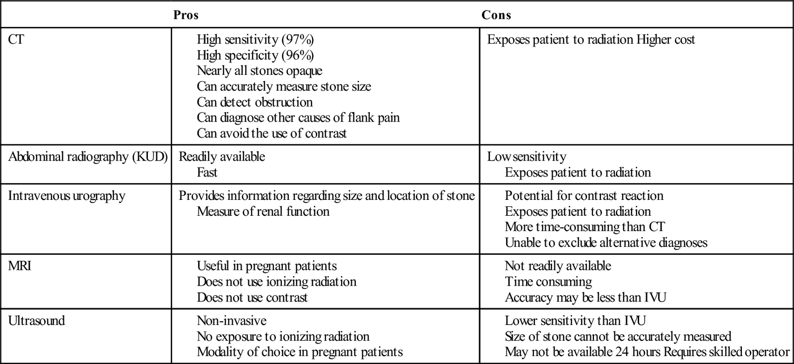
Most stones (90%) are radiopaque and theoretically should be visible on plain X-ray; if seen, they are irregularly shaped densities on abdominal radiography (KUB). However, a KUB alone is not usually sufficient to make the diagnosis of nephrolithiasis as it has poor sensitivity of around 60% [9]. Phlebitis in the pelvic veins and calcified mesenteric lymph nodes may add confusion and many small stones may be obscured by the bony density of the sacrum. Thus, plain X-ray should only be used in conjunction with another imaging modality, such as ultrasound, in the setting of renal colic.
Computed tomography (CT), with or without contrast, is the first-line test in most centres and has become the adopted gold standard with high sensitivity (97%) and specificity (96%) for ureterolithiasis [10]. Nearly all stones are opaque on CT and thus the size of the stone and its position can be accurately measured. Other positive findings include perinephric stranding, dilatation of the kidney (hydronephrosis) or ureter and low density of the kidney, suggesting oedema. Non-contrast CT is equivalent to intravenous urography (IVU) in the diagnosis of obstruction and is more reliable in the detection of ureterolithiasis [11]. It is also useful in the exclusion or confirmation of the other intra-abdominal differential diagnoses, such as appendicitis, abdominal aortic aneurysm or diverticulitis. As no contrast is used, there is not the risk of contrast reaction that is associated with IVU. It is more rapid than IVU and does not depend on the technical expertise required by other imaging modalities, such as ultrasound, but does subject the patient to a larger dose of radiation than IVU.
The intravenous pyelogram had been the standard investigation for the evaluation of renal colic until the widespread adoption of CT. It establishes the diagnosis of calculus disease in 96% of cases and determines the severity of obstruction [12]. Classic findings of acute obstruction include a delay in the appearance of one kidney, a dilated ureter and a dilated renal pelvis [13]. IVU is useful in estimating the size of the stone, in identifying extravasation of dye and in evaluating renal function. Its main disadvantage is the use of ionizing radiation, although less than in CT. In addition, administration of intravenous iodinated contrast media could precipitate a contrast reaction. Compared with CT, it is time-consuming and is not useful in confirming alternative diagnoses.
Ultrasonography is a useful, safe and a non-invasive alternative when renal function is impaired, risk from radiation is high (e.g. pregnancy) or contrast media contraindicated. It can identify the stone, its location and demonstrate proximal obstruction, such as hydroureter or a dilated pelvis, as well as the size and configuration of each kidney but, unfortunately, not size of the stone. Ultrasound has significantly lower sensitivity than IVU and misses more than 30% of stones [14].
Magnetic resonance imaging (MRI) can easily depict a dilated ureter and demonstrate the level of obstruction without using ionizing radiation or contrast, but the accuracy of MRI for stones may be lower than IVU as its spatial resolution is often not high enough to detect small stones. When used in combination with ultrasound, it may have a role in the evaluation of loin pain, especially in the pregnant patient; however, it is expensive, time-consuming and usually not readily available to most emergency departments.
Management
As 90% of stones are passed spontaneously, the most urgent therapeutic step is relief of pain, along with provision of adequate hydration and antiemetics. Opioid analgesics and non-steroidal anti-inflammatory drugs (NSAIDs) remain the mainstay of treatment.
Intravenous narcotics provide rapid analgesia, are titratable to effect and relieve anxiety in most cases. However, prolonged use may cause dependence and tolerance. Side effects are common and include nausea, vomiting, drowsiness, constipation and, with larger doses, precipitation of respiratory depression and hypotension. The data are very variable with regards to the effect of opioids on ureteric tone. Results indicate an increase in ureteric tone or no effect at all [15]. Options include morphine and fentanyl
Codeine, a less potent opioid than morphine, is effective for relieving mild to moderate pain associated with renal colic. Constipation is a significant side effect and limits its long-term use. Another option for analgesia is tramadol, an opioid-like agent, but with fewer side effects. One study showed that 100 mg of tramadol, when used for treating renal colic, was as effective as 50 mg pethidine [16], but more research is needed before adopting tramadol as an alternative to conventional opioids.
NSAIDs appear to be equally effective when compared with opioids [17]. A double-blind study comparing diclofenac and an opioid demonstrated a better effect with diclofenac and fewer side effects, but slower onset of action [18]. There are many NSAIDs available, differing in preparation and route of administration, the major differences between them being the incidence and nature of side effects, predominantly gastric irritation, ulceration and precipitation of renal failure. Ibuprofen has the fewest side effects and the lowest risk of gastrointestinal effects, but the weakest analgesic action. Naproxen and diclofenac provide stronger analgesia and a relatively low incidence of side effects. Oral diclofenac and oral/rectal indomethacin have both been shown to be effective in reducing the number of new renal colic episodes as well as further admission to hospital, but have no effect on spontaneous stone passage rates [19,20]. It has been suggested that one should give both a rapidly acting titratable opioid and a slower acting NSAID, which may result in earlier discharge from the ED [21]. Intravenous preparations of NSAIDs have limited availability in Australian EDs and have been reported to have a faster onset of action but a higher incidence of side effects, with considerably high cost and therefore, if available, should be used with caution. Commonly available alternative options for patients unable to tolerate oral medications include IM ketorolac and PR indomethacin
Buscopan, an antimuscarinic agent used for treating smooth muscle spasm, has been shown to decrease ureteric activity to some degree in 80% of the subjects studied [22]. However, one study comparing its use to an NSAID found that buscopan was less effective [23] and was associated with significant side effects, including dry mouth, photophobia, urgency, urinary retention and constipation, significantly limiting its use in renal colic.
Recently, the use of alpha-blockers in renal colic has been reported, with a number of studies showing that patients treated with alpha-blockers as well as standard therapy achieve stone clearance more often and in less time than controls [24–27].
Intravenous crystalloid should be administered to ensure a urine volume of 100–200 mL/h in those unable to tolerate oral fluids.
The size, shape and site of the stone at initial presentation are factors that determine whether a stone passes spontaneously or requires removal. Stones less than 5 mm in patients without associated infection or anatomic abnormality pass within 1 month in 90% of cases, stones 4–6 mm pass 50% of the time but only 5% of stones larger than 7 mm pass and hence usually require elective surgical removal [8]. The overall passage rate for ureteral stones is:


Disposition
Most patients with renal colic can be discharged with oral analgesia (codeine, paracetamol and NSAIDs), hydration and a referral for outpatient urology. Rectal administration of indomethacin is particularly effective if tolerated by the patient.
Indications for admission to hospital are listed in Table 10.3.3.
Table 10.3.3
Indications for hospital admission in renal colic
Presence of infection
Deteriorating renal function
Persistent pain requiring parenteral narcotics
Stone greater than 5 mm in diameter
Extravasation of dye (uncommon)
Further intervention is required if obstruction with hydronephrosis is present, the stone is a large stag horn calculus or the patient continues to have pain and no stone is passed within 2–3 days. A percutaneous nephrostomy allows drainage of an obstructed kidney until the blockage can be removed, either by ureteroscopic procedures for low stones or by open surgery for large or infected stones. Extracorporeal shockwave lithotripsy is preferred for single or small (>2 cm) otherwise uncomplicated stones as it has minimal complications and morbidity.
Urology follow up is essential for all patients, for elective removal of stones when complications have not ensued and for the prevention of recurrence. Indications for stone removal include stone diameter>7 mm, stone obstruction associated with infection, single kidneys with obstruction and bilateral obstruction.
Precautions
Renal colic, with its minimal findings on examination, is a commonly used presentation for those seeking narcotics or with Munchausen’s syndrome and treating physicians should be aware of this. However, it is essential to give analgesia to those patients suffering from renal colic and it is preferable to give patients analgesia unnecessarily than cause unnecessary suffering. Features suggesting narcotic seeking are discussed in Chapter 21.5.
Conclusion
Renal colic is an acutely distressing medical condition that requires a careful evaluation of symptoms and signs to ensure timely analgesia, recognition of other causes of acute abdominal pain and avoidance of inappropriate narcotic usage.
Controversies and future directions


[/level-membership-for-emergency-medicine-category][not-level-membership-for-emergency-medicine-category]
Genitourinary Emergencies
Edited by George Jelinek
10.1 Acute kidney injury
Nicholas Adams and Linas Dziukas
Introduction
The basic process in acute kidney injury (AKI) is a rapid (hours to days) reduction in the glomerular filtration rate (GFR) due to renal hypoperfusion, damage to glomeruli, tubules, interstitium or blood vessels, or obstruction to urine flow. The GFR is inversely related to the serum creatinine (SCr) concentration and the diagnosis of AKI is made when there is an acute increase in the SCr concentration, with or without a decrease in the urine output. A simple definition of AKI is an acute and sustained (lasting for 48 hours or more) increase in the SCr of 44 μmol/L if the baseline is less than 221 μmol/L, or an increase in the SCr of more than 20% if the baseline is more than 221 μmol/L. A more comprehensive definition (the RIFLE system) is used to classify persons with acute impairment of renal function (Table 10.1.1) [1].
Table 10.1.1
RIFLE classification of acute renal failure
| Stage | Serum creatinine (SCr) concentration | Urine output |
| RISK | Increase of 1.5 times the baseline | <0.5 mL/kg/h for 6 h |
| INJURY | Increase of 2.0 times the baseline | <0.5 mL/kg/h for 12 h |
| FAILURE | Increase of 3.0 times the baseline or SCr is 355 μmol/L or more when there has been an acute rise of greater than 44 μmol/L for 24 h or anuria for 12 h | <0.3 mL/kg/h |
| LOSS | Persistent acute renal failure; complete loss of kidney function for longer than 4 weeks | |
| END-STAGE RENAL DISEASE | End-stage renal disease for longer than 3 months |
Aetiology and pathogenesis
The causes of AKI are grouped according to the source of renal injury: prerenal (hypoperfusion), renal (parenchymal) and post-renal (obstructive). More than one cause can be present simultaneously.
Prerenal acute kidney injury
Prerenal AKI is initially an adaptive response to severe volume depletion and hypotension in structurally intact nephrons. Prerenal AKI that is prolonged or inadequately treated can be followed by parenchymal renal damage (acute tubular necrosis). Prerenal AKI is a potentially reversible cause of acute renal failure (ARF).
Reductions in renal blood flow (RBF) and GFR occur in the setting(s) of hypovolaemia, hypotension, oedematous states with a reduced ‘effective’ circulating volume (cardiac failure, hepatic cirrhosis, nephrotic syndrome) or impaired renal perfusion (renal artery stenosis, hepatorenal syndrome). Drugs that interfere with renal autoregulation (e.g. prostaglandin inhibitors, angiotensin-converting enzyme [ACE] inhibitors or angiotensin II receptor antagonists) can reduce glomerular perfusion [2]. The physiological responses to volume depletion and hypotension, and the link to prerenal AKI are shown in Figure 10.1.1.
Renal acute kidney injury
Ischaemic, cytotoxic or inflammatory processes may damage the renal parenchyma. The causes of the damage can be grouped according to the major structures that are damaged: vessels, glomeruli, renal tubules or renal interstitial tissue.
Vascular causes involving the larger vessels include acute thrombosis of the renal artery, embolism of the renal arteries, renal artery dissection and renal vein thrombosis. Microvascular causes include vasculitis, malignant hypertension and thrombotic microangiopathy.
The glomeruli are the site of injury in acute glomerulonephritis which can cause proteinuria, haematuria, nephrotic syndrome or nephritic syndrome. A number of different forms of glomerulonephritis have been described, generally diagnosed by the histological changes seen on renal biopsy. The distinction between these forms is not of direct concern for the emergency practitioner.
Acute tubular necrosis (ATN) is the most common pathological process that causes AKI. While the terminology suggests that the main cause is tubular damage, the actual pathophysiology is more complex: impaired autoregulation and marked intrarenal vasoconstriction (the main mechanism for the greatly reduced GFR), tubular damage (with cytoskeleton breakdown), increased tubuloglomerular feedback, endothelial cell injury, fibrin deposition in the microcirculation, release of cytokines, activation of inflammation and activation of the immune system [3].
ATN is often classified as ischaemic ATN or cytotoxic ATN but both processes may be present in some patients. Ischaemic ATN represents an advanced form of prerenal AKI, but the distinction between these two entities is based on histopathological changes and is of little use to the clinician. Important causes of cytotoxic ATN are listed in Table 10.1.2. Non-steroidal anti-inflammatory drugs (NSAIDs), ACE inhibitors and angiotensin receptor blockers (ARBs) often cause a gradual and asymptomatic decrease in the GFR, but can also cause AKI. NSAIDs do not impair renal function in healthy persons, but can reduce the GFR in the elderly with atherosclerotic cardiovascular disease, in persons with chronic renal failure, when chronic prerenal hypoperfusion is present (e.g. cardiac failure, cirrhosis) or in persons using diuretics and calcium channel blockers [4]. AKI may occur after the administration of intravenous or intra-arterial radiocontrast agents. A number of risk factors have been identified for this, the most important being pre-existing renal impairment, hypovolaemia, a large contrast load and the use of hyperosmolar contrast agents [5]. Drugs that alter angiotensin levels (ACE inhibitors and ARBs) reduce renal perfusion by their antihypertensive effects or by impairing vasoconstriction of the efferent arteriole when renal perfusion is reduced by renal artery stenosis. The nephrotoxicity of haem pigments (myoglobin and haemoglobin) is enhanced by volume depletion, low urine flow rates and possibly low urine pH.
Table 10.1.2
Causes of toxic acute tubular necrosis
Exogenous agents
Radiocontrast
Non-steroidal anti-inflammatory drugs
Antibiotics: aminoglycosides, amphotericin B
Antiviral drugs: acyclovir, foscarnet
Immunosuppressive drugs: ciclosporin
Organic solvents: ethylene glycol
Poisons: snake venom, paraquat, paracetamol
Chemotherapeutic drugs: cisplatin
Herbal remedies
Heavy metals
Endogenous agents
Haem pigments: haemoglobin, myoglobin
Uric acid
Myeloma proteins
Correct intravascular volume depletion
Maintain perfusion pressure
Choice of resuscitation fluid
Diuresis in rhabdomyolysis
Avoid nephrotoxins
Use derived GFR or creatinine clearance when calculating drug doses
Abnormalities of renal interstitial structure and function are only one feature of ATN, but represent the primary abnormality in acute tubulointerstitial nephritis (ATIN). The damage in ATIN is due to immunological mechanisms, the most important involving cell-mediated immunity. ATIN is usually due to an allergic reaction to a drug, commonly antibiotics (β-lactam antibiotics, sulphonamides, fluoroquinolones), NSAIDs, cyclooxygenase-2 inhibitors, proton pump inhibitors, diuretics, phenytoin, carbamazepine and allopurinol.
Post-renal acute kidney injury
Obstructive uropathy refers to the functional or structural processes in the urinary tract that impede the normal flow of urine and obstructive nephropathy is the renal damage caused by the obstruction. Hydronephrosis and hydroureter refer to dilatation of the renal urinary collecting system and the ureters, respectively. They may occur in the absence of obstruction and, conversely, may be absent in some patients with obstruction.
Casts or crystals within the renal tubular lumen can cause intrarenal obstruction. Extrarenal obstruction can develop in the urethra, bladder, ureter or the pelvi-ureteric junction. Obstructive uropathy in adults is commonly caused by prostate disease or retroperitoneal neoplasm (cancer of the cervix, uterus, bladder, ovary or colon). Metastatic cancer, lymphomas or inflammatory processes in the retroperitoneum (appendicitis, diverticulitis, Crohn’s disease) or a neurogenic bladder can also cause obstructive uropathy. Bilateral renal stones are an uncommon cause of obstructive uropathy.
Obstructive nephropathy usually develops gradually and can cause chronic renal failure if the obstruction involves the urethra, the bladder or both ureters. Unilateral ureteric obstruction will cause AKI only if it involves a single functioning kidney.
Epidemiology
Studies of the pathogenesis of community-acquired ARF have produced conflicting results. In one study, the major processes were identified as prerenal in 70% of cases, renal in 11% of cases and post-renal in 17% of cases [6]. There are geographical differences in the causes of ATN. In Africa, India, Asia and Latin America, ATN is usually caused by infections (e.g. diarrhoeal illnesses, malaria, leptospirosis), ingestion of plants or medicinal herbs, envenomation, intravascular haemolysis due to glucose-6-phosphate dehydrogenase deficiency or poisoning.
Prevention
Maintaining intravascular volume and renal perfusion
The rate and volume of intravenous fluid given to hypovolaemic persons depends on the nature of the intravascular depletion, the blood pressure and heart rate, the (estimated) volume of fluid lost, cardiac function and ongoing circulatory losses. The response to treatment is evaluated by simple bedside measurements (heart rate, blood pressure, urine output).
Rhabdomyolysis
Most studies on the prevention of ATN after rhabdomyolysis have been in persons with crush injury after earthquakes, where the incidence of AKI is about 50%. In this situation, fluid resuscitation should, if possible, begin before the crush is relieved. These patients may require massive amounts of fluid because of fluid sequestration in the injured muscles. The goal of intravenous fluid treatment is to produce a urine output of 200–300 mL/h while myoglobinuria (discoloured urine) persists. There is no evidence to support this rate of fluid replacement in persons who have rhabdomyolysis and AKI without crush injury, although a urine output of 100 mL/h would be reasonable while the urine is discoloured. The intravenous administration of mannitol and sodium bicarbonate to produce an alkaline diuresis as a means of preventing ATN in severe rhabdomyolysis has not been shown to be effective [7].
Radiocontrast nephropathy
The incidence of radiocontrast nephropathy can be reduced by saline infusion to produce intravascular volume expansion and by using low osmolar contrast agents. N-acetyl cysteine administration before and after radiocontrast administration does not appear to be effective [5].
Clinical features
The diagnosis of AKI should be considered when there is a decrease in urine output or an elevated SCr concentration. The clinical features depend on the pre-existing conditions that increase the risk of developing AKI, the initiating factor(s) and the effects of AKI (Fig. 10.1.2). The history should include a detailed drug history, enquiry about recent invasive vascular or radiological procedures and any family history of renal disease. This is followed by clinical examination and evaluation of investigations. A number of key issues then need to be resolved (Table 10.1.3).
Table 10.1.3
Evaluation of acute kidney injury
Assess the intravascular volume
Look for renovascular disease
Look for symptoms or signs of obstruction to urine flow
Systematic search for presence of infection or sepsis
Evaluate for pre-existing renal disease or chronic renal failure
Obtain a detailed history of medication or drug use
Consider possibility of glomerulonephritis
Evaluation of prerenal (intravascular volume) status
Imprecise terminology, such as ‘dry’ or ‘dehydrated’, should be avoided. ‘Dehydration’ refers to situations where more water than electrolyte(s) has been lost, shrinking body cells and increasing the serum sodium concentration and osmolality. In other words, ‘dehydration’ means water depletion. Hypovolaemia is a decrease in the intravascular volume due to loss of blood (haemorrhage, trauma) or loss of sodium and water (e.g. vomiting, diarrhoea, sequestration of fluid in the bowel, etc.).
The (bedside) assessment of the (extracellular) volume status determines the initial resuscitation strategy. This involves evaluation of heart rate and blood pressure, the state of the skin and mucous membranes and the jugular venous pulse. The examination also includes auscultation of the lungs (for pulmonary crackles), abdominal examination (for ascites or masses) and examination of the legs (for peripheral oedema).
The ‘typical’ features of intravascular volume depletion (tachycardia or hypotension or both, in the supine position, or postural hypotension) are not as consistent or reliable as implied by many textbook descriptions. The presence of (supine) tachycardia has low sensitivity as a diagnostic feature of increasing hypovolaemia in healthy persons. An increase in the pulse rate of 30 beats per minute or more between the supine and standing positions is a highly sensitive and specific sign of hypovolaemia after phlebotomy of large volumes (600–1100 mL) of blood, but the sensitivity is much less after phlebotomy of smaller volumes. The inability to stand long enough for vital signs to be measured because of severe dizziness is a sensitive and specific feature of acute large blood loss. A systolic blood pressure of 95 mmHg or less in the supine position has high specificity but low sensitivity for hypovolaemia. Postural hypotension is present in 10% of normovolaemic people younger than 65 years and in up to 30% of normovolaemic people older than 65 years [8].
The textbook descriptions of the signs of saline depletion in adults (dry mucous membranes, shrivelled tongue, sunken eyes, decreased skin turgor, weakness, confusion) are neither specific nor sensitive compared to laboratory tests for hypovolaemia. The presence of a dry axilla argues somewhat for the presence of saline depletion; the absence of tongue furrows and the presence of moist mucous membranes argue against the presence of saline depletion.
The central venous pressure (CVP) is an indicator of the vena caval or right atrial pressure. A vertical distance greater than 3 cm between the top of the jugular venous pulsation (using the external jugular vein or internal jugular vein) and the sternal angle indicates that the CVP is elevated. An elevated venous pressure in persons with pulmonary crackles or peripheral oedema means that the intravascular volume is greater than normal.
The absence of visible venous pulsation in the neck veins when the patient is supine or in a head down position indicates significant intravascular volume depletion. The presence of visible venous pulsations in the neck at or below the level of the sternal angle that is seen only when the patient is supine indicates that the intravascular volume is below normal.
Evaluation of the renovascular state
Acute renal infarction is caused by dissection of the aorta or renal artery, embolism, renal artery thrombosis, renal vein thrombosis or renal artery aneurysm. Acute arterial occlusion is usually symptomatic, with the development of pain (loin, abdominal or back pain), haematuria, proteinuria, nausea and vomiting. Vascular occlusion of a single functioning kidney produces anuria.
Atheromatous disease of the renal arteries is common in persons older than 50 years with widespread atherosclerosis. Persons with stenosis or occlusion of one or both renal arteries can develop an elevation in SCr concentration after starting treatment with ACE or ARB drugs or develop acute on chronic renal failure.
Exclusion of thrombotic microangiopathy (TMA)
TMA is a syndrome of microangiopathic haemolytic anaemia, thrombocytopaenia and varying degrees of organ injury caused by platelet thrombosis in the microcirculation. There are two clinically distinct entities: haemolytic uraemic syndrome (HUS) and thrombotic thrombocytopaenic purpura (TTP). HUS affects young children and causes AKI with absent or minimal neurological abnormalities. TTP occurs in adults and causes severe neurological involvement in most cases and variable degrees of renal damage. Both conditions are rare.
Pre-existing renal disease or chronic renal failure
It can be difficult to distinguish between chronic and acute renal impairment. The following features suggest the presence of chronic renal failure: documented renal impairment in the past, family history of renal disease, polyuria or nocturia, uraemic pigmentation, normochromic and normocytic anaemia or small kidneys on ultrasound or computed tomography (CT) scans. Renal size may be normal or increased in chronic renal failure associated with diabetes, polycystic kidney disease or amyloidosis.
Exclusion of urinary obstruction
The symptoms and signs of urinary tract obstruction depend upon the site, the cause and the rapidity with which it develops. Pain is more common in acute obstruction and is felt in the lower back, flank or suprapubic region, depending on the level of the obstruction. Chronic obstruction is usually painless. Symptoms of prostatic obstruction include frequency, nocturia, hesitancy, post-void dribbling, poor urinary stream and incontinence. Bladder neck obstruction usually results in an enlarged (and palpable) bladder.
Recognition of rhabdomyolysis
Muscle necrosis releases intracellular contents into the circulation. This causes red-brown urine (that tests positive for haem in the absence of visible red cells on microscopy or tests positive for myoglobin with specific tests), pigmented granular casts in the urine, elevated serum creatine kinase (CK) levels that are five times or more above the upper limit of normal and clear serum (serum is reddish in haemolysis). The severity of the rhabdomyolysis ranges from asymptomatic elevations of muscle enzymes in the serum to AKI and life-threatening electrolyte imbalances.
Urine dipstick findings may be normal because myoglobin is renally cleared from the serum more rapidly than CK, thus myoglobinuria may be absent in patients with renal failure or those who present later in the illness. Muscle pain is absent in about 50% of cases and muscle swelling is an uncommon finding. Muscle weakness occurs in those with severe muscle damage. Fluid sequestration in muscles can cause hypovolaemia. Marked muscle swelling can cause a compartment syndrome.
Other blood test abnormalities include hyperkalaemia, AKI with rapid and marked elevation in SCr (e.g. 220 μmol/L per day), hypocalcaemia (which occurs early and is usually asymptomatic), hyperuricaemia, hyperphosphataemia, metabolic acidosis and disseminated intravascular coagulopathy [7].
Acute kidney injury and acute renal failure
The early stages of AKI are usually asymptomatic and the diagnosis is based on an elevated SCr concentration. It may take 24 hours or more for an initially normal SCr concentration to show a definite increase and up to 48 hours after the event(s) that caused the AKI to distinguish between the early stages of AKI (risk and injury) and the development of renal failure.
The urine output usually decreases and the patient may be oliguric (urine output less than 400 mL per day) or anuric (urine output less than 100 mL per day). Persons with AKI and oliguria have more severe kidney impairment than those without oliguria. Only a few conditions cause complete anuria: total obstruction, vascular lesions, severe ATN or rapidly progressive glomerulonephritis. The clinical features caused by ARF are shown in Table 10.1.4.
Table 10.1.4
Clinical features of acute renal failure
1. Anorexia, fatigue, confusion, drowsiness, nausea and vomiting, and pruritus
2. Signs of salt and water retention in the intravascular and interstitial spaces: an elevated jugular venous pressure, peripheral oedema, pulmonary congestion, acute pulmonary oedema
3. Abnormal plasma electrolyte concentrations, particularly hyperkalaemia
4. Metabolic acidosis
5. Anaemia
6. Uraemic syndrome: ileus, asterixis, psychosis, myoclonus, seizures, pericardial disease (pericarditis, pericardial effusion, tamponade)
Differential diagnosis
The diagnosis of AKI requires synthesis of data from the patient’s history, physical examination, laboratory studies and urine output. The category of AKI (Risk, Injury or Failure) may be difficult to determine in the emergency department (ED) if the baseline SCr is unknown. The reversibility of the AKI may be inferred if there is a marked increase in urine output after correction of prerenal problems, but a reduction in SCr (due to an increase in GFR) may not be seen for 12–24 hours.
Criteria for diagnosis
Serum biochemistry
The following are measured: serum concentration of electrolytes (sodium, potassium, bicarbonate, chloride, calcium, phosphate), serum urea and SCr concentrations, random blood glucose, liver function tests, coagulation tests and CK concentration.
AKI causes acute elevation in the SCr concentration or serum urea concentrations or both. In prerenal AKI, the low urine flow rate favours urea reabsorption out of proportion to decreases in GFR, resulting in a disproportionate rise of serum urea concentration or blood urea nitrogen (BUN) concentration relative to the SCr concentration. However, serum urea concentrations depend on nitrogen balance, liver function and renal function. Severe liver disease and protein malnutrition reduce urea production, resulting in a low serum urea concentration. Increased dietary protein, gastrointestinal haemorrhage, catabolic states (e.g. infection, trauma) and some medications (corticosteroids) increase urea production and increase serum urea concentration without any change in GFR.
The SCr concentration is the best available guide to the GFR. Acute reductions in GFR produce an increase in the SCr concentration. The changes in SCr concentration lag behind the change in GFR and can be affected by the dilutional effect of intravenous fluid. Correct interpretation of the SCr concentration extends beyond just knowing the normal values (Fig. 10.1.3). Creatinine is a metabolic product of creatine and phosphocreatine, which are found almost exclusively in skeletal muscle. The SCr concentration is affected by the muscle mass, meat intake, GFR, tubular secretion (which can vary in the same individual and increases as the GFR decreases) and breakdown of creatinine in the bowel (which increases in chronic renal failure). The GFR decreases by 1% per year after 40 years of age, yet the SCr concentration remains unchanged because the decrease in muscle mass with age reduces the production of creatinine. The GFR (corrected for body surface area) is 10% greater in males than females, but men have a higher muscle mass per kilogram of body weight. The SCr concentration in men is thus greater than in women.
The creatinine clearance (CCr) or GFR are estimated indirectly using formulae (Cockcroft–Gault formula or the modification of diet in renal disease (MDRD) study equation) based on the SCr concentration (Fig. 10.1.4) [9]. These equations assume a steady-state SCr concentration and are inaccurate if the GFR is changing rapidly. They will also be less accurate in amputees, very small or very large persons or persons with muscle-wasting diseases.
Knowledge of a patient’s baseline SCr concentration is important in assessing the severity and progression of AKI. Small changes when the baseline SCr concentration is low are more important than larger changes when the baseline SCr concentration is high. Major decreases in GFR can occur in the normal range of SCr concentration. If the previous SCr concentration is not known, the MDRD equation can estimate the expected (normal) SCr concentration (using a value for the GFR at the lower range of normal).
Hyperkalaemia is a common complication, with the serum K+ usually rising by 0.5 mmol/L/day in ARF. The serum Ca2+ concentration may be normal or reduced in ARF. Both hypocalcaemia and hypercalcaemia may occur at different stages of ARF in rhabdomyolysis. Rhabdomyolysis is characterized by a very high blood CK concentration. Abnormal liver function tests invariably accompany the hepatorenal syndrome associated with hepatic cirrhosis.
Full blood examination
Anaemia develops rapidly in ARF, but its presence or the degree of anaemia does not reliably distinguish between acute and chronic renal failure. Leucocytosis is usually seen if sepsis is the cause of ARF. Eosinophilia is often present in acute interstitial nephritis, polyarteritis nodosa and atheroembolic disease. Anaemia and rouleaux formation suggest a plasma cell dyscrasia. Disseminated intravascular coagulation can complicate ARF due to rhabdomyolysis. A microangiopathic blood film associated with ARF occurs in vasculitis or thrombotic thrombocytopenic purpura.
Serological tests
Tests for the detection of antinuclear antibody (ANA) or antineutrophil cytoplasmic antibody (ANCA) or measurement of complement concentration are indicated in suspected cases of vasculitis or glomerulonephritis.
Urine tests
The results of urine analysis may be normal in AKI. A positive test for leucocytes, nitrates or both is found in urinary tract infections. A positive test for blood, protein or both suggests a renal inflammatory process. The presence of red cell casts on microscopy is diagnostic of glomerulonephritis.
The measurement of the concentration of electrolytes in the urine and the calculation of their fractional excretion is of intellectual interest in understanding the pathophysiological responses of the nephron to different types of AKI. The calculations are cumbersome, the results are inconsistent and the information obtained does not alter the patient’s immediate treatment.
Imaging
A chest X-ray is taken to assess the heart size and the presence of cardiac failure, infection, malignancy or other abnormalities. Ultrasound can define renal size and demonstrate calyceal dilation and hydronephrosis, but the findings depend on the expertise of the operator. Obtaining adequate images is difficult in obese patients, in ascites or where there is a large quantity of gas within the bowel. Ultrasound also provides information about bladder size and can detect prostamegaly.
A normal ultrasound examination can occur in the very early stages of obstruction or if ureteric obstruction is due to retroperitoneal fibrosis or to infiltration by tumour. Hydronephrosis not due to obstruction occurs in pregnancy, vesicoureteric reflux or in diabetes insipidus.
Doppler scans are useful for detecting the presence and nature of renal blood flow in thromboembolism or renovascular disease; however, because renal blood flow is reduced in prerenal or intrarenal AKI, test findings are of little use in the diagnosis of AKI. CT scans of the urinary tract evaluate renal size and renal position, renal masses, renal calculi, the collecting system and the bladder. Non-contrast CT is the examination of choice in persons with suspected renal calculi and can be used to assess the urinary tract in persons at risk of radiocontrast AKI. Injection of intravenous contrast is used for CT urography, CT angiography and CT venography, which may be necessary in some circumstances. Radionuclide can be used to assess renal blood flow and tubular function.
Renal biopsy
A renal biopsy provides a tissue diagnosis of the intrarenal cause of AKI and is indicated if the findings will identify a treatable condition. A renal biopsy is also valuable when renal function does not recover after several weeks of ARF and a prognosis is required for long-term management.
Treatment
The basis of emergency management is recognizing that AKI is present, correcting reversible factors, providing haemodynamic support and treating life-threatening complications. This is followed by treatment (if available) of the specific cause of AKI and management of ARF by supportive measures and (if required) renal replacement treatment.
Correction of hypovolaemia
[/not-level-membership-for-emergency-medicine-category]
