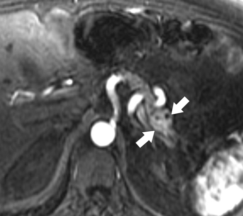
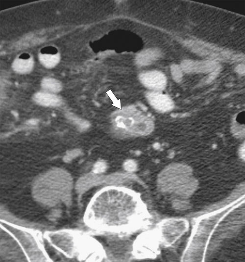
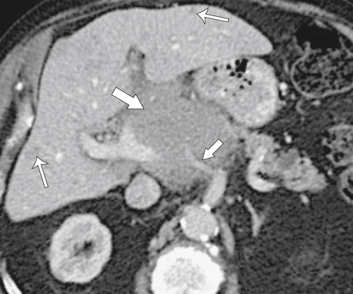
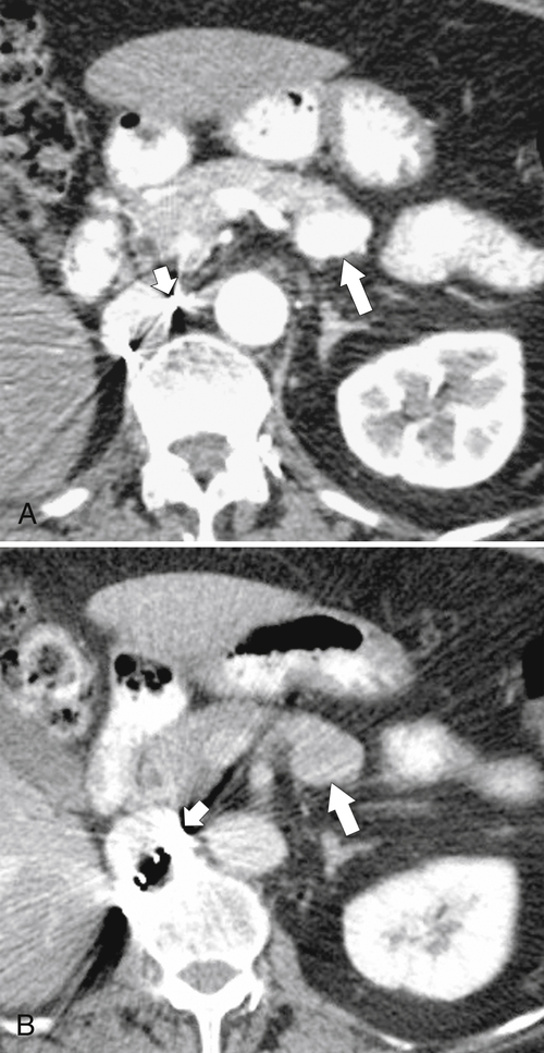
Pancreas

Published on 10/05/2015 by admin

Filed under Gastroenterology and Hepatology

Last modified 10/05/2015

Print this page

rate 1 star rate 2 star rate 3 star rate 4 star rate 5 star
Your rating: none, Average: 0 (0 votes)

This article have been viewed 10318 times

). The midgut (which includes the duodenum) then rotates clockwise, and the ventral bud rotates along with it and fuses with the dorsal pancreatic bud from behind. The fused pancreas now lies completely to the left of the duodenum. The dorsal duct (duct of Santorini) from the original dorsal bud now drains the tail, body, and upper head and for a while drains into the minor papilla. The ventral duct (from the original ventral bud), also known as duct of Wirsung, drains the lower head and uncinate into the major papilla along with the common bile duct. Normally, the ventral and proximal dorsal ducts fuse to form the main pancreatic duct, which drains into the major papilla (papilla of Vater). The remaining distal remnant of the dorsal duct usually regresses and drains into the accessory duct of Santorini.
image
Figure 9-1 Schematic representation of embryological development of the pancreas. The ventral and dorsal buds are originally separate (A). The ventral pancreas then rotates clockwise to fuse with the dorsal pancreas (B and C).

Annular Pancreas

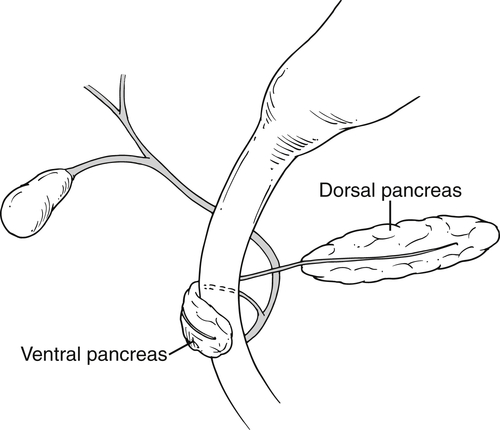
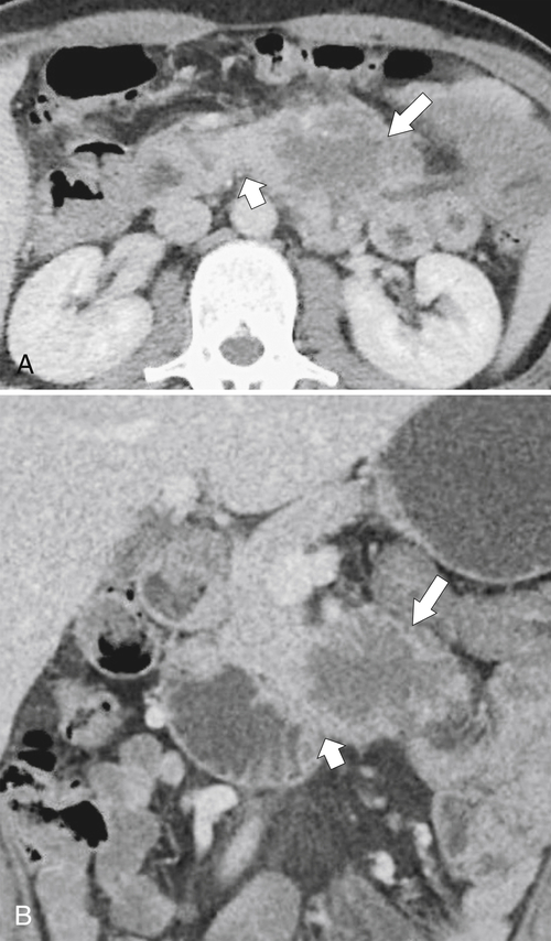
Annular pancreas is a rare anomaly resulting from abnormal or failed rotation of the ventral bud and/or rotation of the duodenum embryologically (Fig. 9-2) such that the bud fails to achieve its position to the left of the duodenum and instead forms a partial or complete ring of pancreatic tissue around the second part of the duodenum. It can also result when the bifid ventral ducts encircle the duodenum rather than fuse together. Many patients are asymptomatic, but annular pancreas can present early in childhood because it is commonly associated with other congenital atretic anomalies, including those of the esophagus, duodenum, and anus. Furthermore, the resulting ring can cause duodenal strictures, which if severe can be recognized in the neonate because of copious vomiting from duodenal obstruction, often requiring surgery to remove the stricture. Presentation in adults is usually after repeated episodes of epigastric pain, early satiety, and vomiting from subacute duodenal obstruction.
image
Figure 9-2 Schematic representation of an annular pancreas. The ventral pancreas rotates abnormally to variable degrees and either partially or whole envelops the second part of the duodenum.
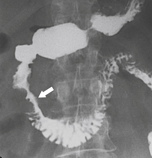
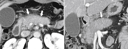
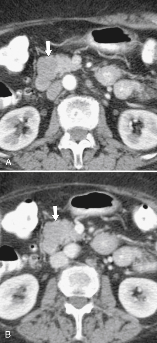
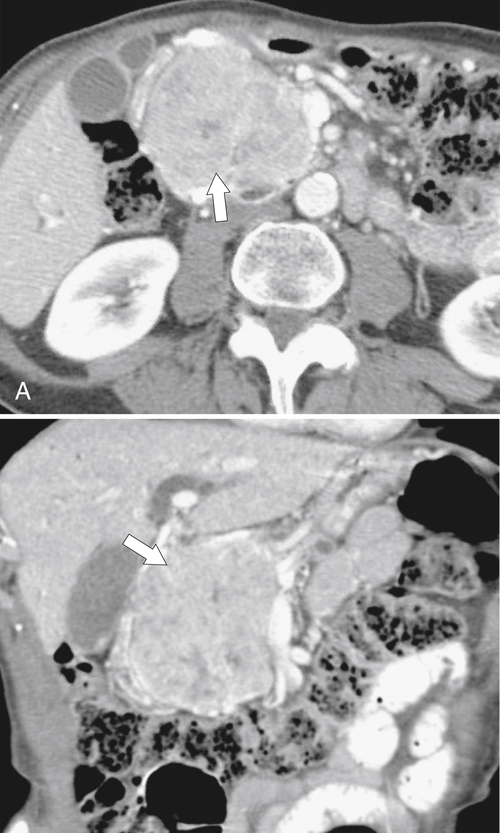
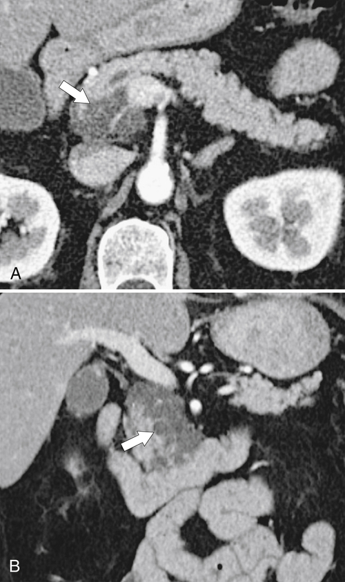
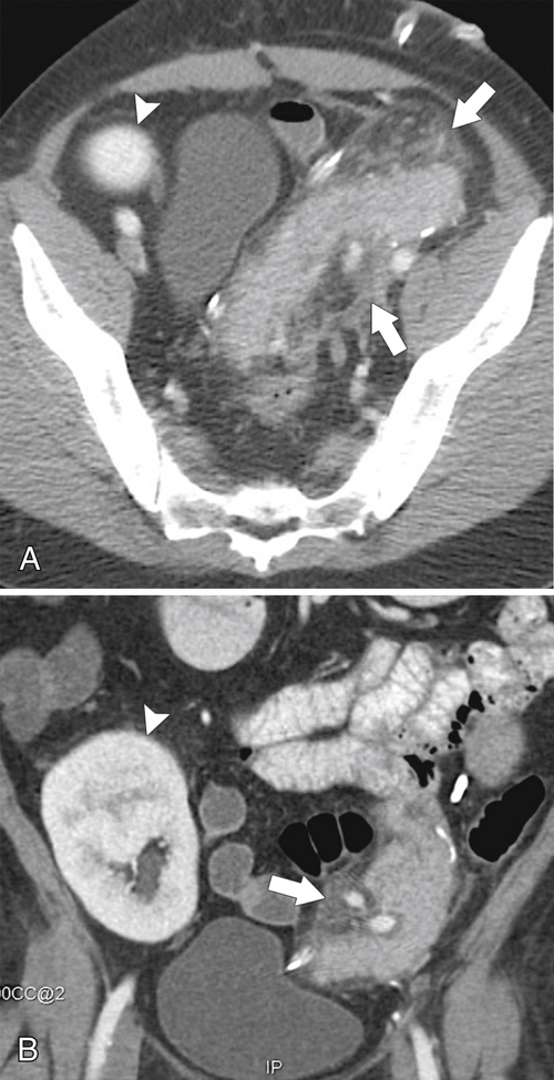
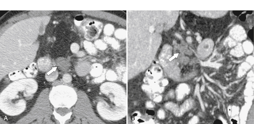
The diagnosis was formerly made with an upper gastrointestinal (GI) series, in which the second part of the duodenum appears strictured to a variable degree depending on the degree of the anomaly and associated with loss of the normal duodenal fold pattern (Fig. 9-3). Depending on the degree of stricture formation, the proximal duodenum may be distended or dilated. However, annular pancreas is now detected primarily by CT because of its increased use. The CT findings are characteristic of pancreatic tissue passing to the right and encircling the second part of the duodenum, which is sometimes better visualized on multiplanar imaging (Fig. 9-4). The diagnosis can also be made with ERCP that shows the ventral duct (Wirsung) completely encircling the duodenum. Annular pancreas is associated with an increased incidence of pancreatitis and peptic ulcer disease, both of which may be identified by CT.
image
Figure 9-3 Upper GI series in a 44-year-old woman with narrowing of the second part of the duodenum (arrow) caused by an annular pancreas.
image
Figure 9-4 Axial (A) and coronal (B) CT in a 71-year-old man with an annular pancreas. A cuff of pancreatic tissue surrounds the second part of the duodenum (arrows).

Agenesis of the Dorsal Pancreas

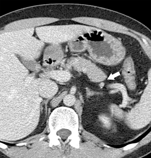
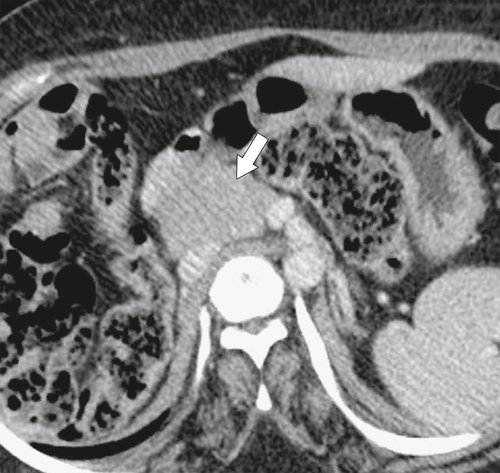
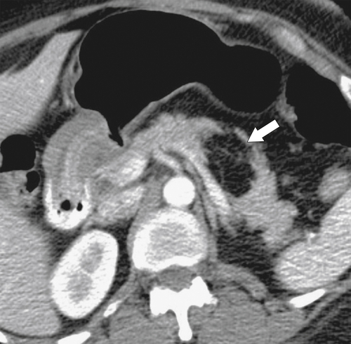
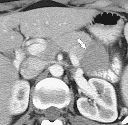
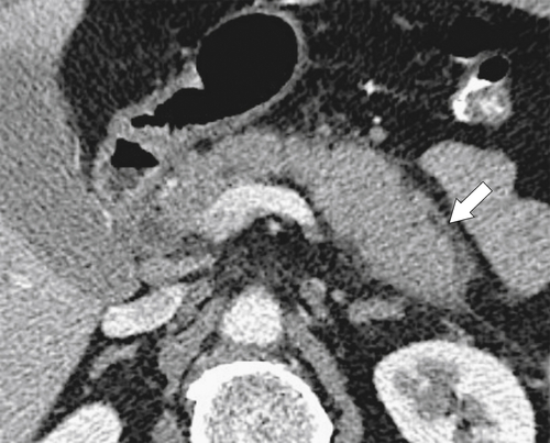
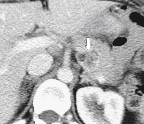
Complete pancreatic agenesis, defined as absence of the neck, body, tail, duct of Santorini, and minor papilla, is extremely rare, particularly because many with this condition are stillborn or die of multiple other congenital anomalies as neonates. More commonly the agenesis is partial, with the neck, body, or tail missing (Figs. 9-5 and 9-6). Many patients are asymptomatic, but if present, symptoms include abdominal pain and an increased incidence of pancreatitis. Depending on the degree of agenesis, patients can also present with pancreatic insufficiency (diabetes mellitus and steatorrhea).
image
Figure 9-5 Axial contrast-enhanced CT in a 38-year-old woman with a congenitally short pancreas and missing tail (arrow).
image
Figure 9-6 Axial contrast-enhanced CT in a 41-year-old woman with a pancreatic uncinate (arrow) but no body or tail because of partial agenesis.
On imaging, pancreatic agenesis can be identified by a short pancreas, but the findings may be subtle. ERCP or MCRP, which will identify a ventral (Wirsung) duct but little or no accessory (Santorini) duct, confirms the diagnosis.

Pancreas Divisum

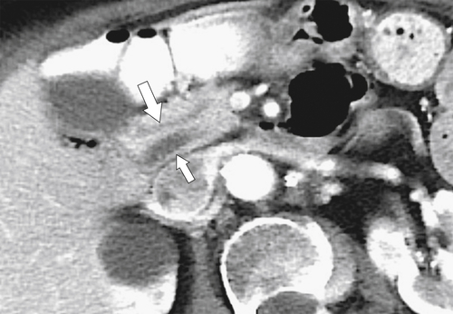
Pancreas divisum is far more common than other pancreatic anomalies, with up to 10% of the population showing some variant, depending on the degree of ductal fusion anomalies. Essentially, despite normal rotation of the ventral bud, its duct (Wirsung) fails to fuse appropriately with the dorsal duct (Santorini), so a single pancreatic duct is not formed (Fig. 9-7). With complete lack of fusion, the dorsal duct now drains exclusively into the minor papilla, which remains patent, and the body and upper pancreatic head drain through the minor papilla. The ventral duct continues to drain into the major papilla, draining the uncinate and lower half of the pancreatic head.
image
Figure 9-7 Axial and coronal contrast-enhanced CT in a 76-year-old man with pancreas divisum. The minor pancreatic duct (Santorini; large arrow) remains anterior to the bile duct and major pancreatic duct (Wirsung; small arrow).
Many patients are asymptomatic, but others have intermittent pain, which usually appears between 30 and 50 years of age and results from mild and occasionally severe pancreatitis, possibly because of the restricted drainage of the bulk of pancreatic fluid through the smaller minor papilla. Therefore pancreatitis, when present, is usually of the neck, body, and tail, although pancreatitis of the uncinate caused by reflux of bile into a short ventral duct can also occur.
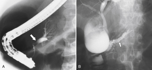
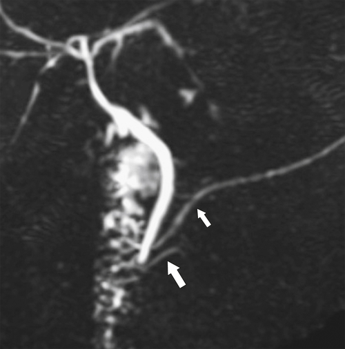
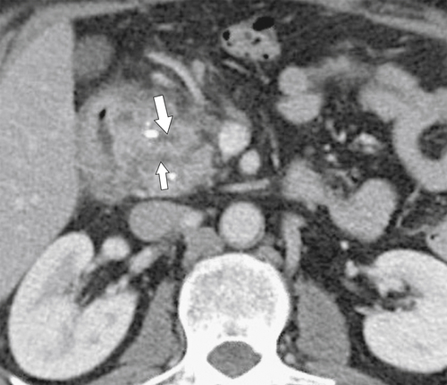
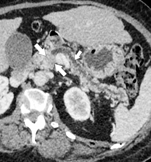
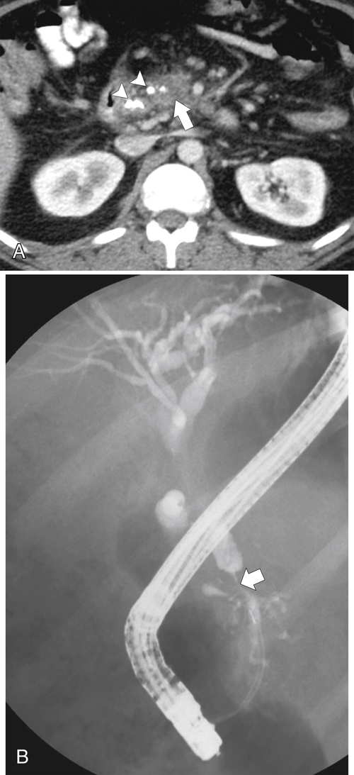
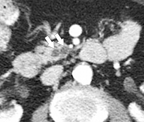
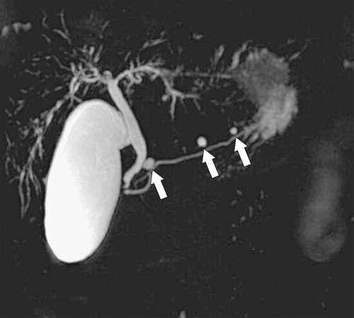
The diagnosis of pancreas divisum can be challenging. At ERCP, injection of contrast material into the major papilla opacifies only the duct of Wirsung (Fig. 9-8). Ideally the minor papilla is also cannulated, but because of its small size, this sometimes fails. Should it prove successful, a long dorsal, noncommunicating duct is outlined. Imaging with MRCP, which demonstrates the anatomical anomaly in most patients noninvasively, may therefore be preferable (Fig. 9-9). Imaging with CT is less sensitive, and the anomaly is often missed unless the pancreatic ductal anatomy is closely scrutinized. The pancreatic head may be enlarged, and sometimes there is a subtle fatty cleft between the uncinate process and the remaining pancreas, but this is rarely observed and nonspecific. With thin-section and multiplanar imaging, the two distinct noncommunicating ducts can be observed (Fig. 9-10). Evidence of pancreatitis, a relatively common feature of pancreas divisum, may also be present (Fig. 9-10).
image
Figure 9-8 A and B, ERCP in a 53-year-old man with initial injection into the major duodenal papilla outlining only the duct of Wirsung (arrow). Subsequent cannulation of the minor papilla fills a dilated duct of Santorini (small arrow) owing to chronic pancreatitis resulting from pancreatic divisum. There are changes of chronic pancreatitis with a dilated irregular duct of Santorini.
image
Figure 9-9 MRCP in a 43-year-old woman demonstrating drainage of the duct of Santorini (small arrow) via the minor papilla duct of Wirsung into the common bile duct and major papilla (large arrow).
image
Figure 9-10 Axial contrast-enhanced CT in a 43-year-old woman with pancreatic divisum and pancreatic head enlargement, peripancreatic inflammation, and calcification caused by an acute flare-up of chronic pancreatitis from pancreatic divisum. The ventral duct (small arrow) is just identifiable separate from the dorsal duct (large arrow).

Secretin Stimulation Test

The secretin stimulation test is designed to determine any functional obstruction of the pancreatic duct. Secretin is a powerful hormonal stimulant for pancreatic bicarbonate production (required for duodenal neutralization of gastric acid). When injected, it causes a temporary increase in the production and therefore volume of pancreatic fluid, which should pass normally into the duodenum. Typically, in the healthy patient, after 0.4 µg/kg is injected, the duct dilates slightly (from a normal 1 to 3 mm to between 4 and 6 mm) but returns rapidly to normal. Conversely, the duct remains dilated for up to 30 minutes in the presence of a functional obstruction. Measurements are made using US or, more commonly, coronal MRCP (Fig. 9-11).
image
Figure 9-11 Secretin MRCP in a 63-year-old woman with papillary dysfunction. The pancreatic duct (small arrow) measured 3 mm at baseline (A), which increased to 5 mm at 5 minutes (B) and returned to 3 mm at 10 minutes (C). The distal duct is tortuous (large arrow) due to previous episodes of pancreatitis.

Ectopic Pancreas

The true incidence of ectopic pancreas is unknown, but some figures put the incidence surprisingly at 10% of the population, although the condition is rarely seen in clinical practice. Ectopic pancreas is also known as pancreatic rest or aberrant pancreas. Small areas of pancreatic tissue come to “rest” in an ectopic position, usually in the distal gastric antrum along the greater curvature or near the pancreatic ampulla in the second portion of the duodenum. They can, however, be detected in more disparate locations, including other intestinal locations and even the pelvis, liver, and spleen.
Patients are usually asymptomatic, but ectopic pancreas can cause gastric or duodenal hemorrhage and, if large enough, biliary obstruction or even small bowel intussusception. Rests that are identified at imaging are usually in the expected location of the stomach or duodenum and found mostly with upper GI series because they are too small to be identified by cross-sectional imaging techniques (although they may be inferred from the presence of a small hypodense and cystic mass). The characteristic appearance is of a 1- to 2-cm submucosal mass, either round or eccentric in shape, sometimes lobular, with a central pit or depression that fills with contrast material in the dependent position.

Fatty Pancreatic Replacement

image
Figure 9-12 Axial noncontrast CT in a 79-year-old woman with an age-related atrophic fatty-replaced pancreas (small arrows) that has tiny punctate calcifications (arrows).

Cystic Fibrosis

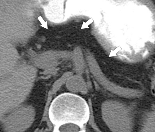
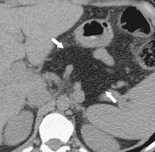
Cystic fibrosis is an autosomal-recessive disease with a number of abdominal complications, including diffuse pancreatic insufficiency when severe. The pancreatic tissue becomes largely fatty replaced, sometimes with small cystic change. On US the fatty change is hyperechoic, but on CT the pancreas is partially replaced by fat (Fig. 9-13) or may not be seen at all, the fatty replacement merging into the surrounding retroperitoneal fat (Fig. 9-14). Other features may include small bowel dilatation and cecal thickening (see discussion in Chapters 4, on the small bowel, and 5, on the colon). The pancreas in cystic fibrosis can also become diffusely calcified or undergo cystosis (see later in the chapter).
image
Figure 9-13 Axial contrast-enhanced CT in a 24-year-old woman with a partially fatty-replaced pancreas (arrow) resulting from cystic fibrosis. The pancreatic head and uncinate are normal (small arrow).
image
Figure 9-14 Axial noncontrast CT in a 39-year-old man with cystic fibrosis and complete pancreatic fatty replacement (arrows).

Uneven Lipomatosis

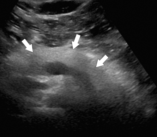
Fatty pancreatic replacement can be either diffuse (Fig. 9-15) or focal, termed “uneven” (Fig. 9-16). It is of little clinical significance, except that it has been associated with pancreas divisum and can be confused with a pancreatic neoplasm. Fat-suppressed MRI can be helpful in distinguishing between the two.
image
Figure 9-15 Transverse US of the pancreas in a 44-year-old woman with diffuse echogenicity (arrows) secondary to a fatty pancreas.
image
Figure 9-16 Axial contrast-enhanced CT in a 65-year-old man demonstrating a nondeforming hypodense anterior pancreatic head (arrows) consistent with uneven lipomatosis.

Pancreatic Lipomatous Pseudohypertrophy

Pancreatic lipomatous pseudohypertrophy is of unknown but possibly congenital origin, causes pancreatic hypertrophy with focal or diffuse fatty replacement, and is readily identified with CT or MRI (Fig. 9-17). It is, however, often associated with chronic liver disease (i.e., cirrhosis), which may therefore be contributory. It is also associated with cystic fibrosis but should be distinguished from diffuse pancreatic fatty replacement, which is also seen in cystic fibrosis but shows no pancreatic hypertrophy.
image
Figure 9-17 Axial noncontrast CT in a 28-year-old woman with cystic fibrosis and diffusely enlarged fatty pancreas (arrows) caused by pancreatic lipomatous pseudohypertrophy.

Pancreatic Lipoma

Lipomas are very rare in the pancreas, being much more common in the intestinal tract. In the pancreas, as elsewhere, lipomas are relatively straightforward to diagnose with CT because of their fat content, which is confined to the pancreas and is well circumscribed (Fig. 9-18).
image
Figure 9-18 Axial contrast-enhanced CT in a 44-year-old woman with a 4-cm pancreatic hypodense fatty mass (arrow) that represents a pancreatic lipoma.

Pancreatitis

Pancreatitis represents acute or chronic inflammation of the pancreas caused by release of pancreatic enzymes, mainly trypsin, into the pancreatic parenchyma rather than the duodenum. Pancreatitis has numerous precipitating factors (Box 9-1), but gallstones cause the majority of cases of acute pancreatitis, whereas alcohol is mainly responsible for chronic pancreatitis. The diagnosis is based on the clinical history, elevated serum amylase and lipase levels, and imaging. However, clinical symptoms and presentation vary widely and range from mild to severe. Patients with milder forms present with pain, vomiting, and abdominal tenderness, whereas those with severe forms present with shock, organ failure, and hemorrhage. The severe hemorrhagic clinical signs have been well recognized for more than a century. Grey Turner sign is flank bruising, and Cullen sign is periumbilical bruising, resulting from acute pancreatitis. Clinical severity is ranked by either Ranson or APACHE II criteria.
 
Box 9-1   Causes of Pancreatitis
Gallstones
Alcohol
Hyperparathyroidism
Hypercalcemia
Hypertriglyceridemia
Hypothermia
Pregnancy
Pancreas divisum
Viral infections: mumps
Pancreatic duct stones
Trauma
Postpump pancreatitis
Post-ERCP
Drugs: thiazide diuretics, sulfonamides, AIDS medication, valproic acid, azathioprine, statins
Hereditary pancreatitis (trypsinogen activation)
Porphyria
AIDS, Acquired immunodeficiency syndrome; ERCP, endoscopic retrograde cholangiopancreatography.
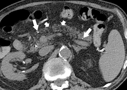
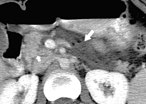
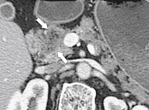
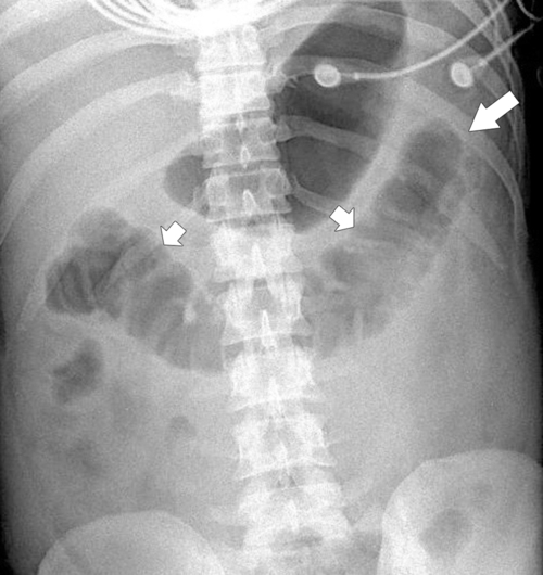
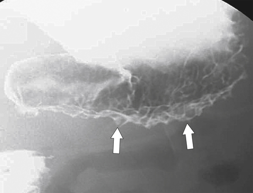
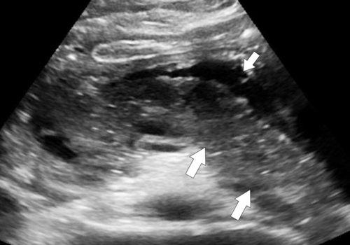
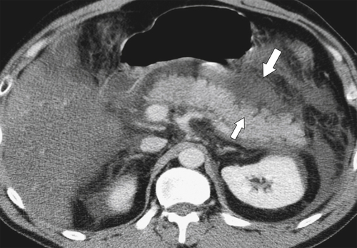
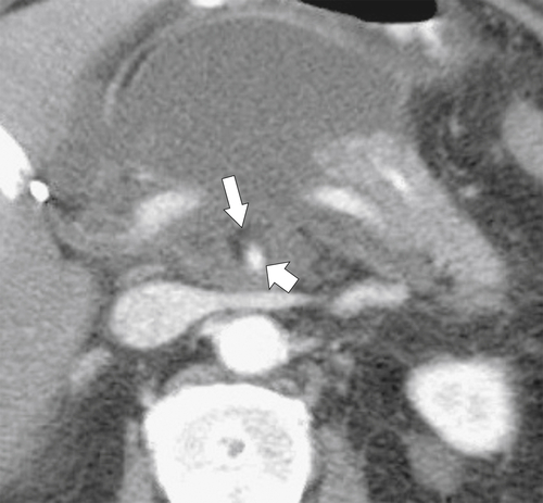
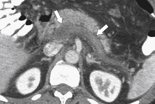
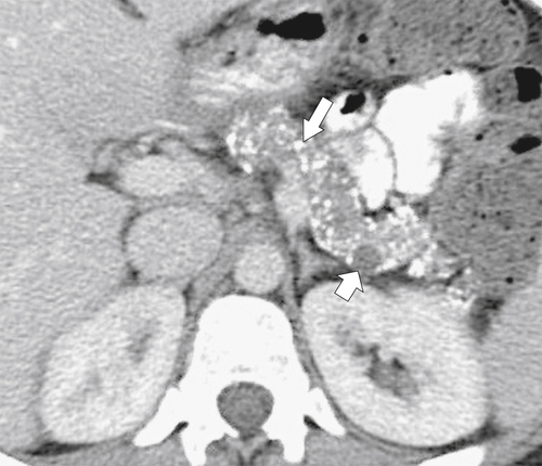
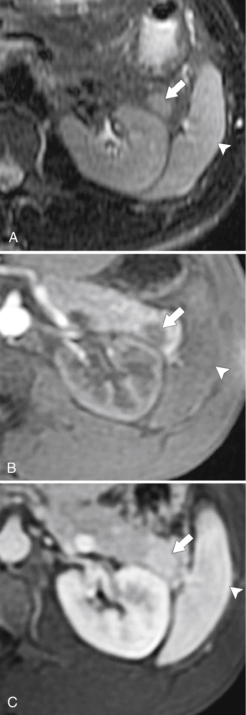
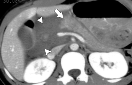
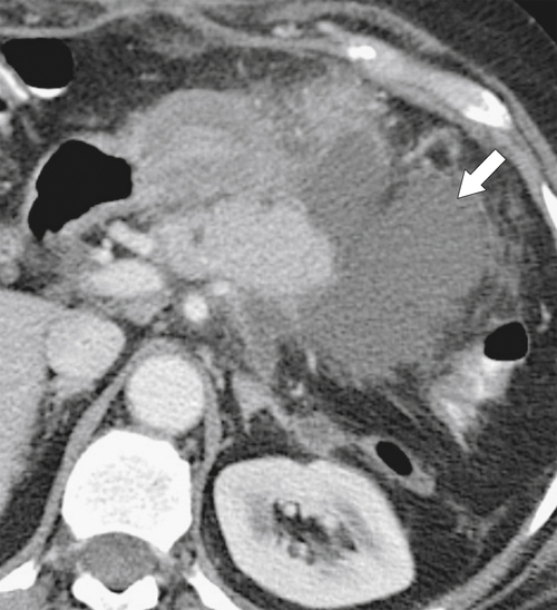
Many patients with pancreatitis have no imaging abnormalities. When pancreatitis is more severe, however, plain radiographs may demonstrate ileus, particularly a sentinel loop, representing a distended loop of small bowel immediately adjacent to the inflamed pancreas. A colon “cut-off” sign represents a distended transverse colon, with paucity of air from the splenic flexure distally because of colonic spasm (Fig. 9-19). Upper GI studies often demonstrate thickened and spiculated duodenal and gastric folds representing secondary edematous transmural enteric changes (Fig. 9-20). On US the pancreas can appear enlarged with a heterogeneous echo texture mixed with prominent hypoechoic regions (Fig. 9-21), although contrast-enhanced CT is usually performed to evaluate pancreatitis. Not all patients with clinical and biochemical pancreatitis have abnormal CT findings, although the majority of patients show abnormalities to a variable degree. The pancreas is typically enlarged, sometimes focally, with effacement of the normal indented pancreatic border combined with peripancreatic edematous changes (Fig. 9-22). The latter can be severe with widespread edema throughout the retroperitoneal space, producing single or multiple acute fluid collections (Fig. 9-23). Sometimes pancreatic adenocarcinoma has a similar appearance, and a useful CT sign is preservation of perivascular fat planes (unlike pancreatic adenocarcinoma, in which they are often obliterated), which strongly favors a benign diagnosis (Fig. 9-24). The reverse is not always true in that the peripancreatic inflammation in severe pancreatitis can sometimes obliterate this fat plane. Unless contraindicated, contrast-enhanced CT is administered to evaluate the vascular viability of the pancreatic parenchyma. Pancreatic necrosis is recognized by single or multiple focal hypodensities (or diffuse depending on the severity) surrounded by normally enhancing pancreas (Fig. 9-25). These hypodensities represent the hypovascularized or nonvascularized pancreatic parenchyma, which has been destroyed by the acute inflammatory process. Depending on the degree of pancreatic necrosis, it may be termed necrotizing pancreatitis if more than 50% of the gland is involved.
image
Figure 9-19 CT scout view in a 43-year-old man with distended transverse colon (small arrows) and a colon cut-off sign (arrow).
image
Figure 9-20 Upper GI series in a 66-year-old man with a “puckered” appearance to the greater curvature of the stomach (arrows) caused by inflammation from pancreatitis.
image
Figure 9-21 Transverse US in a 17-year-old male with acute pancreatitis, demonstrating an edematous, heterogeneously enlarged pancreas (arrows) with anterior peripancreatic fluid (small arrow).
image
Figure 9-22 Axial contrast-enhanced CT in a 37-year-old man with hypertriglyceridemia-induced pancreatitis. The liver is markedly fatty, and pancreatic enlargement without necrosis (small arrow) and diffuse peripancreatic inflammation (large arrow) are present.
image
Figure 9-23 Axial contrast-enhanced CT in a 27-year-old man with acute pancreatitis demonstrating evolution of acute pancreatitis. A, Enlarged and inflamed pancreas (arrows) with diffuse peripancreatic inflammation (small arrows). B, Axial contrast-enhanced CT 1 month later demonstrating partial necrosis and partial loss of pancreatic tail (small arrow) and an organizing peripancreatic collection (arrows). C, Axial contrast-enhanced CT 2 months after B, demonstrating pancreatic body and tail of formation of a capsule (arrow) around the peripancreatic collection as a result of pseudocyst formation.
image
Figure 9-24 Axial contrast-enhanced CT in a 38-year-old man with acute pancreatitis and preservation of the fat plane (large arrow) surrounding the superior mesenteric artery (small arrow).
image
Figure 9-25 Transverse US (A) and axial (B) and coronal (C) contrast-enhanced CT in an 80-year-old man with acute pancreatitis. The pancreas is enlarged (arrows) and markedly heterogeneous at US with an anterior peripancreatic acute fluid collection (small arrows) confirmed by CT. CT also demonstrates almost complete pancreatic necrosis (arrowheads), except for the pancreatic tail (thin arrows), and a large peripancreatic acute fluid collection (small arrows).
Documentation of pancreatic necrosis is critical because it places the patient in a much higher risk category than those with simple fluid collections. Necrotic areas can also become infected, which has an even higher mortality rate. Surgical debridement is the treatment of choice. Necrosis is usually associated with pancreatic and peripancreatic fluid collections (Figs. 9-23 through 9-25). There may be evidence of acute fluid collections in the retroperitoneum, which may also become infected and form an abscess. If contrast material is not administered, the pancreas is typically diffusely hypodense. MRI also demonstrates an enlarged gland with areas of necrosis that fail to enhance after IV administration of contrast medium. The terminology used in pancreatitis is defined in Table 9-1.

Table 9-1

Definition of Findings in Pancreatitis

Pancreatic necrosis Single or multiple nonenhancing intrapancreatic regions (necrotizing pancreatitis with >50% necrosis).
Exudative pancreatitis Predominantly peripancreatic fluid collections with normally enhancing pancreas. It is synonymous with peripancreatic fat necrosis.
Acute fluid collection Intrahepatic or extrahepatic fluid collections without defined wall in acute phase. Caused by inflammation or peripancreatic fat necrosis. May or may not be infected.
Pseudocyst Fluid collection with defined fibrous capsule; develops 4-6 weeks after acute event. May or may not be infected.
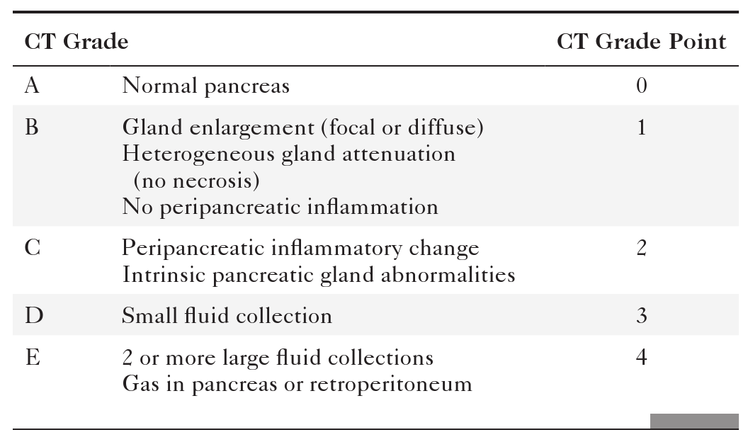
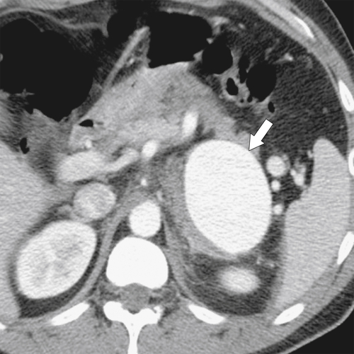
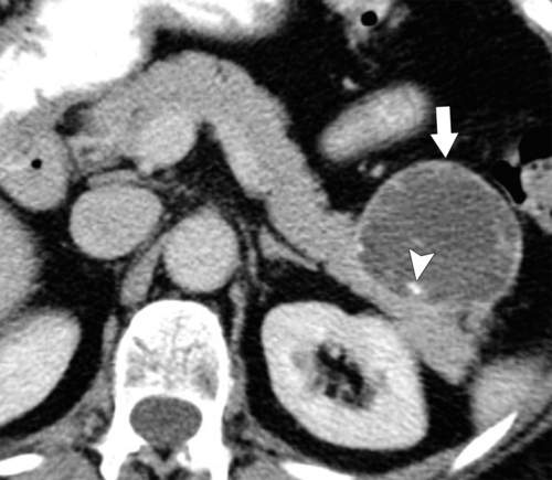
The severity of acute pancreatitis can be staged by contrast-enhanced CT (Table 9-2) and corresponds reasonably closely to the clinical course. When the percentage of gland necrosis is also included (0 to 30%, 30% to 50%, >50%), a CT severity index can be calculated, which corresponds even more closely to the clinical course (Table 9-3). However, the CT severity index does not always match the clinical course, and therefore its use in clinical practice is variable. The most important CT finding is pancreatic necrosis, which markedly increases morbidity and mortality rates associated with disease. Acute hemorrhagic pancreatitis is a particularly severe form of pancreatitis with, as its name suggests, hemorrhage within the pancreas (which might not be recognized if IV contrast medium has been administered) along with diffuse pancreatic inflammation, enlargement, and necrosis. Splenic and portal vein thromboses (Fig. 9-26) and aneurysm formation, particularly of the splenic artery (Fig. 9-27), are other recognized complications of severe pancreatitis.

Table 9-2

CT Staging of Pancreatitis

image

Table 9-3

CT Severity Index

Index Point Necrosis
0 None
2 0-30%
4 30%-50%
6 >50%
image
Figure 9-26 Axial contrast-enhanced CT in a 53-year-old woman with complete thrombosis of the splenic and superior mesenteric veins (arrows) secondary to acute pancreatitis.
image
Figure 9-27 Axial contrast-enhanced CT in a 44-year-old woman with a 6-cm hyperenhancing left upper quadrant mass due to a splenic artery aneurysm (arrow) as a complication of acute pancreatitis.
Mild or moderate pancreatitis usually involves pancreatic inflammation and enlargement that may or may not be focal (grades B and C). With more severe forms, peripancreatic fluid collections develop acutely, representing pancreatic fluid collections. Unlike chronic fluid collections (pseudocyst), they have no encapsulation or wall (Fig. 9-23). These collections can also occur within the pancreas or in other retroperitoneal locations. Many resolve spontaneously, but approximately 50% evolve into pseudocysts (see below).

Pancreatic Pseudocyst

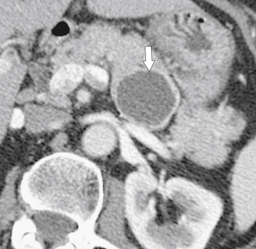
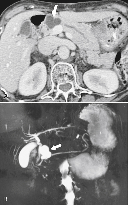
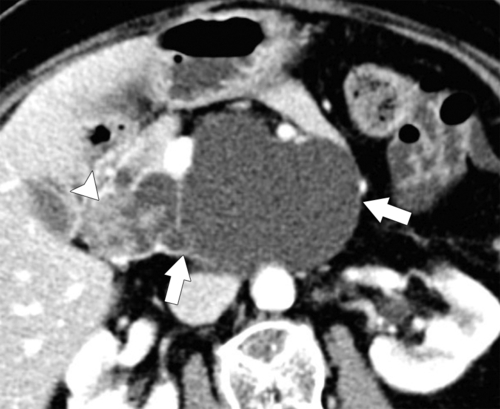
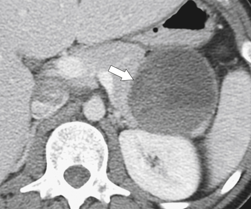
Pancreatic pseudocyst is a complication of pancreatitis (usually acute but also chronic) and is defined as a well-circumscribed, rounded, and fibrous encapsulated pancreatic or peripancreatic fluid collection, rich in blood, necrotic material, and pancreatic enzymes. A pancreatic pseudocyst can be hard to differentiate from acute fluid collections, so the diagnosis is usually made by temporally relating the finding to the onset of symptoms. There is no radiological definition as to when an acute fluid collection becomes a pseudocyst, although the surgical definition is 6 weeks from the onset of symptoms. If ERCP is performed, a connection with the pancreatic duct is seen in up to 50% of patients.
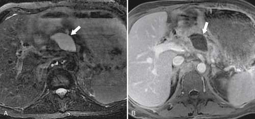
Patients often have upper abdominal pain (radiating to the back) and, because pseudocysts can be quite large, a palpable mass. About 50% of pseudocysts resolve spontaneously with no treatment. Approximately 20% remain without further complications, but about 30% cause local complications, including erosion or dissection into adjacent organs, hemorrhage (erosion into vessels), aneurysm formation, rupture and peritonitis, and duodenal or biliary obstruction. Pseudocysts can also become infected and develop into pancreatic abscesses. These can be suitably drained percutaneously, unlike infected necrosis, which requires surgical debridement.
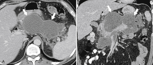
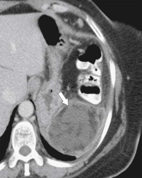
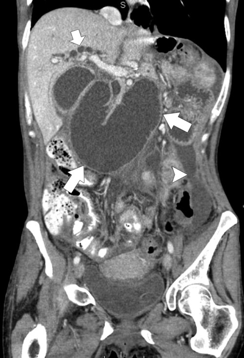
At imaging by CT, a rounded homogeneous hypodense and encapsulated mass is seen (acute fluid collections are not encapsulated) (Fig. 9-28). If the pseudocyst has become secondarily infected (with or without gas) or undergone hemorrhage, the contents of the mass may be heterogeneous. The fibrous rim usually shows fine enhancement on contrast-enhanced CT. MRI features match the CT findings, with hypointense contents on T1-weighted imaging and hyperintense contents on T2-weighted imaging (Fig. 9-29). Less commonly, US is performed, the results of which usually show the cyst filled with complex echoes because of the internal debris and blood.
image
Figure 9-28 Axial (A) and coronal (B) contrast-enhanced CT in a 44-year-old woman with a 10-cm well-contained encapsulated fluid collection (arrows) representing a pancreatic pseudocyst.
image
Figure 9-29 Axial T2-weighted (A) and coronal T1-weighted (B) fat-saturated postcontrast MRI in a 53-year-old man with a 5-cm pancreatic pseudocyst (arrows).

Chronic Pancreatitis

Chronic pancreatitis, as its name suggests, is secondary to repeated bouts of acute pancreatitis, most often in unremitting alcoholics, although many of the causes of acute pancreatitis are also responsible for chronic pancreatitis (Box 9-1). The episodes of pancreatitis range from mild to severe, but the repeated injury to the pancreas ultimately takes its toll with irreversible parenchymal damage. Patients usually admit to episodes of acute pancreatitis and have recurrent or chronic abdominal pain, sometimes jaundice, and signs of pancreatic insufficiency (diabetes mellitus, steatorrhea) with malabsorption and weight loss. Serum amylase and lipase levels are typically elevated, although not usually to the degree seen in acute pancreatitis. The secretin stimulation test by ERCP has high sensitivity for the diagnosis but is not often performed, having been replaced by US or an MRCP technique (Fig. 9-11). Because of pancreatic dysfunction, pancreatic duct bicarbonate production is decreased.
image
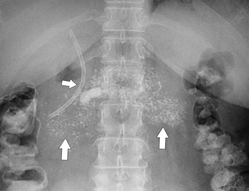
Figure 9-30 Plain abdominal radiograph and axial contrast-enhanced CT in a 47-year-old woman with chronic pancreatitis and diffuse pancreatic calcification (arrows). A plastic biliary stent (small arrow) is present because of an inflammatory biliary stricture.
image
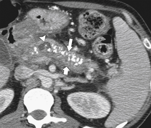
Figure 9-31 Axial contrast-enhanced CT in a 42-year-old man with an acute episode of chronic pancreatitis, demonstrating diffuse pancreatic calcification (large arrow), peripancreatic inflammatory changes (small arrow), and a focally enlarged pancreatic duct (arrowhead). Splenomegaly is caused by chronic splenic vein thrombosis.

image
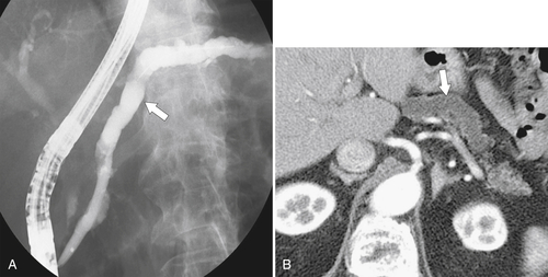
Figure 9-32 ERCP (A) and axial contrast-enhanced CT (B) in a 55-year-old man with a diffusely dilated and irregular pancreatic duct (arrows) from chronic pancreatitis.

Autoimmune Pancreatitis

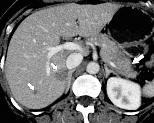
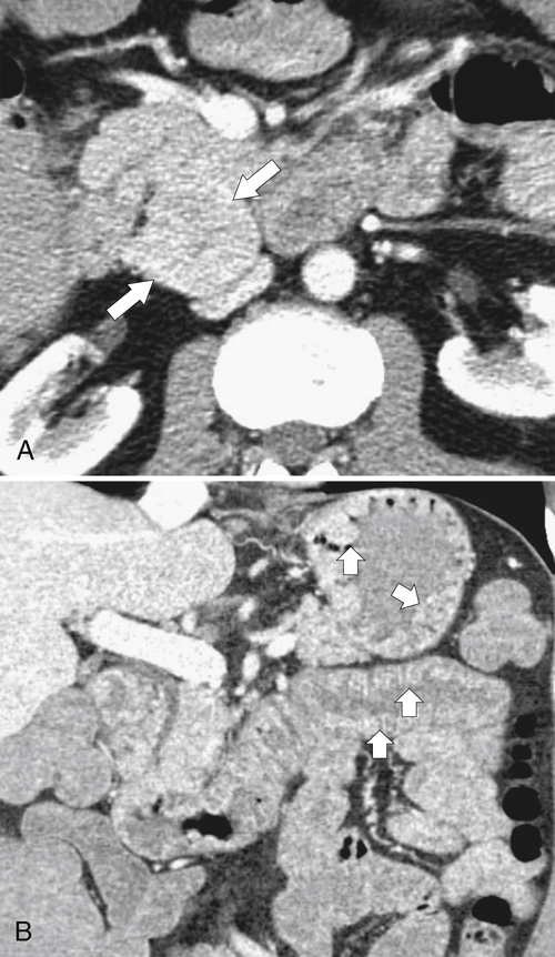
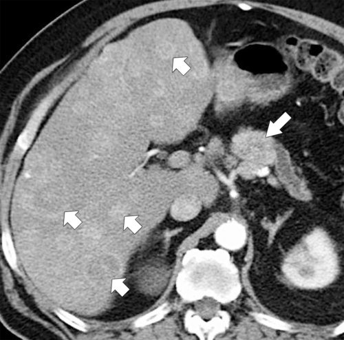
Autoimmune pancreatitis is a poorly understood entity that is also a form of chronic pancreatitis but with no discrete cause. Most patients have elevations of immunoglobulin 4 (IgG4) levels (a specific sign for the disease) and antinuclear antibody levels, which are associated with many other autoimmune diseases, hence the term autoimmune pancreatitis. The pancreas is filled with a lymphoplasmocytic infiltrate. Characteristically, the normal lobulated pancreatic external contour is effaced, producing a more tubular or sausage-shaped outline (Fig. 9-33). Usually the whole gland is affected, but the disease can be focal (Fig. 9-34). After IV contrast administration at CT, capsule-like enhancement that surrounds a low-density band or halo is demonstrated (Fig. 9-35). Peripancreatic adenopathy or inflammatory lesions elsewhere are sometimes observed (Fig. 9-36). The pancreatic duct, if evaluated with ERCP or MRCP, is typically irregularly narrowed and strictured, which may also affect the intrahepatic ducts and resemble primary sclerosing cholangitis.
image
Figure 9-33 Axial (A) and coronal (B) contrast-enhanced CT in a 32-year-old man with a diffusely enlarged pancreas (arrows) and subtle peripancreatic inflammation caused by autoimmune pancreatitis. The pancreas is sometimes described as “sausage” shaped.
image
Figure 9-34 Axial contrast-enhanced CT in a 29-year-old woman with smooth enlargement of the pancreatic tail caused by autoimmune pancreatitis with a sharp margination from the normal pancreas (arrow).
image
Figure 9-35 Axial contrast-enhanced CT in a 62-year-old man with raised IgG4 levels showing a smoothly enlarged body and tail of the pancreas with a peripancreatic halo (arrow) characteristic of autoimmune pancreatitis.
image
Figure 9-36 Axial contrast-enhanced CT in a 46-year-old woman with an enlarged and heterogeneous pancreatic body and tail (large arrow) and hypodense hepatic IgG4 inflammatory pseudotumors (small arrows) concomitant with autoimmune pancreatitis.

Chronic Segmental Pancreatitis (Groove Pancreatitis)

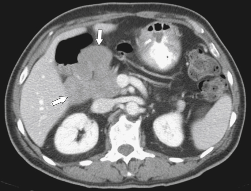
Chronic segmental pancreatitis is a specific form of chronic pancreatitis confined to the pancreaticoduodenal groove (hence its common name “groove pancreatitis”), defined as the area between the head of the pancreas and the second portion of the duodenum. Its exact cause is unknown, but it is associated with excessive and chronic alcohol abuse (similar to chronic pancreatitis), minor papilla strictures (either congenital or caused by neoplasm), and chronic inflammation of the lower common bile duct resulting from prior surgery. Most patients present between 30 and 50 years of age with postprandial pain and vomiting and sometimes with weight loss and mild jaundice. In the correct clinical setting the imaging features at CT are highly suggestive, with separation between the head of the pancreas and the duodenal C-loop because of an intervening hypodense inflammatory mass (or hypointense on T1-weighted MRI) that is often associated with cystic change (Fig. 9-37).
image
Figure 9-37 Axial contrast-enhanced CT in a 47-year-old woman with diffuse inflammation (arrows) in the duodenal pancreatic groove caused by “groove” pancreatitis.

Solid Pancreatic Mass Lesions (Box 9-2)

Pancreatic Adenocarcinoma

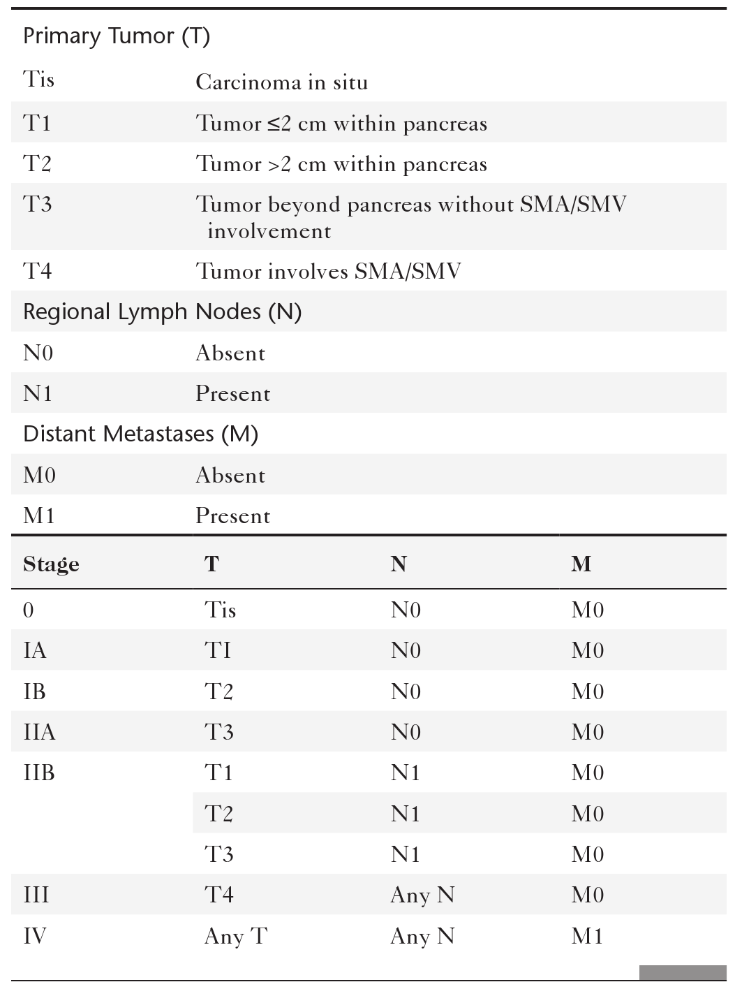
Pancreatic adenocarcinoma is the most common pancreatic neoplasm (90% of malignant pancreatic tumors) and the fourth most common cause of cancer death in the United States. Patients with this type of cancer still have a dismal prognosis (20% 5-year survival rate with surgery and 5% without). The tumor arises from the ductal epithelium of the exocrine pancreas, with two thirds in the head and the remainder in the body and tail. Patients are typically in their seventies and generally present with central abdominal or back pain, weight loss, and jaundice. The Courvoisier sign is painless jaundice in the presence of a distended and palpable gallbladder secondary to biliary obstruction, an ominous sign. Rarer presentations include GI bleeding, diabetes mellitus, and pancreatitis. Pancreatic adenocarcinoma is staged according to the TNM classification (Table 9-4).
 
Box 9-2   Solid Pancreatic Masses
Malignant

Adenocarcinoma
PNET (islet cell, VIPoma, gastrinoma, glucagonoma)
Acinar cell carcinoma
Colloid carcinoma
Anaplastic carcinoma
Giant cell carcinoma
Small cell carcinoma
Pancreaticoblastoma
Lymphoma
Metastases
Sarcoma
Benign

PNET (islet cell, VIPoma, gastrinoma, glucagonoma)
SPEN
Intrapancreatic spleen
Lipoma
PNET, Pancreatic neuroendocrine tumor; SPEN, solid and papillary epithelial pancreatic neoplasm of the pancreas.

Table 9-4

TNM Classification of Pancreatic Adenocarcinoma

image

SMA, Superior mesenteric artery; SMV, superior mesenteric vein.

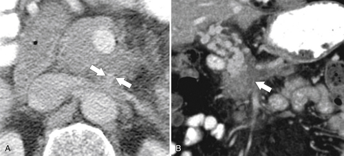
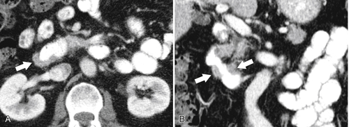
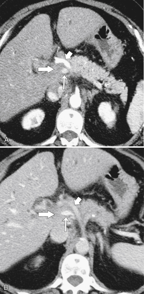
A primary reason for the grim prognosis is that most patients present late, when the tumor is already advanced and inoperable. This is compounded by the difficulty in detecting small lesions at imaging. Lesions less than 2 cm are notoriously difficult to detect, even with a dedicated pancreatic CT protocol. However, contrast-enhanced CT is still the initial imaging investigation of choice. The key to maximizing lesion detection and assessing resectability is pancreatic-phase scanning, which maximizes normal pancreatic parenchymal enhancement while the pancreatic tumor remains relatively unenhanced. This increase in tumor conspicuity is best evaluated with a 40- to 45-second scan delay after the initiation of IV contrast material. At that time, celiac and superior mesenteric arteries are well enhanced, so that local encasement by tumor can also be assessed (Fig. 9-38). Furthermore, sufficient delay for the mesenteric veins to be opacified has usually occurred, so any vascular encasement can be evaluated at the same time. Portal venous–phase scanning at 60 to 70 seconds adds additional diagnostic information about the mesenteric veins and, more important, about the presence of any potential hepatic metastases that were not optimally imaged during the earlier pancreatic-phase scanning.
image
Figure 9-38 Axial pancreatic phase (A) and portal venous phase (B) contrast-enhanced CT in a 70-year-old woman with a pancreatic head adenocarcinoma. On pancreatic-phase imaging, the tumor (large arrows), celiac axis (small arrows), and superior mesenteric vein (thin arrows) are all visualized to better effect.

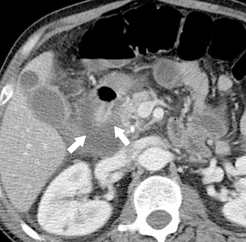
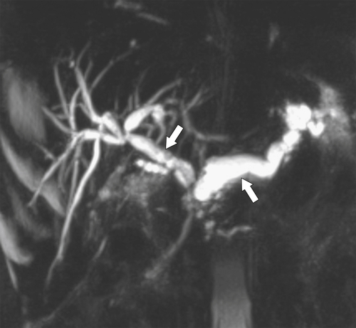
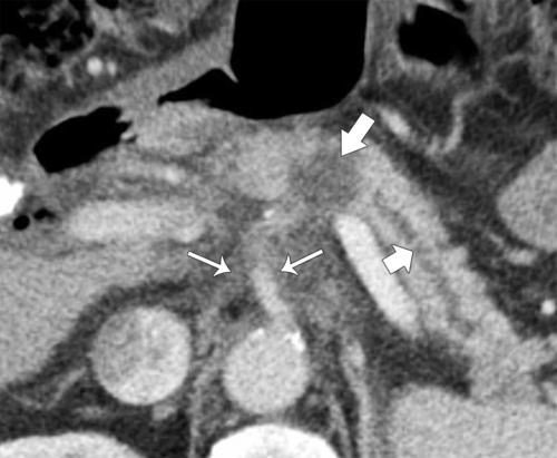
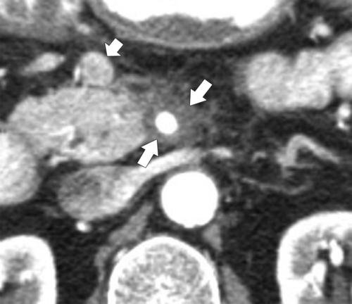
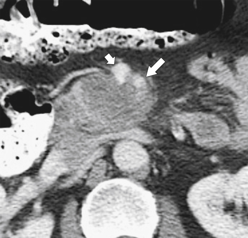
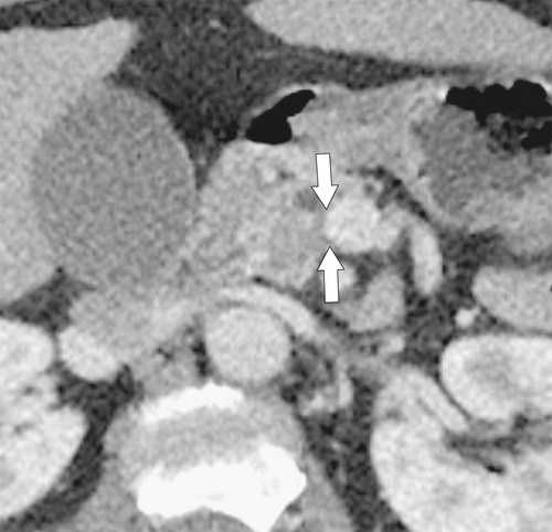
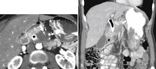
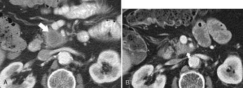
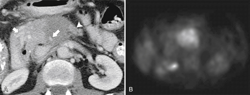
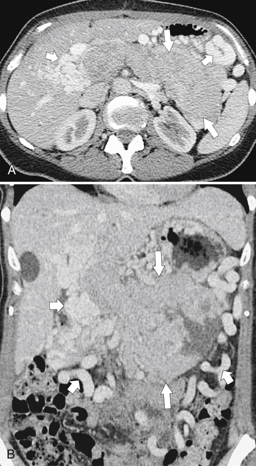
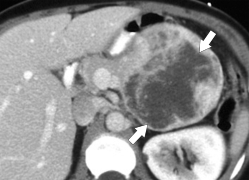
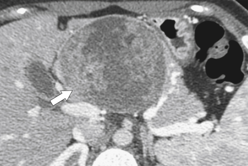
When a tumor is detected at contrast-enhanced CT, there may or may not be mass effect, depending on the size of the tumor. Lack of a pancreatic mass effect should not deter the diagnosis, but larger tumors will inevitably distort the normal pancreatic outline. Tumors are typically hypodense (particularly during the pancreatic phase) because of the central hypovascularity of the tumor (Fig. 9-38). As the tumor enlarges, recognition of hypodensity becomes easier, and large tumors may show frank necrosis (Fig. 9-39). As the mass grows in size, pancreatic duct dilatation becomes more evident, particularly if the tumor is in the head, resulting in upstream ductal dilatation (Fig. 9-40). Smaller tumors in the uncinate may cause little ductal dilatation because of their distance from the main pancreatic duct. Ultimately, the common bile duct may also become obstructed with large pancreatic head tumors (Fig. 9-41), which may progress to a “double-duct” sign when both the pancreatic and biliary ducts are dilated. This is best visualized by ERCP or MRCP (Fig. 9-42). The double-duct sign indicates that the likelihood of a pancreatic adenocarcinoma in the head of the pancreas is very high, even if the tumor is not visualized. Uncommonly, chronic pancreatitis can also cause a double-duct sign, but there should be other signs of chronic pancreatitis at CT and a prolonged clinical history. As the tumor extends beyond the pancreas, the peripancreatic fat becomes invaded or obliterated, which is recognized by increased (sometimes hazy) inflammatory change (i.e., stranding) (Fig. 9-43). Often the tumor causes local pancreatitis, further adding to the peripancreatic inflammation so that it is sometimes difficult to differentiate changes due to tumor from those due to pancreatitis. The local extension into the peripancreatic fat can abut and then surround key vascular structures, particularly the superior mesenteric artery (Fig. 9-44) or vein and sometimes both (Fig. 9-45). The superior mesenteric vein (SMV), however, is often compressed by tumor mass effect as it courses through the pancreas rather than becoming invaded and encased (Fig. 9-46). Multiplanar images can offer better depiction of the relationship of the tumor to the adjacent vessels (Fig. 9-47) and are often used to determine surgical resectability of the mass. Local invasion of splenic, celiac, and mesenteric vessels may preclude operability but will depend on the local surgical expertise. Tumors with more than 50% encasement of the SMV or portal vein (Figs. 9-46 through 9-48) are generally unresectable, although some surgeons are more aggressive and have shown some success with up to 75% circumferential encasement. Splenic vein invasion is not a contraindication to surgery. Celiac axis (celiac or hepatic artery or both) and superior mesenteric artery encasement greater than 25% is generally unresectable (Figs. 9-45 through 9-47). Splenic and gastroduodenal artery encasement is not a contraindication to surgery. Tumors with distant metastases, which are common in pancreatic cancer because of its late presentation, are unresectable. However, it is becoming increasingly apparent that some newer neoadjuvant therapies (e.g., FOLFIRINOX with or without radiation therapy) cause a dramatic reduction in tumor size and residual perivascular soft tissue changes now represent fibrosis rather than active tumor. Radiologists should therefore hesitate before assuming inoperability in these patients (Fig. 9-49). Contrast-enhanced CT has a high predictive value (close to 100%) for unresectability but only 75% to 80% for tumor resectability. Hence, when the CT features suggest unresectability, the radiologist is usually correct. The converse is not true, as a number of tumors deemed resectable by CT turn out not to be. Pancreatic positron emission tomography (PET)/CT does not usually add value to the diagnosis of the primary tumor because of variable fluorodeoxyglucose uptake (Fig. 9-50), but it can sometimes be useful to evaluate extrapancreatic disease, particularly regional adenopathy.
image
Figure 9-39 Axial (A) and coronal (B) contrast-enhanced CT in a 76-year-old man with a large pancreatic adenocarcinoma having a central hypodense necrotic area (large arrows). The tumor also obstructs the duodenum (small arrows).
image
Figure 9-40 Axial reformatted contrast-enhanced CT in a 60-year-old man with a subtle 2-cm uncinate mass (large arrow) that obstructs the pancreatic duct (small arrow).
image
Figure 9-41 Axial contrast-enhanced CT (A) and ERCP (B) in a 53-year-old man with chronic pancreatitis and a new pancreatic head mass caused by adenocarcinoma (large arrow). A stricture is present in the lower bile duct (small arrow), as well as calcification (arrowheads).
image
Figure 9-42 MRCP in a 61-year-old with dilated pancreatic and biliary ducts (double-duct sign) (arrows) resulting from an obstructing pancreatic adenocarcinoma.
image
Figure 9-43 Axial pancreatic-phase contrast-enhanced CT in a 78-year-old man with an adenocarcinoma of the midpancreatic body (large arrow) with upstream pancreatic duct dilatation (small arrow) and peripancreatic extension of tumor. The tumor has extended to encase the celiac axis (thin arrows), rendering the patient inoperable.
image
Figure 9-44 Axial pancreatic-phase contrast-enhanced CT in a 73-year-old woman with soft tissue encasement by pancreatic adenocarcinoma of the superior mesenteric artery (arrows), but the superior mesenteric vein is unaffected (small arrow). This patient was inoperable.
image
Figure 9-45 Axial contrast-enhanced CT in a 71-year-old with pancreatic adenocarcinoma and almost complete encasement of the superior mesenteric artery (large arrow) and approximately 50% of the superior mesenteric vein (small arrow), which has lost its normal ovoid shape. This patient proved inoperable.
image
Figure 9-46 Axial pancreatic-phase contrast-enhanced images in a 66-year-old woman with a 1.5-cm subtle hypodense mass in the junction of the head and uncinate that abuts the superior mesenteric vein between the 6 and 11 o’clock positions with effacement of the normal fat plane (arrows) but preservation of vessel caliber. This patient was operable.
image
Figure 9-47 Axial (A) and coronal (B) contrast-enhanced CT in a 54-year-old man with inoperable pancreatic adenocarcinoma and complete encasement of the superior mesenteric artery (arrows).
image
Figure 9-48 Axial (A) and coronal (B) contrast-enhanced CT in a 63-year-old woman with pancreatic adenocarcinoma showing complete encasement of the superior mesenteric vein (arrows).
image
Figure 9-49 Axial contrast-enhanced CT in a 49-year-old woman with what was initially thought to be inoperable pancreatic cancer by imaging (A) as evidenced by encasement of the superior mesenteric artery (large arrow). After treatment with FOLFIRINOX (B), the mass has significantly reduced in size (small arrow). Even though there is some residual tissue around the superior mesenteric artery, this was proven to be fibrosis and no residual tumor at Whipple operation.

image
Figure 9-50 Axial contrast-enhanced CT (A) and PET (B) in a 63-year-old woman with pancreatic adenocarcinoma. A 5-cm hypodense mass in the body of the pancreas (arrowheads) completely surrounds the superior mesenteric artery (large arrow). There are also superior mesenteric vein occlusion and collateral formation (small arrow). PET shows increased fluorodeoxyglucose uptake in the pancreatic mass (arrowhead).

Further extension of pancreatic tumor may involve local organs, including the stomach, small bowel, spleen, and even the kidneys. More regional extension frequently involves local lymphadenopathy, and further distant metastases often involve the liver and peritoneum and less frequently the lungs.
image
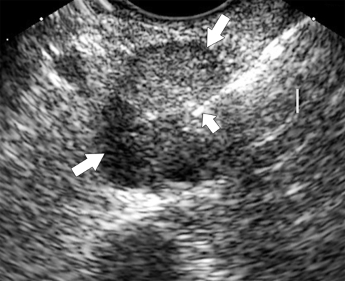
Figure 9-51 Endoscopic US in a 65-year-old woman with an ill-defined 2-cm hypoechoic mass (arrows) in the head of the pancreas that proved to be ductal adenocarcinoma at biopsy (small arrow).

Pancreatic Neuroendocrine Tumor

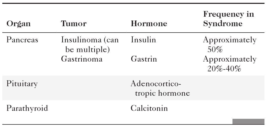
Pancreatic neuroendocrine tumors (PNETs) are also known as islet cell tumors or amine precursor and uptake decarboxylation tumors. They can hypersecrete various hormones (insulin, glucagon, gastrin, and somtatostatin), producing unusual but classic clinical findings. The amine precursor and uptake decarboxylation cells are embryonic and migrate from the neural crest to the pancreas and also to the intestinal tract, thyroid, parathyroid, and adrenal gland. They are classified as either functioning (85% of all islet cell tumors) or nonfunctioning (15%) if no excessive hormones are produced. If hyperfunctioning, they may be part of a multiple endocrine neoplasia syndrome 1 (MEN 1), an autosomal-dominant disease associated with endocrine hyperplasia or neoplasia in disparate organs (Table 9-5). They can be benign or malignant (Table 9-6), although the most common type of tumor, insulinoma, is usually benign.

Table 9-5

MEN 1 (Wermer Syndrome)

image

Paul Wermer (1898-1975), American physician.

Table 9-6

Features of Pancreatic Neuroendocrine Tumors

image

WDHA, Watery diarrhea, hypokalemia, and achlorhydria.

Insulinoma

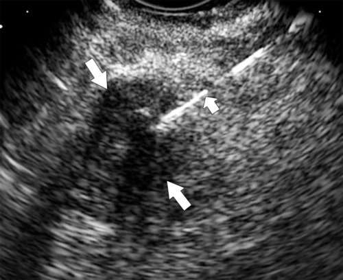
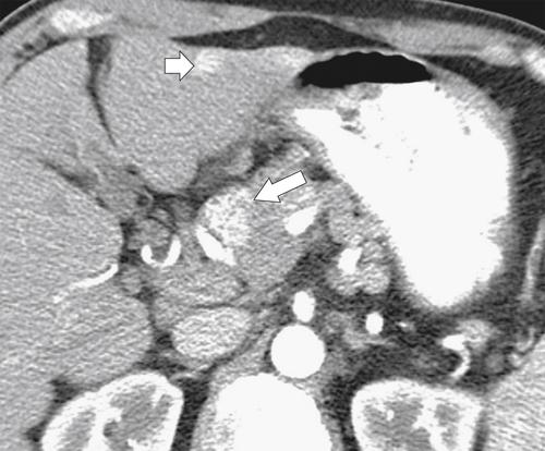
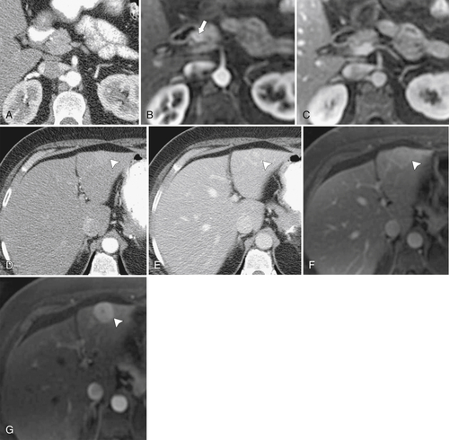
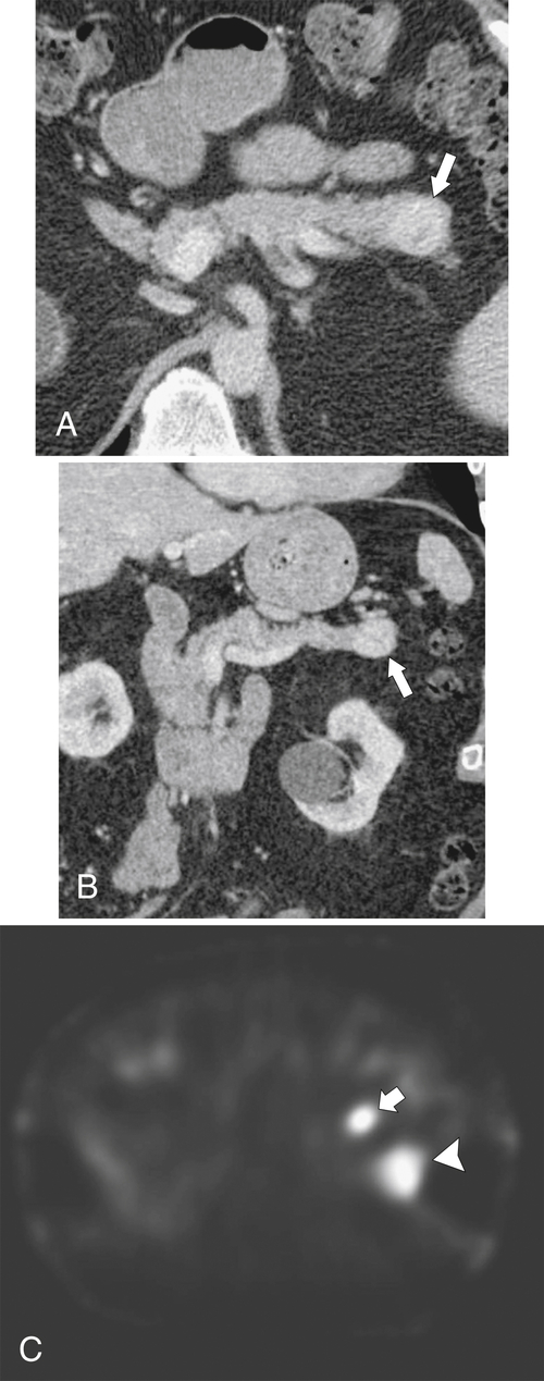
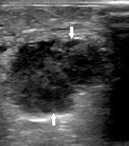
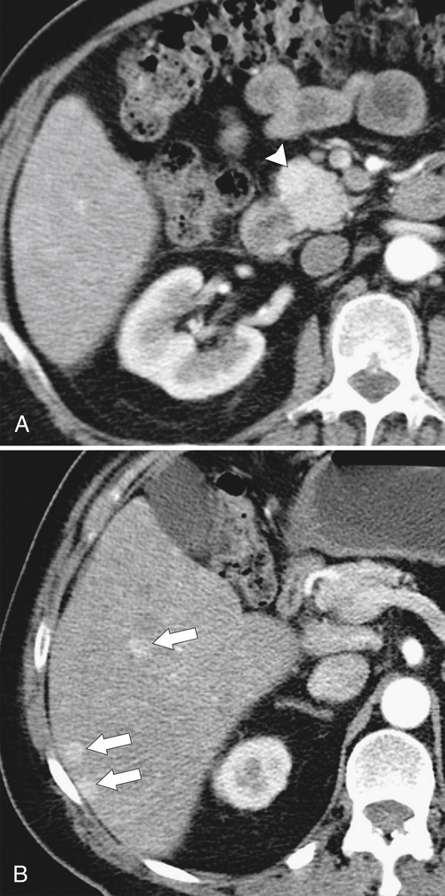
Insulinomas are the most common islet cell tumors that are either functioning, producing insulin from hyperactivity of alpha cells, or nonfunctioning, without significant insulin elevation. Most tumors are small (<2 cm), isolated (70%), benign (90%), and hypervascular (70%). The resulting hyperinsulinemia leads to classic symptoms when florid, but milder forms can go clinically undetected for many years. Typical features are those of hypoglycemia with weakness, confusion, loss of consciousness, and even seizures. Because insulinomas are hypervascular, anyone suspected of having this tumor should undergo dedicated arterial-phase (rather than pancreatic-phase) multidetector CT pancreatic imaging. The lesion, particularly if small (as they frequently are), is often detected only during the arterial phase. Many are missed if the timing of the IV bolus is suboptimal. Classically, optimal CT scanning shows a small, uniform, hyperenhancing mass in the pancreas (Fig. 9-52), which rapidly washes out on portal venous–phase or later imaging, so much so that the sometime-lesion disappears. Dedicated contrast-enhanced MRI has slightly higher accuracy for islet cell tumor detection (especially with fat-saturated techniques) (Fig. 9-53), but if it is still not detected when there is a very high index of suspicion, selective angiography can delineate the hypervascular mass or excessive insulin at pancreatic venous sampling. Alternatively, endoscopic US has proved more accurate than either CT or MRI for the detection of small lesions (Fig. 9-54). Because lesions have high levels of somatostatin receptors, the somatostatin analogue indium-111 pentetreotide (octreotide) can help locate and characterize these pancreatic lesions (Fig. 9-55) as neuroendocrine in origin (insulinoma or otherwise). Ultimately, an exploratory laparotomy with intraoperative US may be required if the index of suspicion remains very high and noninvasive imaging tests fail to identify the mass (Fig. 9-56).
image
Figure 9-52 Axial arterial-phase contrast-enhanced CT in a 54-year-old man with a 2-cm enhancing mass in the pancreatic head (large arrow) that represents a functioning insulinoma. An additional hypervascular liver mass is in segment II of the liver (small arrow).
image
Figure 9-53 Axial arterial-phase CT (A) and T1-weighted fat-saturated arterial phase MRI (B) and portal venous–phase T1-weighted fat-saturated MRI (C) in a 50-year-old woman with a 1.2-cm pancreatic head neuroendocrine tumor (arrow) identified only on arterial-phase MRI (B). A segment II hepatic metastasis (arrowheads) is barely visible on arterial-phase CT (D) and portal venous–phase CT (E) and fat-saturated PVP MRI (F) but is clearly visible on arterial-phase T1-weighted fat-saturated MRI (G).
image
Figure 9-54 Endoscopic US in a 44-year-old woman with a normal CT and a 1.4-cm hypoechoic mass (large arrows) in the pancreatic tail that represents an insulinoma. A biopsy needle is inserted into the mass (small arrow).

image
Figure 9-55 Axial (A) and coronal (B) contrast-enhanced CT and pentetreotide study (C) in a 66-year-old man with a 1.5-cm hypervascular neuroendocrine tumor in the pancreatic tail (large arrows). The lesion is hyperactive on pentetreotide imaging (small arrow). Other activity (arrowhead) is normal in the left kidney.
image
Figure 9-56 

Buy Membership for Gastroenterology and Hepatology Category to continue reading. Learn more here